
Study tip: This chapter follows the KMU learning outcomes in a logical sequence. First understand the anatomy, ischemic mechanism and clinical pattern; then revise the drug logic and final high-yield points.
📖 AIM Learning Material
Coronary Anatomy, Ischemic Heart Disease and Angina
CVS Module — anatomy of coronary circulation, myocardial ischemia, chronic stable angina, coronary vasospasm and the pharmacological basis of antianginal treatment.
Topic Introduction
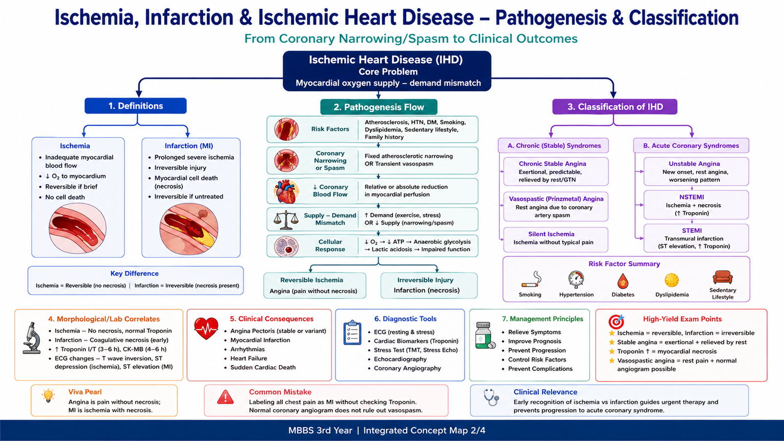
Ischemic heart disease develops when the myocardium receives less oxygenated blood than it needs. The commonest reason is narrowing of the coronary arteries by atherosclerosis. If the shortage is brief, myocardial cells remain alive and the process is called ischemia. If it is severe and prolonged, the cells die and an infarction develops. This chapter first reviews the heart surface, valves and coronary circulation, then explains the main forms of ischemic heart disease and the clinical pattern of chronic stable angina. It also covers vasospastic and microvascular angina and shows how nitrates, β-blockers, calcium channel blockers and ranolazine correct the myocardial oxygen supply–demand mismatch.
A. Heart Surface Anatomy, Valves and Coronary Circulation
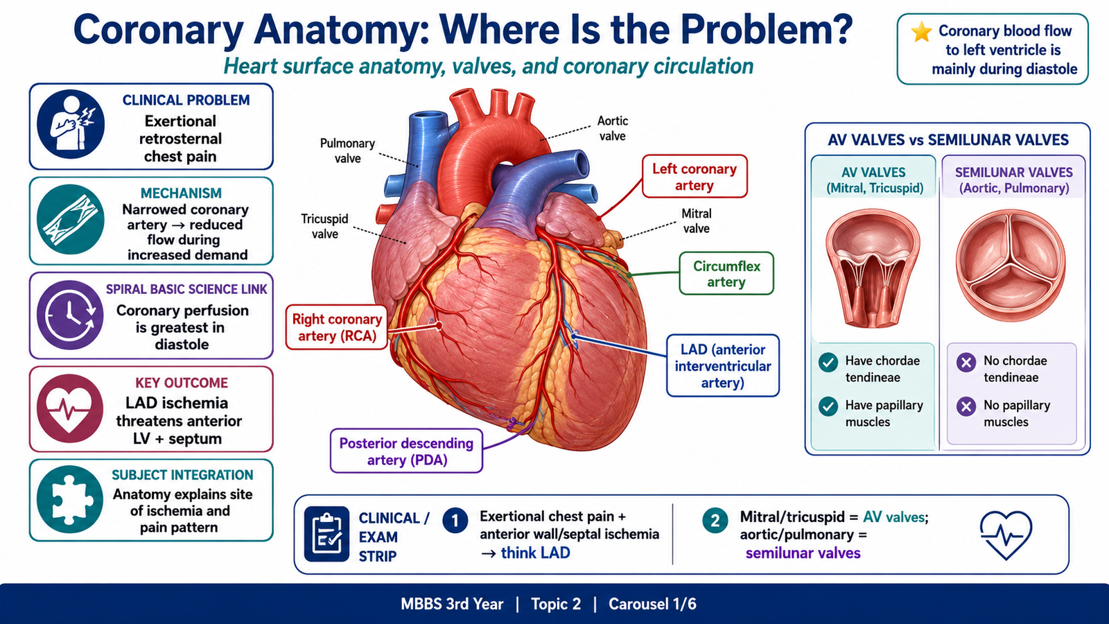
The heart lies in the middle mediastinum behind the sternum. Its apex points downwards and to the left. Anginal pain is usually felt behind the sternum and may radiate to the left arm, shoulder, neck, jaw or epigastrium because cardiac sensory fibres enter spinal cord segments that also receive sensation from these areas.
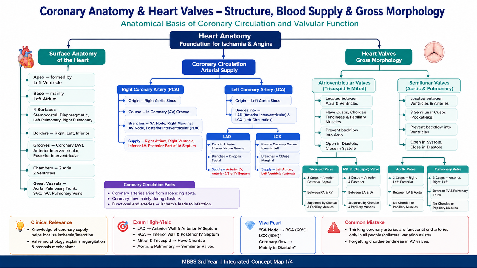
The heart contains four valves that maintain one-way blood flow. The atrioventricular valves have cusps attached to chordae tendineae and papillary muscles, whereas the semilunar valves do not have chordae.
| Valve | Type | Gross morphology |
|---|---|---|
| Tricuspid | Right atrioventricular | Three cusps with chordae tendineae and papillary muscles |
| Mitral | Left atrioventricular | Two cusps with chordae tendineae and papillary muscles |
| Pulmonary | Semilunar | Three semilunar cusps; no chordae tendineae |
| Aortic | Semilunar | Three semilunar cusps; no chordae tendineae |
The right and left coronary arteries arise from the ascending aorta. The left anterior descending artery (LAD) supplies the anterior wall of the left ventricle, the apex and the anterior interventricular septum. The circumflex artery mainly supplies the lateral and, depending on coronary dominance, part of the posterior left ventricular wall. The right coronary artery supplies much of the right atrium and ventricle, commonly the SA and AV nodes, and often gives the posterior descending artery in right-dominant circulation.
Coronary flow to the left ventricle occurs mainly during diastole. During systole, contracting myocardium compresses intramyocardial vessels. When heart rate rises, diastole becomes shorter. A narrowed coronary artery therefore has less time and less capacity to increase blood flow, making exertional ischemia more likely.
B. Ischemia, Infarction and the Main Forms of Ischemic Heart Disease
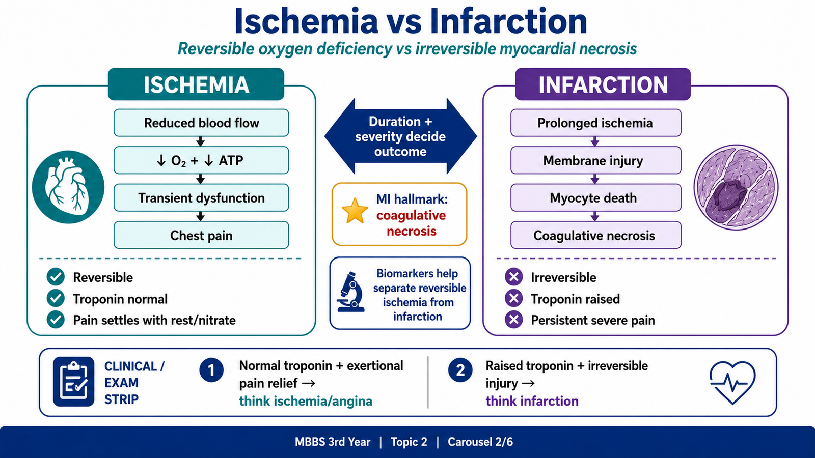
Ischemia is inadequate blood supply to a tissue. In the myocardium, reduced oxygen delivery lowers aerobic ATP production. The cells shift towards anaerobic glycolysis, metabolites such as lactate accumulate, contraction becomes less efficient and chest discomfort may occur. If blood flow returns soon, these changes are potentially reversible.
Infarction is tissue death caused by prolonged ischemia. Persistent ATP depletion, membrane damage and calcium entry produce irreversible injury and coagulative necrosis of cardiac muscle. Troponin enters the blood because necrotic myocardial cells lose membrane integrity.
| Feature | Myocardial ischemia | Myocardial infarction |
|---|---|---|
| Cell injury | Usually reversible if brief | Irreversible necrosis |
| Troponin | Usually normal in stable angina | Raised because cells die |
| Pain pattern | Brief and often relieved by rest or nitrate | Usually longer and may occur at rest |
| Morphology | No permanent necrotic lesion | Coagulative necrosis followed by inflammation and scar |
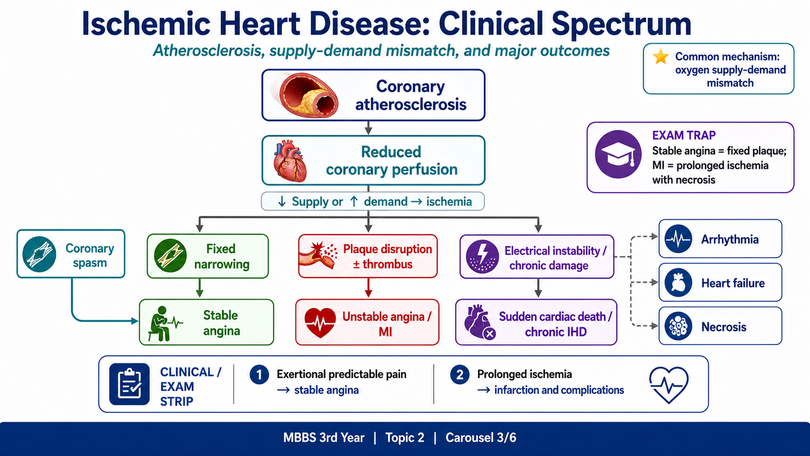
Ischemic heart disease includes several clinical forms:
- Chronic stable angina: predictable exertional ischemia, usually from a fixed atherosclerotic narrowing.
- Unstable angina: new, worsening or rest pain, usually related to plaque disruption and a non-occlusive thrombus.
- Myocardial infarction: prolonged ischemia with myocardial necrosis.
- Sudden cardiac death: often caused by a fatal arrhythmia related to ischemia.
- Chronic ischemic heart disease with heart failure: progressive ventricular dysfunction after repeated or extensive ischemic injury.
- Vasospastic and microvascular angina: ischemia caused by transient epicardial spasm or dysfunction of small coronary vessels.
The usual underlying process is coronary atherosclerosis. A plaque narrows the lumen and reduces coronary flow reserve, meaning that the artery cannot increase flow sufficiently when demand rises. If a plaque ruptures or erodes, platelet activation and thrombosis can suddenly reduce flow further.
When infarction occurs, early gross changes may be subtle. Later the affected myocardium becomes pale or yellow-tan. Microscopically, coagulative necrosis is followed by neutrophils, macrophage removal of dead tissue, granulation tissue and finally a fibrous scar. Major complications include arrhythmia, heart failure, cardiogenic shock, papillary muscle dysfunction, mural thrombosis and myocardial rupture.
C. Chronic Stable Angina: Clinical Features, Assessment and Management
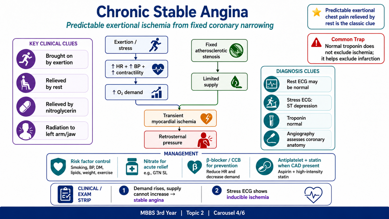
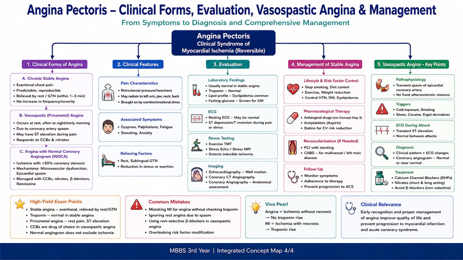
Chronic stable angina is transient myocardial ischemia that usually occurs when a fixed atherosclerotic narrowing prevents coronary blood flow from rising during increased cardiac work. Exertion, emotional stress, cold exposure or a heavy meal increases heart rate, contractility, blood pressure or ventricular wall tension. These changes raise myocardial oxygen demand, but the narrowed artery cannot provide enough additional oxygen.
The pain is typically retrosternal pressure, heaviness, tightness or squeezing. It may radiate to the left arm, shoulder, neck, jaw, back or epigastrium. It usually lasts a few minutes and improves with rest or sublingual nitroglycerin because both measures reduce the oxygen supply–demand mismatch. Troponin remains normal because the ischemia has not caused myocardial necrosis.
Clinical assessment
Diagnosis begins with the history. A predictable relationship with exertion and relief with rest or nitrate strongly supports stable angina. Examination may be normal between attacks, but it should assess blood pressure, pulse, cardiac signs and evidence of associated vascular disease.
- Risk factors: smoking, hypertension, diabetes, dyslipidemia, obesity and family history.
- Resting ECG: may be normal.
- Exercise ECG: may show horizontal or down-sloping ST-segment depression during inducible ischemia.
- Cardiac troponin: normally remains within the reference range in stable angina.
- Echocardiography: assesses ventricular structure, function and possible alternative causes.
- Stress imaging: demonstrates inducible ischemia when appropriate.
- CT coronary angiography or invasive coronary angiography: defines coronary anatomy in selected patients.
Management principles
Management has two linked aims: relieve or prevent anginal attacks and reduce the long-term risk of myocardial infarction and death. Acute discomfort is relieved with a short-acting nitrate. Future attacks are reduced by lowering myocardial oxygen demand or improving coronary supply. Long-term risk is addressed by controlling smoking, blood pressure, diabetes, lipids and body weight, and by using antiplatelet and statin therapy when coronary artery disease is present or strongly suspected. Revascularization is considered when symptoms remain severe or coronary anatomy is high risk.
D. Coronary Vasospasm and Angina with a Normal Coronary Angiogram
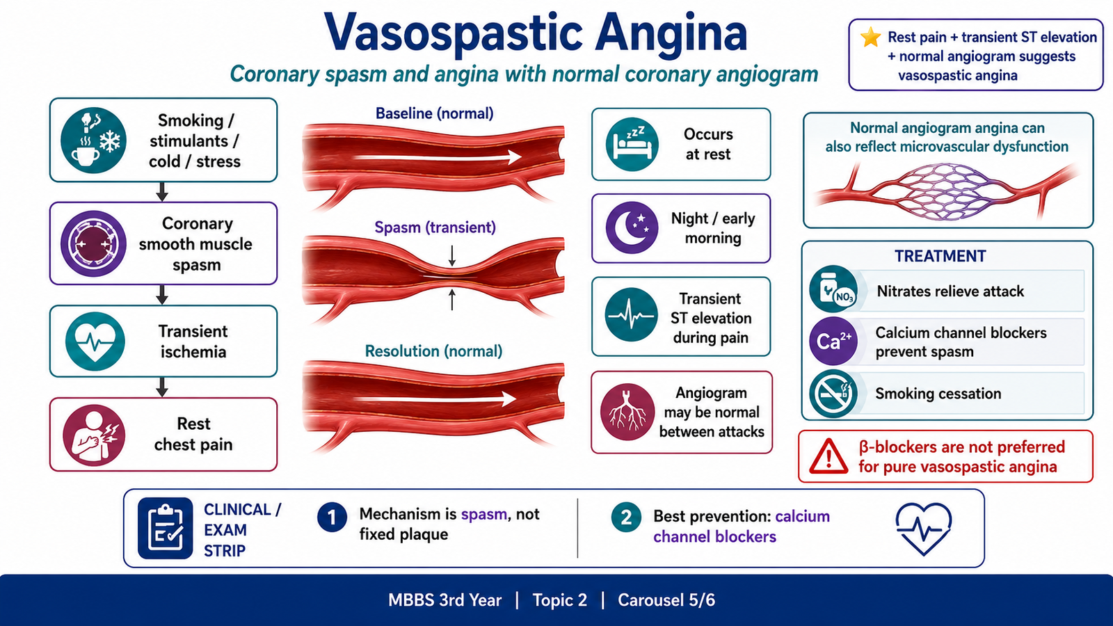
Not all angina is caused by a fixed obstructive plaque. Vasospastic angina, also called variant or Prinzmetal angina, results from temporary contraction of an epicardial coronary artery. The spasm sharply reduces blood flow even when the artery is structurally normal or only mildly diseased.
Attacks commonly occur at rest, often at night or in the early morning, and may show transient ST-segment elevation while pain is present. The angiogram may appear normal between attacks because the spasm has relaxed. Smoking, stimulant drugs, cold exposure, hyperventilation, emotional stress and endothelial dysfunction can promote vasoconstriction.
Some patients have angina despite no important obstruction in the large coronary arteries. In microvascular angina, small coronary vessels fail to dilate normally or undergo spasm. This reduces flow at the tissue level even though the major vessels appear normal on angiography.
Nitrates relieve active coronary spasm, while calcium channel blockers prevent spasm by relaxing vascular smooth muscle. Nonselective β-blockers may worsen pure vasospastic angina because blockade of β-mediated vasodilation can leave α-mediated vasoconstriction relatively unopposed.
E. Organic Nitrates and Calcium Channel Blockers
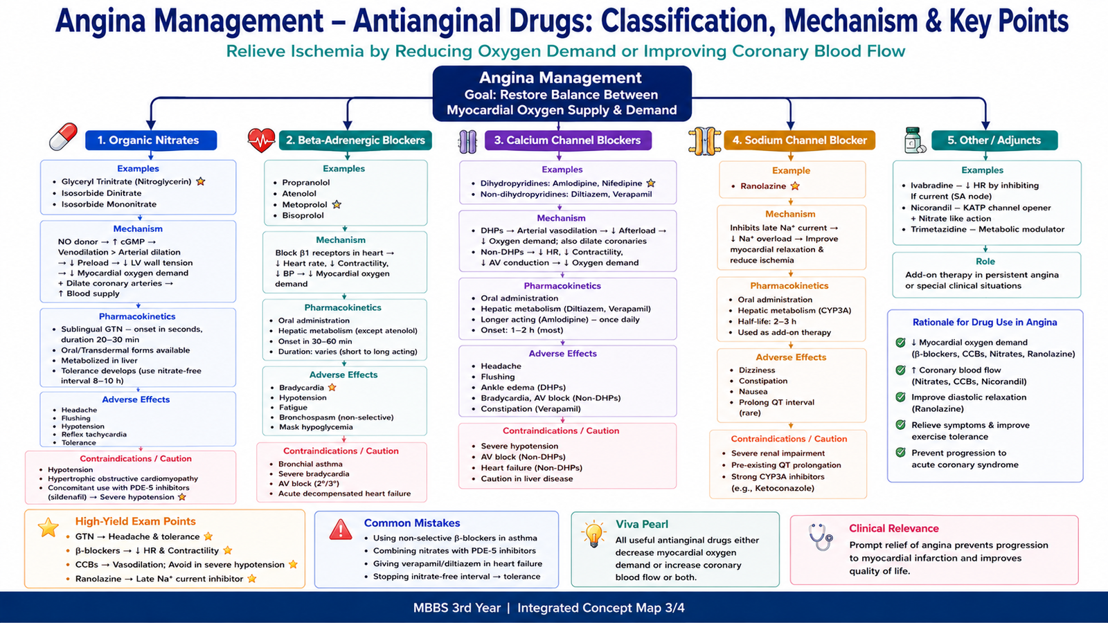
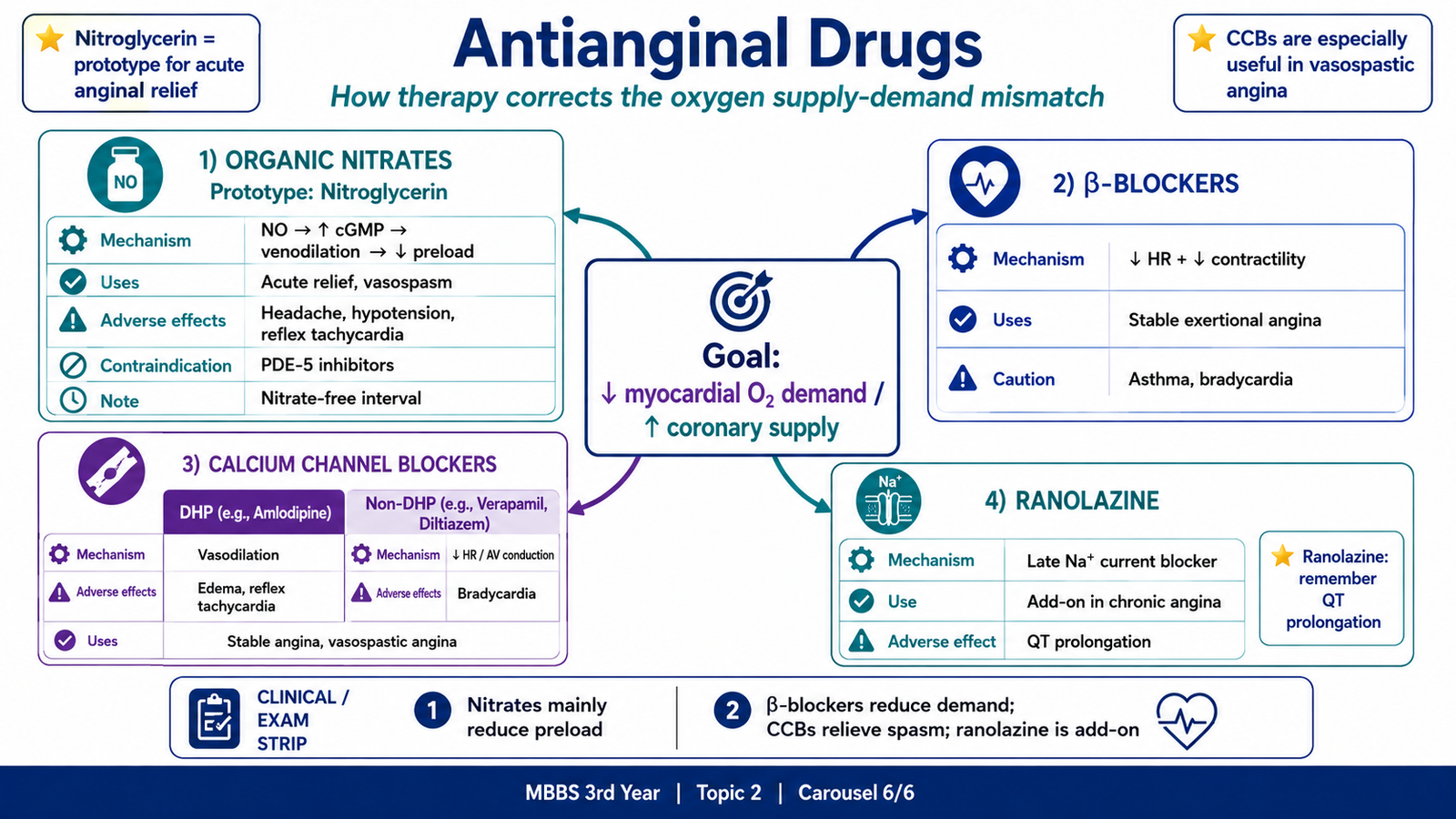
Antianginal drugs work by reducing myocardial oxygen demand, improving coronary oxygen supply, or both. Organic nitrates mainly reduce preload, whereas calcium channel blockers reduce vascular resistance, prevent coronary spasm and, with some agents, slow the heart.
Organic nitrates
Nitroglycerin, isosorbide dinitrate and isosorbide mononitrate release nitric oxide in vascular smooth muscle. Nitric oxide increases cyclic GMP, which causes smooth-muscle relaxation. The strongest effect is venodilation. Venodilation reduces venous return and ventricular filling, so preload and ventricular wall tension fall. The myocardium then requires less oxygen. Nitrates also dilate large coronary arteries and relieve coronary spasm.
Mechanism: Nitrate → nitric oxide → increased cGMP → venodilation → reduced preload and wall tension → reduced myocardial oxygen demand → relief of angina.
- Uses: rapid relief of an acute stable-angina attack, prevention before exertion and relief of vasospastic angina.
- Pharmacokinetics: sublingual nitroglycerin acts rapidly; oral nitrates undergo hepatic first-pass metabolism; isosorbide mononitrate has better oral bioavailability; transdermal forms provide longer prophylaxis.
- Tolerance: continuous exposure reduces the response, so a daily nitrate-free interval is required.
- Adverse effects: throbbing headache, flushing, dizziness, postural hypotension, reflex tachycardia and syncope; methemoglobinemia is rare.
- Dangerous interaction: PDE-5 inhibitors such as sildenafil can produce severe hypotension when combined with nitrates.
Calcium channel blockers
Calcium channel blockers inhibit L-type calcium channels. Their effects differ according to the tissue on which they act most strongly.
- Dihydropyridines such as amlodipine and nifedipine mainly relax arterioles. Reduced systemic resistance lowers afterload, while coronary dilation improves supply and prevents spasm.
- Non-dihydropyridines such as verapamil and diltiazem also reduce heart rate, contractility and AV nodal conduction. These effects reduce myocardial oxygen demand.
Many calcium channel blockers are orally active and undergo hepatic metabolism. Amlodipine has a long duration of action. Verapamil and diltiazem have important conduction effects and interaction potential.
- Dihydropyridine adverse effects: headache, flushing, ankle edema, hypotension and reflex tachycardia, especially with short-acting drugs.
- Verapamil/diltiazem adverse effects: bradycardia, AV block and worsening systolic heart failure; verapamil commonly causes constipation.
- Important caution: combining a β-blocker with verapamil or diltiazem can cause severe bradycardia or AV block.
F. β-Blockers, Ranolazine and Selection of Antianginal Therapy
β-adrenergic blockers are particularly useful in chronic exertional angina because they reduce the rise in cardiac work that normally occurs during activity. β1-receptor blockade lowers heart rate, contractility and blood pressure. The longer diastolic period may also improve coronary perfusion.
Mechanism: β-blockade → reduced heart rate and contractility → reduced myocardial oxygen demand → fewer exertional angina attacks.
- Examples: metoprolol, atenolol, bisoprolol and propranolol.
- Useful situations: stable exertional angina, especially when hypertension or previous myocardial infarction is also present.
- Adverse effects: bradycardia, fatigue, hypotension, AV block, bronchospasm with nonselective agents and worsening of acute decompensated heart failure.
- Cautions: severe bradycardia, AV block, acute decompensated heart failure and severe bronchospastic disease. Abrupt withdrawal may cause rebound tachycardia and ischemia.
- Selection point: they are not preferred for pure vasospastic angina because the main abnormality is coronary spasm.
Ranolazine inhibits the late inward sodium current in myocardial cells. This reduces intracellular sodium and secondary calcium overload, improves myocardial relaxation and reduces ventricular wall tension. It therefore lowers oxygen demand without substantially reducing heart rate or blood pressure.
- Use: add-on treatment for chronic stable angina when standard therapy is insufficient or poorly tolerated.
- Adverse effects: nausea, dizziness, constipation and QT-interval prolongation.
- Cautions: avoid strong CYP3A inhibitors, significant liver disease and combinations that markedly prolong the QT interval.
Drug selection follows the dominant mechanism. A short-acting nitrate is used for rapid symptom relief. β-blockers are logical for exertional angina with increased cardiac workload. Calcium channel blockers are preferred when coronary spasm is important or when β-blockers are unsuitable. Ranolazine is useful when symptoms persist and further reduction of heart rate or blood pressure is undesirable.
Integrated Mechanism Flow
Coronary atherosclerosis or coronary spasm
→ reduced coronary blood flow or reduced coronary flow reserve
→ oxygen supply becomes insufficient for myocardial demand
→ ATP falls and anaerobic metabolism increases
→ impaired contraction, metabolite accumulation and chest pain
→ brief ischemia remains reversible, but prolonged severe ischemia causes coagulative necrosis and myocardial infarction.
AIM High-Yield Review
- Ischemia is reduced blood supply with potentially reversible cell injury; infarction is irreversible tissue necrosis.
- Stable angina usually results from fixed atherosclerotic coronary narrowing and a demand–supply mismatch during exertion.
- Typical stable angina is retrosternal pressure brought on by exertion and relieved by rest or sublingual nitroglycerin.
- Troponin is normally not raised in stable angina because myocardial necrosis has not occurred.
- The LAD supplies the anterior left ventricular wall, apex and anterior interventricular septum.
- Left ventricular coronary flow occurs mainly in diastole; tachycardia shortens diastole and can worsen ischemia.
- Vasospastic angina commonly causes rest pain with transient ST elevation and may have a normal angiogram between attacks.
- Nitrates increase cGMP, mainly dilate veins, reduce preload and rapidly relieve angina.
- Continuous nitrate exposure causes tolerance; a nitrate-free interval is required.
- Nitrates must not be combined with PDE-5 inhibitors because profound hypotension may occur.
- β-blockers reduce heart rate and contractility, making them effective for stable exertional angina.
- Calcium channel blockers are especially useful for coronary vasospasm; verapamil and diltiazem may cause bradycardia and AV block.
- Ranolazine inhibits the late sodium current and may prolong the QT interval.
- Myocardial infarction classically produces coagulative necrosis followed by inflammation, granulation tissue and scar formation.
Concept Not Clear? Ask AIM AI
Use the AIM Learning Assistant to clarify difficult concepts from this topic before proceeding to assessment.
You may ask AIM AI to:
- Explain a difficult concept in simpler words.
- Describe a mechanism step by step.
- Compare two confusing diseases, drugs or findings.
- Provide a clinically relevant example.
- Highlight a high-yield KMU examination point.
- Explain a common MCQ trap related to the topic.
Best use: Ask one focused question at a time. AIM AI supports your understanding but does not replace the complete learning material.
▶ AIM Video Explanation
Coronary Artery Disease, Ischemic Heart Disease and Angina
Watch this video after completing the learning material to strengthen your understanding of coronary blood-flow reduction, myocardial ischemia, angina and progression toward acute coronary syndromes.
While watching, focus on: coronary atherosclerosis, myocardial oxygen supply–demand mismatch, stable versus unstable angina, and the relationship between prolonged ischemia and myocardial infarction.