
AIM Study Tip: This chapter follows the KMU learning outcomes in a logical sequence. First understand how blood pressure is regulated and how hypertension damages organs, then connect these mechanisms with antihypertensive drug actions and the final high-yield review.
📖 AIM Learning Material
Blood Pressure Regulation, Hypertension and Antihypertensive Therapy
CVS Module — This chapter explains normal blood pressure control, the causes and pathology of hypertension, hypertension in pregnancy, antihypertensive drug classes, treatment principles, hypertensive crises and patient counselling.
Topic Introduction
Blood pressure is the force produced when blood moves through the arteries. It must remain high enough to supply the organs, but not so high that it injures blood vessels. The body therefore regulates blood pressure through the heart, arterioles, kidneys, sympathetic nervous system, hormones and vascular endothelium. Hypertension develops when this control remains disturbed and arterial pressure stays elevated. It may remain clinically silent for years while gradually damaging the heart, brain, kidneys, retina and large vessels. Understanding hypertension requires three connected ideas: how blood pressure is normally controlled, how persistent pressure produces vascular and organ injury, and how antihypertensive drugs correct the responsible mechanisms. This chapter also explains hypertension in pregnancy, hypertensive emergencies and the importance of counselling for long-term treatment adherence.
A. Regulation of Arterial Blood Pressure
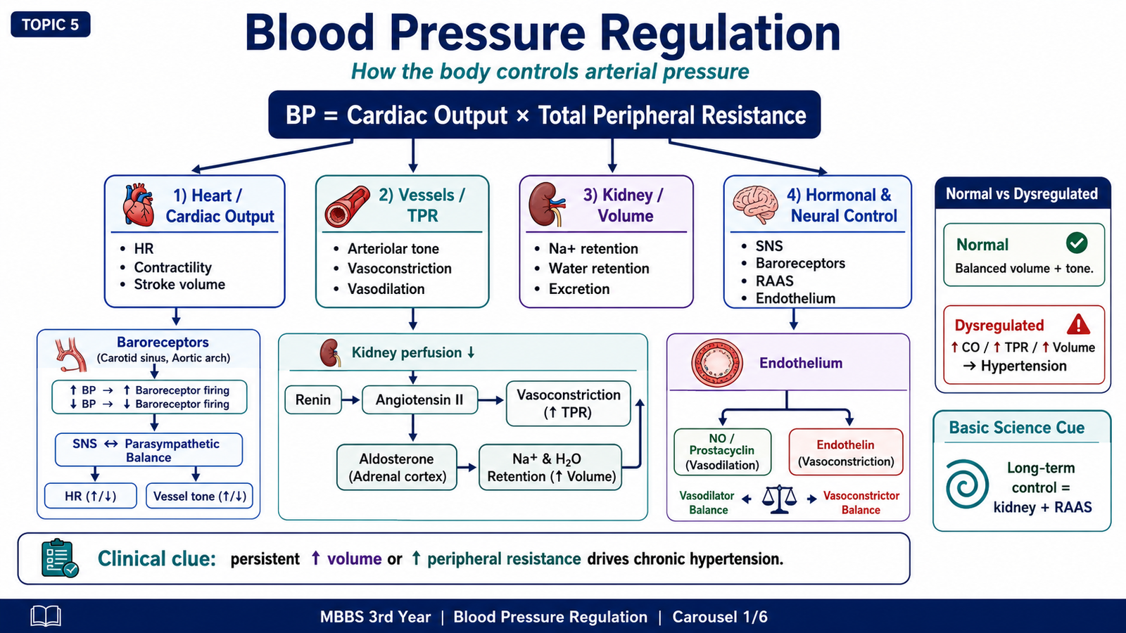
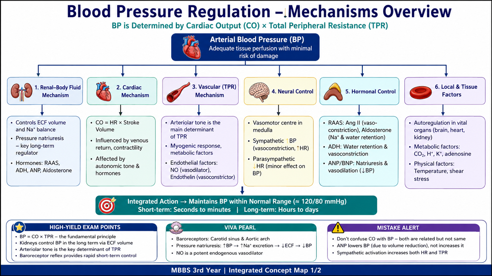
Arterial blood pressure depends mainly on the amount of blood pumped by the heart and the resistance offered by the systemic arterioles. This relationship can be expressed as:
Blood Pressure = Cardiac Output × Total Peripheral Resistance
Cardiac output is the volume of blood pumped by the heart each minute. It depends on heart rate, myocardial contractility and venous return. Total peripheral resistance is determined mainly by the diameter of small arteries and arterioles. A small reduction in arteriolar diameter markedly increases resistance and therefore raises blood pressure.
Short-term neural regulation
Rapid changes in blood pressure are corrected within seconds to minutes by neural reflexes. The most important is the baroreceptor reflex.
Baroreceptors are stretch-sensitive receptors located mainly in the carotid sinus and aortic arch. When arterial pressure rises, the vessel wall stretches more and baroreceptor firing increases. Signals reach the medulla, which reduces sympathetic activity and increases parasympathetic activity. Heart rate and contractility fall, arterioles dilate and blood pressure decreases. When pressure falls, the opposite response occurs.
Raised BP → increased baroreceptor stretch → reduced sympathetic output → lower heart rate, contractility and arteriolar tone → reduced BP
Chemoreceptors in the carotid and aortic bodies respond mainly to reduced oxygen, increased carbon dioxide and reduced pH. Their activation increases sympathetic activity and supports blood pressure during severe circulatory or respiratory disturbance.
Sympathetic nervous system
The sympathetic nervous system can raise blood pressure through several connected actions:
- α1-receptor stimulation contracts arteriolar smooth muscle and increases peripheral resistance.
- β1-receptor stimulation in the heart increases heart rate and contractility, raising cardiac output.
- β1-receptor stimulation in juxtaglomerular cells increases renin release and activates the renin–angiotensin–aldosterone system.
Renin–angiotensin–aldosterone system
The renin–angiotensin–aldosterone system, or RAAS, is activated when renal perfusion falls, sodium delivery to the macula densa decreases or renal sympathetic stimulation increases. Renin converts angiotensinogen to angiotensin I. Angiotensin-converting enzyme then converts angiotensin I to angiotensin II.
Angiotensin II raises blood pressure because it constricts arterioles and stimulates aldosterone secretion. Aldosterone increases sodium reabsorption in the distal nephron, and water follows sodium. Blood volume and cardiac output therefore rise. Angiotensin II also increases sympathetic activity and promotes cardiac and vascular remodelling when its activity remains excessive.
Reduced renal perfusion → renin release → angiotensin II formation → vasoconstriction + aldosterone secretion → sodium and water retention → increased blood pressure
Kidneys and long-term control
The kidneys are the major long-term controllers of blood pressure because they determine how much sodium and water remain in the body. When pressure rises, normal kidneys excrete more sodium and water. This process lowers extracellular fluid volume, venous return and cardiac output. If the kidneys require an abnormally high pressure to excrete sodium, the blood pressure remains elevated.
Impaired sodium excretion causes sodium retention, followed by water retention. Plasma volume expands, venous return increases and cardiac output rises. Over time, autoregulatory vasoconstriction may increase peripheral resistance and maintain hypertension even when cardiac output is no longer markedly elevated.
Endothelial and local vascular factors
The vascular endothelium actively regulates vessel tone. Nitric oxide and prostacyclin produce vasodilation, while endothelin and angiotensin II favour vasoconstriction. Endothelial dysfunction reduces vasodilator activity and shifts the vessel toward vasoconstriction, inflammation, thrombosis and structural remodelling. These changes contribute to persistent hypertension and target-organ damage.
B. Definition, Classification, Causes and Risk Factors of Hypertension
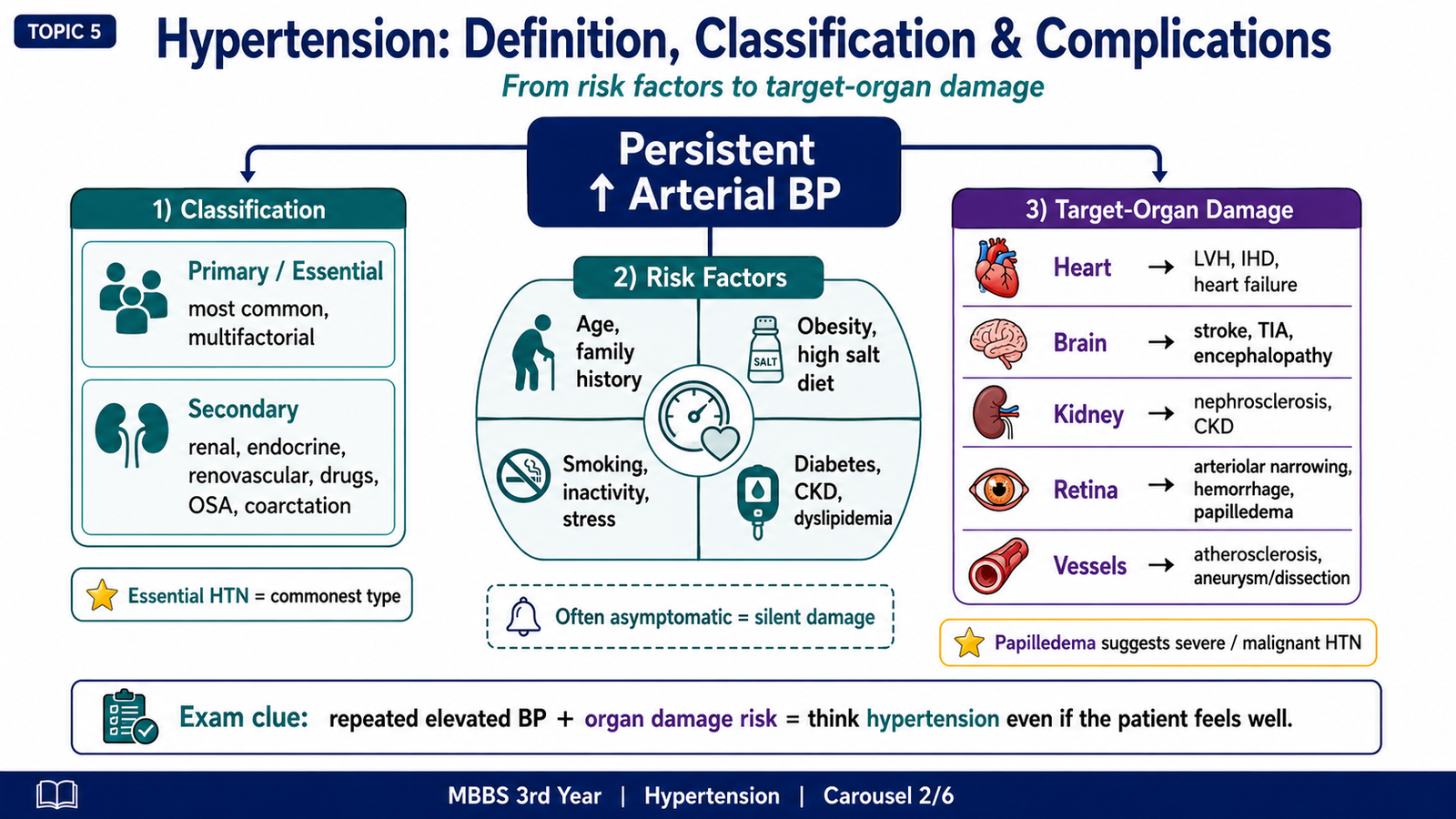
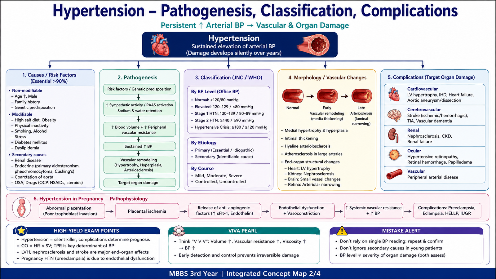
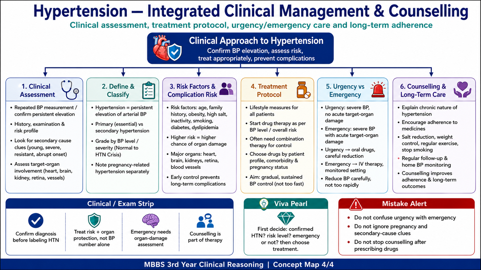
Hypertension is a persistent elevation of arterial blood pressure that increases the risk of cardiovascular, cerebrovascular, renal and retinal injury. A single high reading does not usually establish chronic hypertension because pain, anxiety, exercise, caffeine and incorrect measurement can temporarily raise blood pressure. Diagnosis therefore requires properly performed and repeated measurements, except when severe hypertension is accompanied by acute target-organ damage.
For undergraduate clinical learning, persistent office blood pressure around or above 140/90 mmHg is commonly used to recognize hypertension. Exact categories may vary between guidelines, but the central principle remains that both the degree of elevation and the patient’s total cardiovascular risk influence management.
Classification by cause
Primary or essential hypertension has no single identifiable cause. It results from interaction between genetic susceptibility and environmental factors. Abnormal renal sodium handling, increased sympathetic activity, inappropriate RAAS activity, obesity, endothelial dysfunction and vascular stiffness may all contribute.
Secondary hypertension results from an identifiable disease or drug. It should be considered particularly when hypertension begins at a young age, appears suddenly, becomes very severe, remains resistant to treatment or is associated with clues such as hypokalaemia, renal bruit or endocrine features.
| Category | Core feature | Examples or clues |
|---|---|---|
| Primary hypertension | Multifactorial; no single cause | Family tendency, obesity, salt sensitivity, increasing age |
| Secondary hypertension | Caused by a specific disorder or drug | Young onset, abrupt onset, resistant hypertension or suggestive laboratory findings |
Important causes of secondary hypertension
Secondary causes raise blood pressure by increasing sodium retention, activating the RAAS, increasing catecholamine or corticosteroid activity, or producing structural obstruction to blood flow.
- Renal parenchymal disease: impaired sodium excretion and volume expansion raise blood pressure.
- Renal artery stenosis: reduced renal perfusion activates renin release and the RAAS.
- Primary hyperaldosteronism: excess aldosterone causes sodium retention and often hypokalaemia.
- Pheochromocytoma: excess catecholamines produce episodic or sustained vasoconstriction and tachycardia.
- Cushing syndrome: excess glucocorticoid activity increases vascular responsiveness and sodium retention.
- Thyroid disease: altered metabolic and cardiovascular activity may raise systolic or diastolic pressure.
- Coarctation of the aorta: obstruction increases upper-body pressure and activates renal mechanisms.
- Obstructive sleep apnoea: repeated hypoxia and sympathetic activation contribute to persistent hypertension.
- Drugs: NSAIDs, corticosteroids, oral contraceptives and sympathomimetics may raise blood pressure.
Risk factors
Risk factors either increase the chance of developing hypertension or worsen its complications. Some cannot be changed, while others can be reduced through lifestyle and medical care.
Non-modifiable factors
- Increasing age
- Family history
- Genetic susceptibility
Modifiable factors
- Obesity
- High salt intake
- Physical inactivity
- Smoking
- Excess alcohol intake
- Unhealthy diet
- Diabetes and dyslipidaemia
- Poor treatment adherence
Hypertension is often asymptomatic. A patient may feel well while high pressure is progressively injuring vessels and organs. Symptoms such as headache do not reliably indicate either the presence or severity of chronic hypertension.
C. Pathogenesis, Morphology and Complications of Hypertension
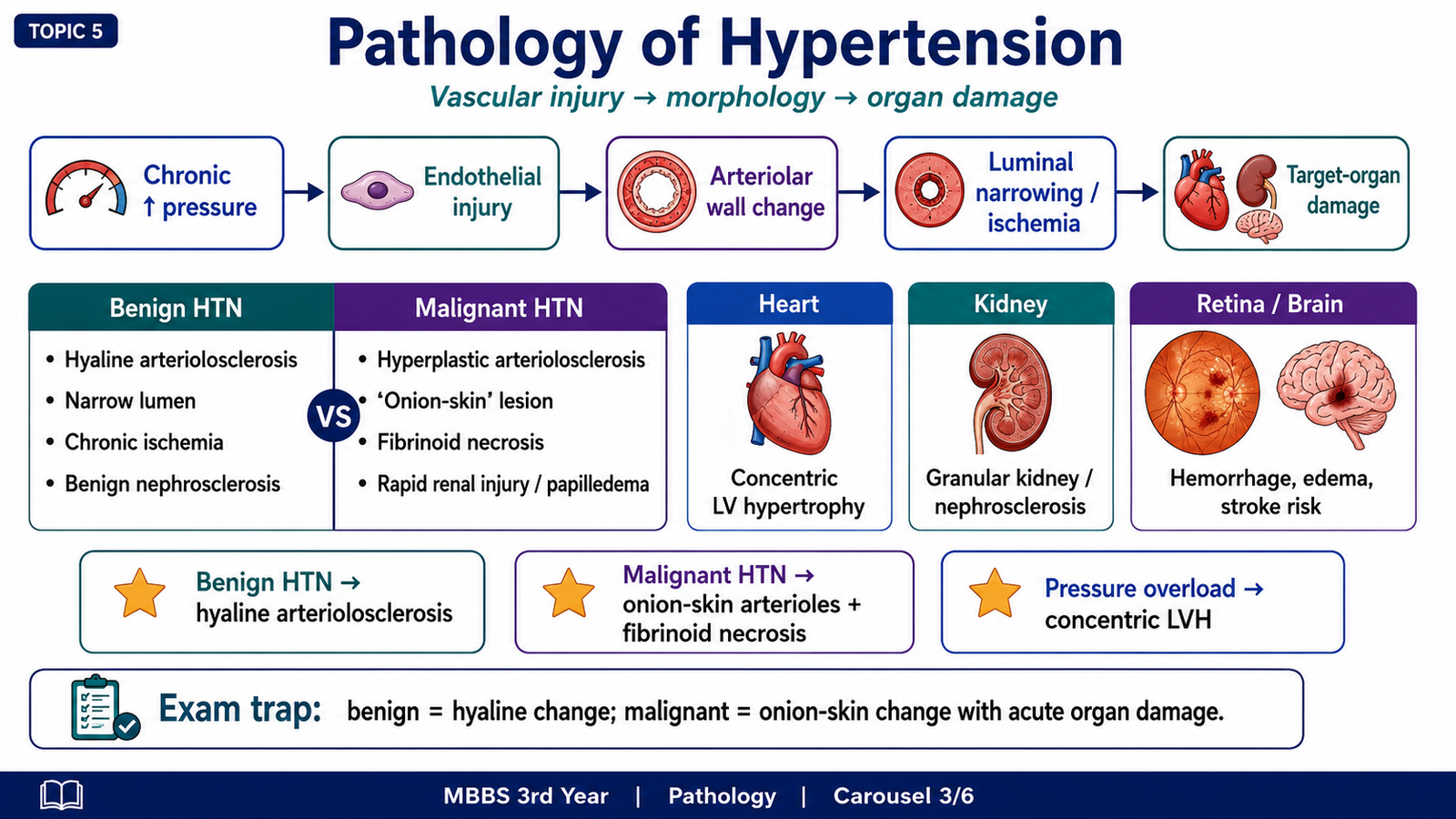
Persistent hypertension first damages arteries and arterioles. High pressure produces endothelial injury, increased movement of plasma proteins into the vessel wall and increased smooth-muscle production of extracellular matrix. The vessel wall becomes thick and the lumen narrows. Reduced blood flow then produces chronic ischaemic injury in the organs supplied by these vessels.
Persistent high pressure → endothelial injury and smooth-muscle response → wall thickening and luminal narrowing → reduced tissue perfusion → target-organ damage
Hyaline arteriolosclerosis
Hyaline arteriolosclerosis is mainly associated with long-standing benign hypertension and diabetes mellitus. Endothelial injury allows plasma proteins to enter the vessel wall, while smooth-muscle cells produce extracellular matrix. The arteriolar wall becomes homogeneously pink and thickened, and the lumen becomes narrow.
- Microscopy: homogeneous hyaline thickening of arteriolar walls with luminal narrowing.
- Functional effect: reduced blood supply and chronic ischaemia.
- Important organ association: benign nephrosclerosis in the kidney.
Hyperplastic arteriolosclerosis and malignant hypertension
Severe or rapidly progressive hypertension causes more intense vascular injury. Smooth-muscle cells proliferate in concentric layers and produce an “onion-skin” appearance. In the most severe injury, the vessel wall undergoes fibrinoid necrosis because endothelial damage allows fibrin and plasma proteins to enter the wall.
- Hyperplastic arteriolosclerosis: concentric smooth-muscle proliferation with marked luminal narrowing.
- Fibrinoid necrosis: necrosis of the vessel wall with deposition of fibrin-like material.
- Clinical effects: acute renal injury, retinal haemorrhages, papilloedema and hypertensive encephalopathy.
- Haematological effect: severe small-vessel injury may produce microangiopathic haemolytic anaemia.
Kidney morphology
Hypertension and kidney disease can reinforce each other. Hypertension narrows renal arterioles and causes ischaemic nephron loss. Reduced renal function then worsens sodium retention and raises blood pressure further.
Benign nephrosclerosis
- Gross appearance: kidneys may become small with a finely granular surface.
- Microscopy: hyaline arteriolosclerosis, ischaemic glomerular collapse, interstitial fibrosis and tubular atrophy.
- Clinical findings: mild proteinuria, gradual rise in serum creatinine and chronic kidney disease.
Malignant nephrosclerosis
- Gross appearance: petechial haemorrhages may produce a “flea-bitten” appearance.
- Microscopy: hyperplastic arteriolosclerosis and fibrinoid necrosis.
- Clinical findings: rapidly worsening renal function, haematuria, proteinuria and severe hypertension.
Cardiac changes
High systemic pressure increases left ventricular afterload. The left ventricle must generate greater force to eject blood, so cardiac myocytes enlarge and the ventricular wall becomes thick. This produces concentric left ventricular hypertrophy.
Initially, hypertrophy helps the heart overcome pressure overload. However, the thickened ventricle becomes stiff and fills poorly during diastole. Oxygen demand also increases, while the relative capillary supply becomes inadequate. The patient may therefore develop diastolic dysfunction, myocardial ischaemia, arrhythmias and eventually heart failure.
- Concentric left ventricular hypertrophy
- Reduced ventricular compliance
- S4 heart sound due to contraction against a stiff ventricle
- Ischaemic heart disease
- Heart failure
- Arrhythmias
Brain, retina and large vessels
Hypertension accelerates atherosclerosis and damages small cerebral vessels. This increases the risk of ischaemic stroke, intracerebral haemorrhage, transient ischaemic attacks and vascular cognitive impairment. In severe hypertension, failure of cerebral autoregulation causes cerebral oedema and hypertensive encephalopathy.
In the retina, chronic arteriolar narrowing reflects vascular wall thickening and vasoconstriction. More severe injury causes haemorrhages and exudates. Papilloedema suggests a dangerous rise in pressure with acute target-organ injury.
In large vessels, hypertension accelerates atherosclerosis and increases mechanical stress on the aortic wall. It therefore increases the risk of aortic aneurysm and aortic dissection.
| Organ | Why injury occurs | Important consequences |
|---|---|---|
| Heart | Increased afterload and coronary vascular disease | LV hypertrophy, ischaemia and heart failure |
| Brain | Small-vessel injury and accelerated atherosclerosis | Stroke, haemorrhage and encephalopathy |
| Kidney | Arteriolar narrowing and chronic ischaemia | Nephrosclerosis and renal failure |
| Retina | Arteriolar injury and leakage | Narrowing, haemorrhages, exudates and papilloedema |
| Aorta | Persistent mechanical stress on the wall | Aneurysm and dissection |
D. Hypertension in Pregnancy
Hypertension in pregnancy is important because it can harm both the mother and the fetus. The major pathological process in pre-eclampsia begins in the placenta but produces widespread maternal endothelial dysfunction. This explains why the disorder affects several organs rather than causing an isolated rise in blood pressure.
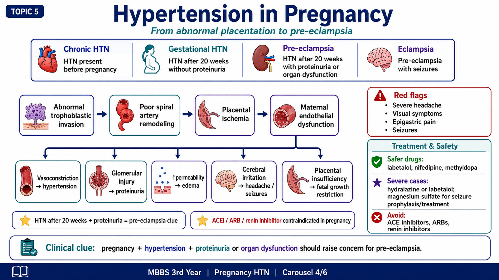
Main hypertensive disorders of pregnancy
- Chronic hypertension: hypertension present before pregnancy or recognized early in pregnancy.
- Gestational hypertension: new hypertension developing during pregnancy without the systemic features of pre-eclampsia.
- Pre-eclampsia: pregnancy-related hypertension accompanied by proteinuria or other evidence of maternal organ dysfunction.
- Eclampsia: pre-eclampsia complicated by seizures that are not explained by another neurological cause.
- Chronic hypertension with superimposed pre-eclampsia: development of pre-eclamptic features in a woman with pre-existing hypertension.
Pathophysiology of pre-eclampsia
During normal pregnancy, trophoblastic cells invade the maternal spiral arteries and convert them into wide, low-resistance vessels. This allows a large and steady blood supply to the placenta. In pre-eclampsia, trophoblastic invasion is incomplete. The spiral arteries remain relatively narrow and reactive, so placental perfusion becomes inadequate.
The poorly perfused placenta releases factors that disturb maternal endothelial function. The endothelium becomes more vasoconstrictor, more permeable and more likely to activate coagulation. Systemic vasoconstriction raises blood pressure, while endothelial leakage causes oedema. Renal endothelial injury increases urinary protein loss. Reduced placental blood flow may restrict fetal growth.
Abnormal trophoblastic invasion → narrow spiral arteries → placental ischaemia → release of endothelial-disrupting factors → maternal vasoconstriction, capillary leakage and coagulation activation → hypertension and organ dysfunction
Major maternal effects
- Hypertension: caused by widespread vasoconstriction and increased vascular sensitivity.
- Proteinuria: caused by glomerular endothelial injury and increased permeability.
- Oedema: caused by increased capillary permeability and reduced plasma oncotic pressure when protein is lost.
- Headache and visual symptoms: may reflect cerebral or retinal vascular disturbance.
- Liver involvement: endothelial injury and microvascular thrombosis may produce right upper abdominal pain and raised liver enzymes.
- Thrombocytopenia: platelets are consumed during widespread endothelial and coagulation activation.
- Seizures: cerebral endothelial dysfunction and oedema may lead to eclampsia.
Placental and fetal effects
Reduced uteroplacental blood flow limits the delivery of oxygen and nutrients. This may produce fetal growth restriction, fetal distress, placental infarction, placental abruption, premature delivery or fetal death in severe cases.
Important clinical warning features
A pregnant patient with hypertension requires careful assessment for proteinuria and organ dysfunction. Severe headache, visual disturbance, epigastric or right upper quadrant pain, reduced urine output, respiratory difficulty, seizures or marked laboratory abnormalities suggest severe disease and require urgent specialist management.
E. Antihypertensive Drugs and Drug-Selection Logic
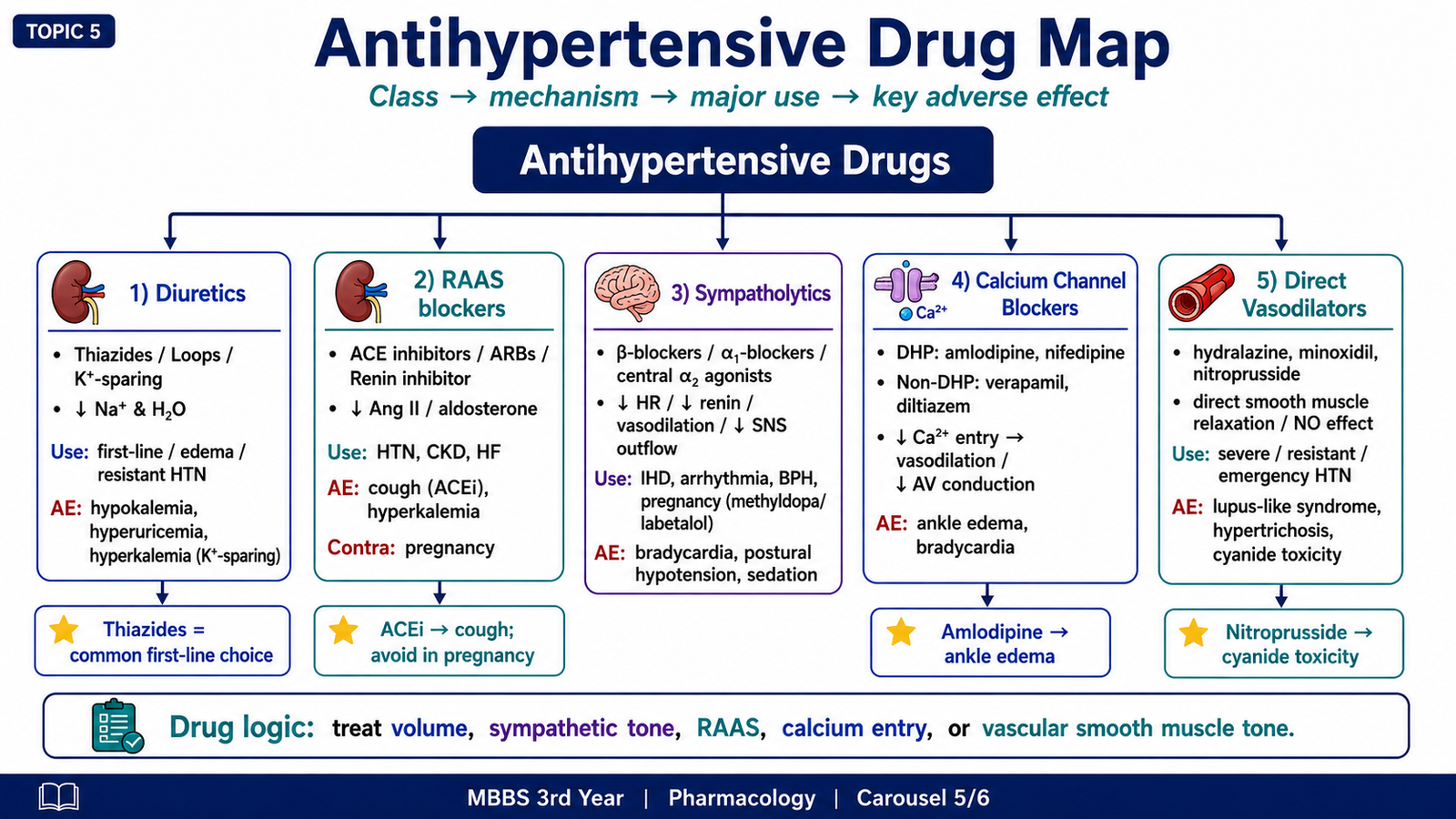
Antihypertensive drugs lower blood pressure by reducing blood volume, cardiac output, sympathetic activity, arteriolar resistance or RAAS activity. Most patients require long-term treatment, and many eventually need more than one drug because different mechanisms contribute to hypertension.
Classification of antihypertensive drugs
| Major class | Examples | Main BP-lowering action |
|---|---|---|
| Diuretics | Hydrochlorothiazide, chlorthalidone, furosemide, spironolactone | Reduce sodium and water, then reduce vascular resistance |
| ACE inhibitors | Captopril, enalapril, lisinopril | Reduce angiotensin II and aldosterone |
| Angiotensin receptor blockers | Losartan, valsartan | Block AT1-receptor effects |
| Direct renin inhibitor | Aliskiren | Reduces formation of angiotensin I |
| Calcium channel blockers | Amlodipine, nifedipine, verapamil, diltiazem | Reduce calcium entry into vascular smooth muscle or heart |
| β-blockers | Propranolol, atenolol, metoprolol | Reduce heart rate, contractility and renin release |
| α1-blockers | Prazosin, doxazosin | Produce arteriolar and venous dilation |
| Central sympatholytics | Clonidine, methyldopa | Reduce sympathetic outflow from the CNS |
| Direct vasodilators | Hydralazine, minoxidil, sodium nitroprusside | Directly relax vascular smooth muscle |
Diuretics
Diuretics lower blood pressure by increasing renal sodium excretion. Water follows sodium, so plasma volume and venous return fall. Cardiac output decreases initially. With continued treatment, peripheral resistance also falls.
Thiazide and thiazide-like diuretics
Thiazides inhibit sodium and chloride reabsorption in the distal convoluted tubule. They are effective in uncomplicated hypertension and are commonly combined with other drug classes.
- Therapeutic benefit: reduced extracellular fluid volume and later reduction in vascular resistance.
- Important adverse effects: hypokalaemia, hyponatraemia, hyperuricaemia, hyperglycaemia and increased calcium retention.
- Clinical caution: hyperuricaemia may precipitate gout.
Loop diuretics
Loop diuretics inhibit sodium, potassium and chloride transport in the thick ascending limb. They produce powerful diuresis and are especially useful when hypertension is accompanied by marked oedema, heart failure or reduced renal function.
- Hypokalaemia and metabolic alkalosis
- Volume depletion
- Ototoxicity
- Hypomagnesaemia and reduced calcium reabsorption
Potassium-sparing diuretics
Spironolactone and eplerenone block aldosterone receptors. They are particularly useful in hyperaldosteronism and resistant hypertension. Because they reduce potassium excretion, hyperkalaemia is the main danger. Spironolactone may also cause endocrine adverse effects such as gynaecomastia.
ACE inhibitors
ACE inhibitors reduce the formation of angiotensin II and decrease aldosterone secretion. Arterioles dilate, sodium retention decreases and harmful cardiac and vascular remodelling is reduced. ACE also normally breaks down bradykinin, so ACE inhibition increases bradykinin levels.
ACE inhibitor → reduced angiotensin II + reduced aldosterone + increased bradykinin → vasodilation and reduced sodium retention → lower BP
- Useful clinical settings: hypertension with heart failure, diabetic kidney disease or other proteinuric renal disease.
- Dry cough: caused by increased bradykinin.
- Angio-oedema: a rare but potentially serious bradykinin-related reaction.
- Hyperkalaemia: caused by reduced aldosterone activity.
- Rise in creatinine: may occur because efferent arteriolar dilation reduces intraglomerular pressure.
- Avoid in pregnancy: RAAS blockade can injure the developing fetal kidneys.
- Avoid in bilateral renal artery stenosis: glomerular filtration may depend on angiotensin-II-mediated efferent arteriolar constriction.
Angiotensin receptor blockers
Angiotensin receptor blockers, or ARBs, block the AT1 receptor and therefore prevent the major vasoconstrictor and aldosterone-stimulating effects of angiotensin II. Their therapeutic effects are similar to those of ACE inhibitors, but they do not significantly increase bradykinin.
- Less likely than ACE inhibitors to cause dry cough.
- May still cause hyperkalaemia and reduced renal function.
- Contraindicated in pregnancy.
- ACE inhibitors and ARBs should not routinely be combined because adverse renal and potassium effects increase.
Direct renin inhibitor
Aliskiren directly inhibits renin and reduces the conversion of angiotensinogen to angiotensin I. This suppresses the RAAS at its first enzymatic step. It may cause hyperkalaemia, hypotension and renal impairment and should not be used in pregnancy. Combining it with other RAAS-blocking drugs increases adverse effects and is generally avoided.
Calcium channel blockers
Calcium channel blockers inhibit L-type calcium channels. Calcium entry is necessary for vascular smooth-muscle contraction and for cardiac conduction and contraction. Their effects depend on whether the drug acts mainly on blood vessels or the heart.
Dihydropyridines
Amlodipine and nifedipine act mainly on vascular smooth muscle. They produce arteriolar dilation and reduce peripheral resistance.
- Headache and flushing due to vasodilation
- Ankle oedema due to increased capillary hydrostatic pressure
- Dizziness and hypotension
- Reflex tachycardia, especially with rapidly acting agents
- Gingival enlargement may occur
Non-dihydropyridines
Verapamil and diltiazem have greater cardiac effects. They slow sinoatrial activity and atrioventricular conduction and reduce cardiac contractility. They may cause bradycardia, heart block and worsening heart failure in susceptible patients. Verapamil commonly causes constipation.
β-adrenoceptor blockers
β-blockers lower blood pressure by blocking β1 receptors in the heart and kidneys. Heart rate and contractility decrease, so cardiac output falls. Renin release also decreases, reducing RAAS activity.
- Useful when another indication is present: ischaemic heart disease, certain tachyarrhythmias or selected patients with heart failure.
- Adverse effects: bradycardia, fatigue, reduced exercise tolerance, sexual dysfunction and sleep disturbance.
- Bronchospasm: non-selective β-blockers may block β2 receptors and should be used cautiously in asthma.
- Metabolic caution: β-blockers may mask adrenergic warning symptoms of hypoglycaemia.
- Withdrawal: abrupt discontinuation can cause rebound sympathetic effects, angina or severe tachycardia.
α1-adrenoceptor blockers
Prazosin and related drugs block α1 receptors in arterioles and veins. This reduces peripheral resistance and venous return. They may be useful when hypertension coexists with benign prostatic enlargement, but they can cause marked postural hypotension, particularly after the first dose.
- First-dose syncope or severe dizziness
- Postural hypotension
- Reflex tachycardia
- Fluid retention
Centrally acting sympatholytic drugs
Clonidine and methyldopa stimulate central α2 receptors and reduce sympathetic outflow from the brainstem. Reduced sympathetic activity lowers heart rate, cardiac output and vascular resistance.
- Clonidine: may cause sedation, dry mouth and bradycardia. Abrupt withdrawal can cause severe rebound hypertension.
- Methyldopa: has an established role in hypertension during pregnancy. Adverse effects include sedation, a positive Coombs test, haemolytic anaemia and liver injury.
Direct vasodilators
Hydralazine
Hydralazine directly relaxes arteriolar smooth muscle and reduces systemic vascular resistance. The fall in pressure activates compensatory sympathetic activity and the RAAS, so tachycardia and sodium retention can occur. It is often combined with a β-blocker and diuretic when used chronically.
- Headache, flushing and palpitation
- Reflex tachycardia
- Fluid retention
- Lupus-like syndrome with prolonged exposure
Minoxidil
Minoxidil opens potassium channels in arteriolar smooth muscle, causing hyperpolarisation and relaxation. It is a powerful vasodilator used mainly for severe or resistant hypertension.
- Marked fluid retention
- Reflex tachycardia
- Hypertrichosis
- Possible pericardial effusion
Sodium nitroprusside
Sodium nitroprusside releases nitric oxide and rapidly dilates both arterioles and veins. It has a very rapid onset and short duration, making it useful for controlled blood-pressure reduction in hypertensive emergencies. Excessive or prolonged exposure may cause cyanide or thiocyanate toxicity.
General drug-treatment approach
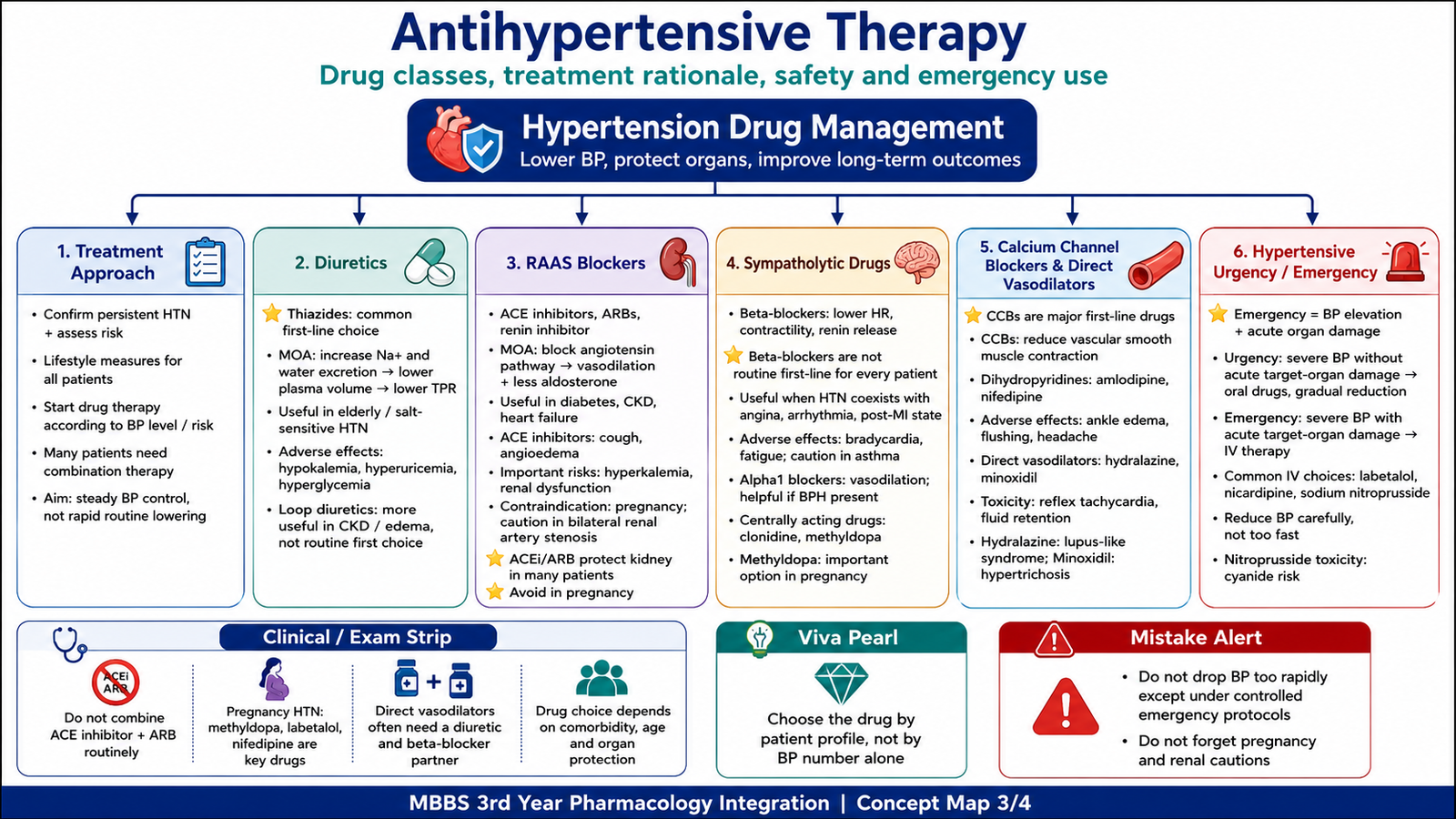
Drug selection should consider the severity of hypertension, target-organ involvement, age, kidney function, pregnancy, comorbid disease and adverse-effect profile. Treatment often begins with a thiazide-type diuretic, ACE inhibitor, ARB or calcium channel blocker, depending on the patient’s clinical features. Markedly elevated blood pressure often requires two complementary drugs.
Effective combinations act through different mechanisms. For example, a RAAS blocker may be combined with a calcium channel blocker or a thiazide. A diuretic may also reduce the sodium retention caused by a direct vasodilator. Drugs that block the same pathway, such as an ACE inhibitor and an ARB, should not routinely be combined because toxicity increases without a proportional benefit.
F. Hypertensive Urgency, Emergency and Patient Counselling
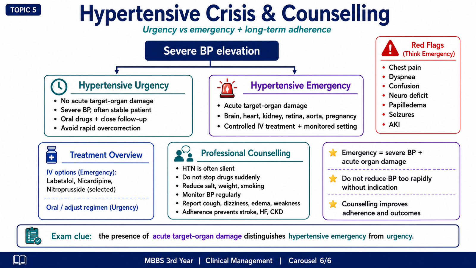
Very high blood pressure becomes an emergency only when it is accompanied by acute target-organ injury. The absolute blood-pressure value is important, but the presence or absence of organ damage determines the immediate management approach.
Hypertensive urgency
Hypertensive urgency refers to severe blood-pressure elevation without evidence of acute, progressive target-organ damage. The patient may have nonspecific symptoms such as headache or anxiety, but there is no acute encephalopathy, myocardial ischaemia, pulmonary oedema, aortic dissection or rapidly worsening renal failure.
Blood pressure is usually reduced gradually with oral treatment and close follow-up. Rapid and excessive reduction is avoided because chronically hypertensive organs have adapted to higher perfusion pressures. A sudden fall can reduce cerebral, coronary or renal blood flow.
Hypertensive emergency
Hypertensive emergency is severe hypertension with acute target-organ damage. The patient requires urgent hospital care, careful monitoring and titratable intravenous treatment. The aim is controlled reduction rather than immediate normalization.
Severe hypertension + acute organ injury = hypertensive emergency
Important forms of acute target-organ damage include:
- Hypertensive encephalopathy with confusion, seizures or altered consciousness
- Intracranial haemorrhage or acute stroke
- Acute coronary syndrome
- Acute left ventricular failure or pulmonary oedema
- Aortic dissection
- Acute kidney injury
- Severe retinopathy with haemorrhages, exudates or papilloedema
- Severe pre-eclampsia or eclampsia
Initial clinical assessment
The clinician should confirm the blood-pressure measurement and rapidly look for symptoms and signs of acute organ injury. Important questions include chest pain, breathlessness, neurological symptoms, visual disturbance, reduced urine output, pregnancy and recent drug use or withdrawal.
Examination should assess mental state, focal neurological signs, fundoscopy, cardiac findings, pulmonary oedema, peripheral pulses and features of aortic disease. Investigations are selected according to the presentation and may include urinalysis, renal function, electrolytes, ECG, cardiac biomarkers, chest imaging, brain imaging and pregnancy-related assessment.
General management principles
- Admit and monitor patients with hypertensive emergency.
- Use a rapidly acting, titratable intravenous drug selected for the affected organ and clinical situation.
- Reduce pressure in a controlled manner to preserve organ perfusion.
- Treat the specific complication, such as pulmonary oedema, acute coronary syndrome, aortic dissection or eclampsia.
- Identify possible causes such as missed medication, renal disease, drug exposure or sympathetic overactivity.
- Once stable, establish a long-term oral regimen and follow-up plan.
Sodium nitroprusside, labetalol, nicardipine and other intravenous agents may be selected according to the clinical situation. Drug choice is not identical for every emergency. For example, aortic dissection requires rapid reduction of both heart rate and arterial pressure, while pregnancy-related emergencies require drugs suitable for pregnancy.
Professional counselling and adherence
Hypertension commonly produces no symptoms, so patients may stop treatment when they feel well. Effective counselling should explain that treatment prevents future organ damage rather than simply relieving current symptoms. The clinician should use clear, respectful language and involve the patient in practical decisions.
- Explain that hypertension may remain silent while damaging the heart, brain, kidneys, retina and blood vessels.
- Explain the purpose of each medicine and how it should be taken.
- Discuss common adverse effects and advise the patient not to stop treatment without consultation.
- Ask about cost, availability, daily routine and other barriers to adherence.
- Encourage reduced salt intake, healthy body weight, regular physical activity and avoidance of smoking.
- Teach correct home blood-pressure measurement when appropriate.
- Encourage regular follow-up even when blood pressure improves.
- Use open questions and confirm understanding rather than giving instructions only.
Professional counselling example: “Your blood pressure may not cause symptoms, but it can still damage your organs over time. These medicines lower that risk. Please tell me if you develop an adverse effect so that we can adjust treatment rather than stopping it suddenly.”
Integrated Mechanism Flow
Genetic and environmental factors or a secondary cause
↓
Abnormal renal sodium handling, sympathetic activation, RAAS activation or increased vascular tone
↓
Increased blood volume, cardiac output or peripheral resistance
↓
Persistent hypertension
↓
Endothelial injury, arteriolar wall thickening and vascular remodelling
↓
Reduced organ perfusion and increased cardiac afterload
↓
Heart, brain, kidney, retinal and large-vessel complications
AIM High-Yield Review
- Blood pressure equals cardiac output multiplied by total peripheral resistance.
- The kidneys control long-term blood pressure mainly by regulating sodium and water balance.
- Angiotensin II causes vasoconstriction, stimulates aldosterone and promotes vascular and cardiac remodelling.
- Primary hypertension is multifactorial; secondary hypertension has an identifiable cause.
- Hypertension may remain asymptomatic while target-organ injury progresses.
- ⭐ Hyaline arteriolosclerosis is associated with long-standing benign hypertension and diabetes.
- ⭐ Hyperplastic arteriolosclerosis produces an onion-skin appearance in severe hypertension.
- Malignant vascular injury may include fibrinoid necrosis and rapidly progressive renal damage.
- Pressure overload produces concentric left ventricular hypertrophy and reduced ventricular compliance.
- Pre-eclampsia begins with abnormal placentation and develops into widespread maternal endothelial dysfunction.
- Thiazides may cause hypokalaemia, hyponatraemia and hyperuricaemia; hyperuricaemia may precipitate gout.
- ACE inhibitors may cause cough, angio-oedema, hyperkalaemia and renal dysfunction and are contraindicated in pregnancy.
- Dihydropyridine calcium channel blockers mainly dilate arterioles and commonly cause ankle oedema.
- Abrupt withdrawal of clonidine can cause severe rebound hypertension.
- ⭐ A hypertensive emergency is severe hypertension with acute target-organ damage; severe pressure alone does not define an emergency.
Hypertension and Antihypertensive Drugs
Watch this video to reinforce blood-pressure regulation, the pathophysiology of hypertension, the renin–angiotensin–aldosterone system and the mechanisms of major antihypertensive drug classes.
Study focus: Relate each drug class to the blood-pressure component it reduces—blood volume, cardiac output, sympathetic activity, peripheral resistance or RAAS activity.