
💡 Study Tip: This chapter follows the KMU learning outcomes in a logical sequence. First understand how lipoproteins, risk factors and arterial injury connect; then use the final high-yield review for revision.
Topic Introduction
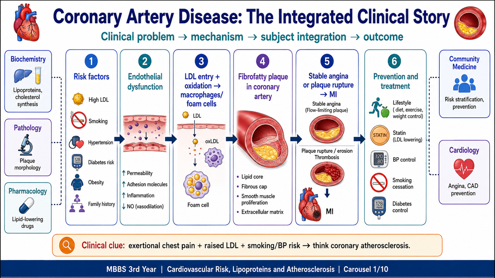
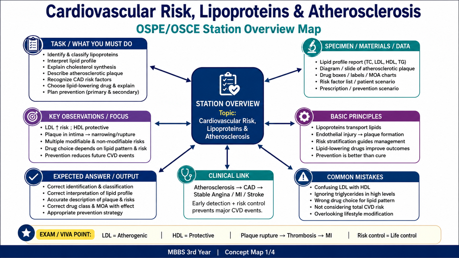
Cardiovascular disease includes disorders of the heart and blood vessels. A major cause is atherosclerosis, a chronic inflammatory process in which lipids accumulate in the inner layer of large and medium-sized arteries. The process develops slowly, often without symptoms, but may later cause coronary artery disease, myocardial infarction, stroke, peripheral arterial disease or aneurysm. To understand this topic, students need to connect normal lipoprotein transport and cholesterol synthesis with dyslipidemia, endothelial injury, plaque morphology, cardiovascular risk stratification, lipid-lowering pharmacology and prevention. These ideas form one continuous pathway from risk exposure to arterial disease and finally to clinical outcomes.
A. Cardiovascular Disease, Lipoproteins and Cholesterol Transport
Cardiovascular disease is a broad term for diseases of the heart and blood vessels. Important forms include coronary heart disease, cerebrovascular disease, peripheral arterial disease, hypertensive heart disease, heart failure, rheumatic heart disease and venous thromboembolism. Coronary artery disease develops when coronary blood flow becomes inadequate, most commonly because atherosclerotic plaques narrow or suddenly obstruct the coronary arteries.
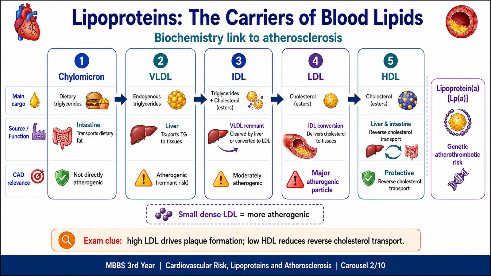
Cholesterol and triglycerides are hydrophobic, meaning that they do not dissolve freely in plasma. They therefore travel inside lipoproteins. Each lipoprotein has a hydrophobic core containing triglycerides and cholesteryl esters, surrounded by phospholipids, free cholesterol and apolipoproteins. The apolipoproteins stabilize the particle and help it interact with enzymes and receptors.
Main Types of Lipoproteins
| Lipoprotein | Main Lipid | Main Role | Clinical Relevance |
|---|---|---|---|
| Chylomicrons | Dietary triglycerides | Carry dietary triglyceride from the intestine to tissues | Marked elevation may cause pancreatitis |
| VLDL | Endogenous triglycerides | Carries triglyceride produced by the liver | Forms IDL and LDL and contributes to atherogenic dyslipidemia |
| IDL | Triglyceride and cholesterol | Intermediate between VLDL and LDL | Atherogenic remnant particle |
| LDL | Cholesterol | Delivers cholesterol to peripheral tissues | Major atherogenic lipoprotein |
| HDL | Cholesterol | Returns cholesterol from tissues to the liver | Associated with lower cardiovascular risk |
| Lipoprotein(a) | LDL-like particle with apo(a) | Genetically determined lipid-transport particle | Inherited atherothrombotic risk |
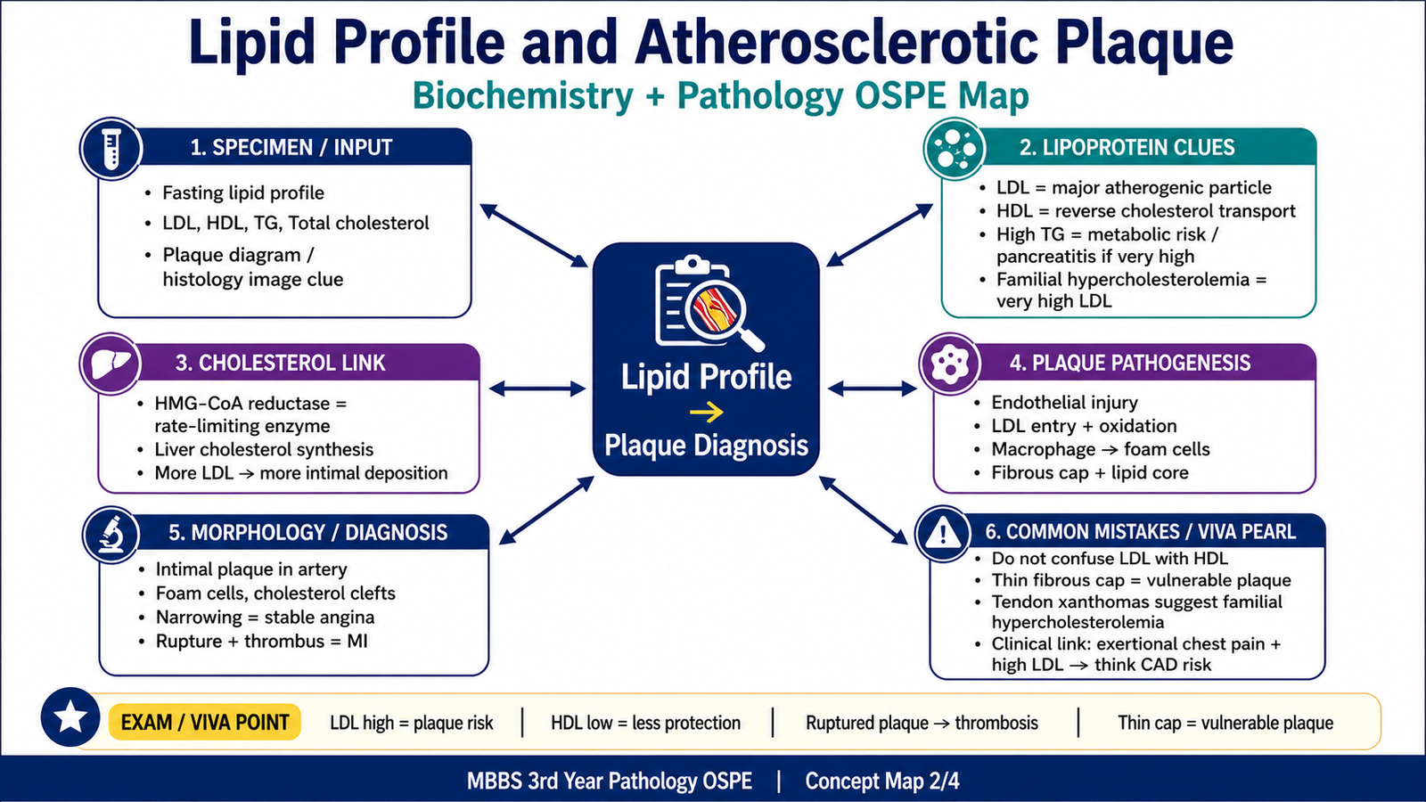
LDL is especially important because excess LDL can enter the arterial intima, become modified and trigger inflammation. HDL participates in reverse cholesterol transport, in which cholesterol is carried from peripheral tissues back to the liver. HDL does not directly dissolve an established plaque.
Cholesterol Synthesis
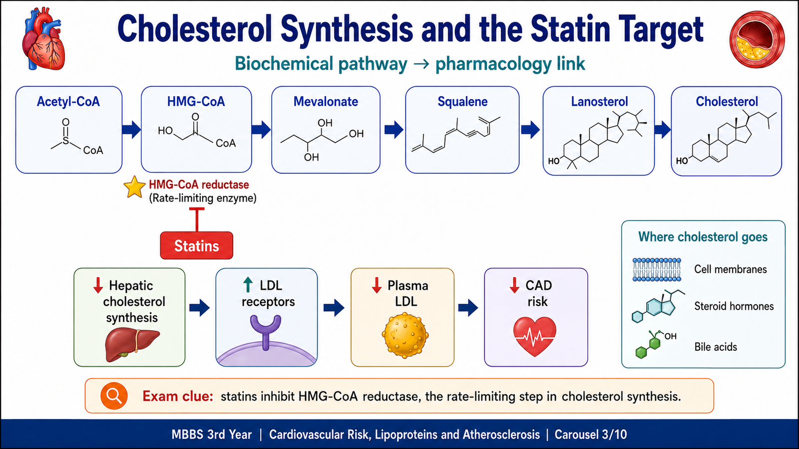
Cholesterol is synthesized mainly in the liver. The rate-limiting step is the conversion of HMG-CoA to mevalonate by HMG-CoA reductase.
When liver cells contain less cholesterol, they increase LDL-receptor expression. More LDL is then removed from blood. This explains why inhibition of HMG-CoA reductase by statins produces a marked fall in serum LDL.
B. Cardiovascular Risk Factors and Dyslipidemia
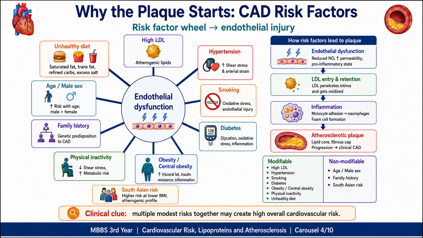
A cardiovascular risk factor is a characteristic or exposure that increases the probability of developing cardiovascular disease. Risk factors often act together. For example, raised LDL provides lipid for plaque formation, while hypertension, smoking and diabetes injure the endothelium and make lipid entry and inflammation more likely.
Non-Modifiable Risk Factors
- Increasing age: arterial injury and lipid accumulation develop over many years.
- Sex: men usually develop coronary disease earlier; risk in women rises after menopause.
- Family history and genetic predisposition: inherited lipid disorders and shared familial risk increase premature disease.
- Ethnic background: South Asian populations commonly show central obesity, insulin resistance and atherogenic dyslipidemia.
Modifiable Risk Factors
- Raised LDL-C: increases the amount of cholesterol entering the arterial intima.
- Low HDL-C: is associated with reduced reverse cholesterol transport and higher cardiovascular risk.
- Hypertension: produces mechanical stress and endothelial dysfunction, accelerating plaque formation and vascular complications.
- Smoking: promotes endothelial injury, inflammation, oxidation and thrombosis.
- Diabetes mellitus: causes endothelial dysfunction and commonly produces high triglycerides, low HDL and small dense LDL.
- Obesity and physical inactivity: promote insulin resistance, hypertension and dyslipidemia.
- Unhealthy diet: excess saturated and trans fats, salt and refined carbohydrates worsen lipids, blood pressure and body weight.
- Other contributors: chronic kidney disease, chronic stress, poor sleep and air pollution may increase total cardiovascular risk.
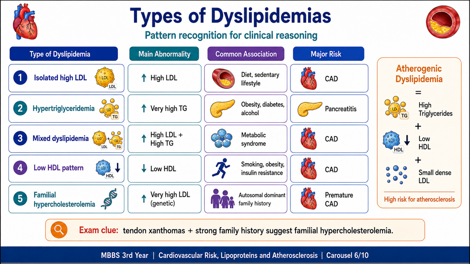
Types of Dyslipidemia
Dyslipidemia means an abnormal concentration or pattern of plasma lipoproteins. It may be primary because of inherited defects or secondary to conditions such as diabetes, obesity, hypothyroidism, renal disease, liver disease, alcohol use or drugs. Clinically, the abnormal pattern is often described by the lipid that is raised or reduced.
| Pattern | Main Abnormality | Importance |
|---|---|---|
| Isolated hypercholesterolemia | High LDL-C | Strong atherosclerotic risk |
| Hypertriglyceridemia | High triglycerides/VLDL | Very high levels may cause pancreatitis |
| Mixed dyslipidemia | High LDL and triglycerides | Common in diabetes and metabolic syndrome |
| Low HDL-C | Reduced HDL | Often accompanies obesity, smoking and diabetes |
| Familial hypercholesterolemia | Very high LDL from early life | Premature CAD and tendon xanthomas |
| Atherogenic dyslipidemia | High TG, low HDL and small dense LDL | Typical of insulin resistance |
A combination of high triglycerides, low HDL and small dense LDL is particularly atherogenic because these particles are linked with insulin resistance and readily contribute to arterial lipid deposition.
C. Atherosclerosis: Pathogenesis, Morphology and Consequences
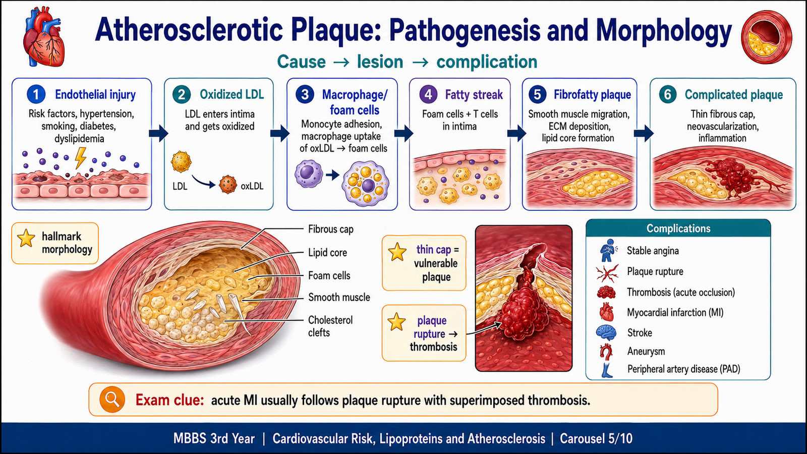
Atherosclerosis is a chronic inflammatory disease of the arterial intima. It affects large and medium-sized arteries and is characterized by lipid accumulation, macrophage foam cells, smooth-muscle proliferation, extracellular-matrix deposition and formation of a fibrous plaque. The lesion is not simply a deposit of cholesterol; it is an active interaction between lipids, endothelial injury, inflammation and tissue repair.
Pathogenesis
The process begins with endothelial dysfunction. Hypertension, smoking, diabetes, hyperlipidemia and inflammation reduce the normal protective functions of endothelium. LDL then enters the intima and becomes oxidized or otherwise modified. Modified LDL activates endothelial cells and attracts monocytes. These monocytes enter the intima, become macrophages and engulf lipid through scavenger receptors, producing foam cells.
Foam cells and activated vascular cells release cytokines and growth factors. Smooth-muscle cells migrate from the media into the intima, proliferate and produce collagen and other extracellular matrix. A fibrous cap forms over a lipid-rich necrotic core. Continuing inflammation may thin and weaken the cap, while healing may produce fibrosis and calcification.
Morphology
Fatty streaks are the earliest visible lesions. They appear as flat yellow areas made mainly of lipid-filled foam cells. They do not usually obstruct blood flow, but they may provide the background from which fibrous plaques develop.
Atheromatous plaques are raised white-yellow intimal lesions. They have a fibrous cap facing the lumen and a softer necrotic lipid core beneath it. Advanced plaques may contain hemorrhage, calcification, ulceration and superimposed thrombosis.
- Yellow fatty streaks in early lesions
- Raised white-yellow plaques
- Irregular luminal narrowing
- Ulceration, hemorrhage, thrombosis or calcification in complicated plaques
- Intimal lipid and foam cells
- Necrotic lipid core and cholesterol clefts
- Smooth-muscle cells and collagen-rich fibrous cap
- Inflammatory cells and later calcification
The hallmark lesion is a fibrous cap over a necrotic lipid core. Plaques with a thick cap and smaller lipid core are relatively stable and tend to cause gradual stenosis. Plaques with a thin inflamed cap, large lipid core and fewer smooth-muscle cells are more vulnerable to rupture.
Consequences and Complications
Clinical effects depend on the artery involved and whether the plaque narrows the lumen gradually or changes suddenly.
- Progressive stenosis: limits blood flow and may cause stable angina, intermittent claudication or chronic ischemia.
- Plaque rupture, erosion or ulceration: exposes thrombogenic material and may produce acute thrombosis, unstable angina or myocardial infarction.
- Plaque hemorrhage: rapidly enlarges the lesion and worsens obstruction.
- Atheroembolism: plaque material may travel downstream and obstruct smaller vessels.
- Weakening of the arterial media: may contribute to aneurysm formation and possible rupture.
- Major outcomes: coronary artery disease, myocardial infarction, sudden cardiac death, stroke, peripheral arterial disease and aortic aneurysm.
A severe fixed stenosis can cause chronic symptoms, but many acute events arise when a vulnerable plaque ruptures. Therefore, the degree of narrowing alone does not fully predict the risk of myocardial infarction.
D. Lipid-Lowering Drugs
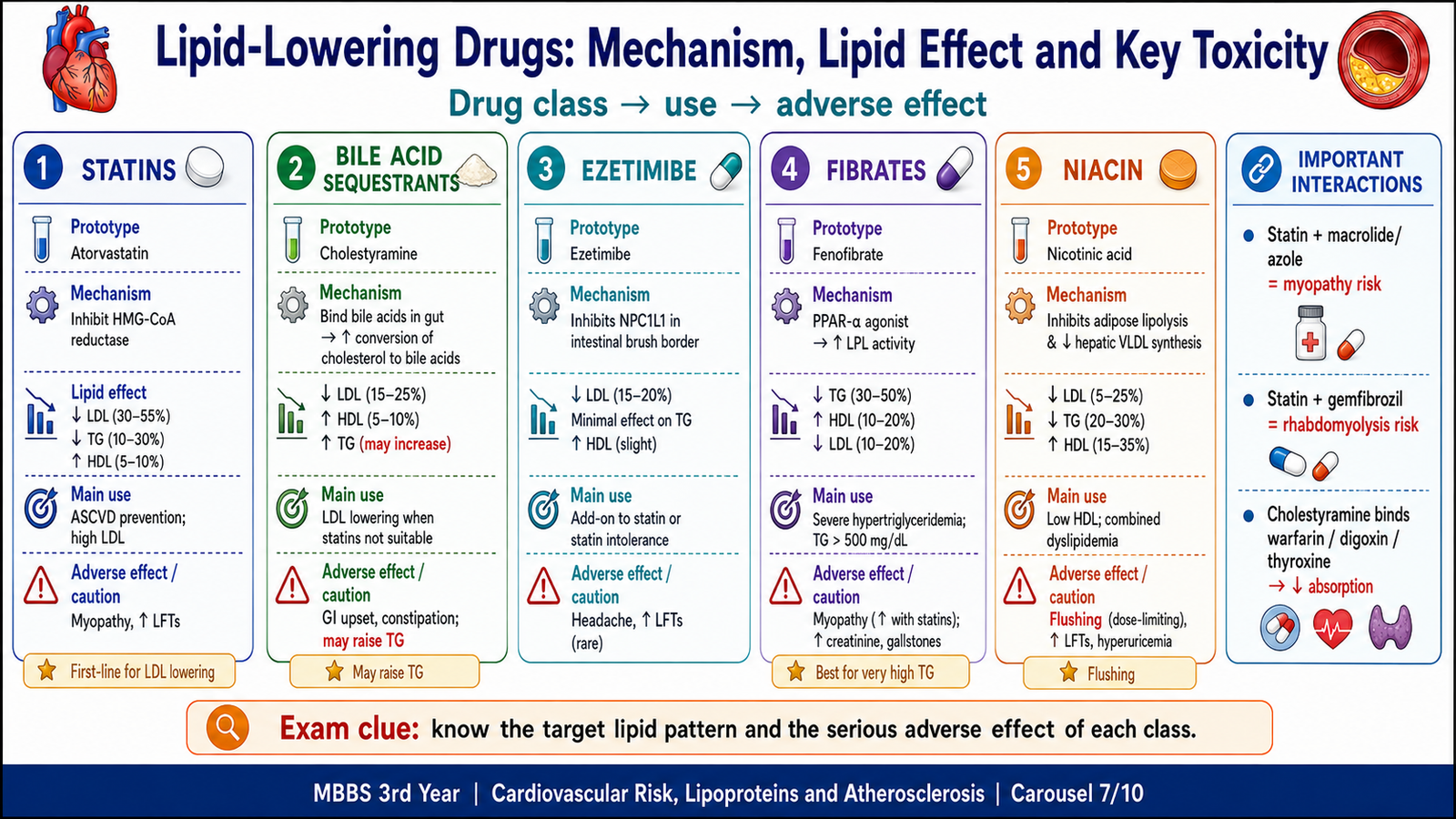
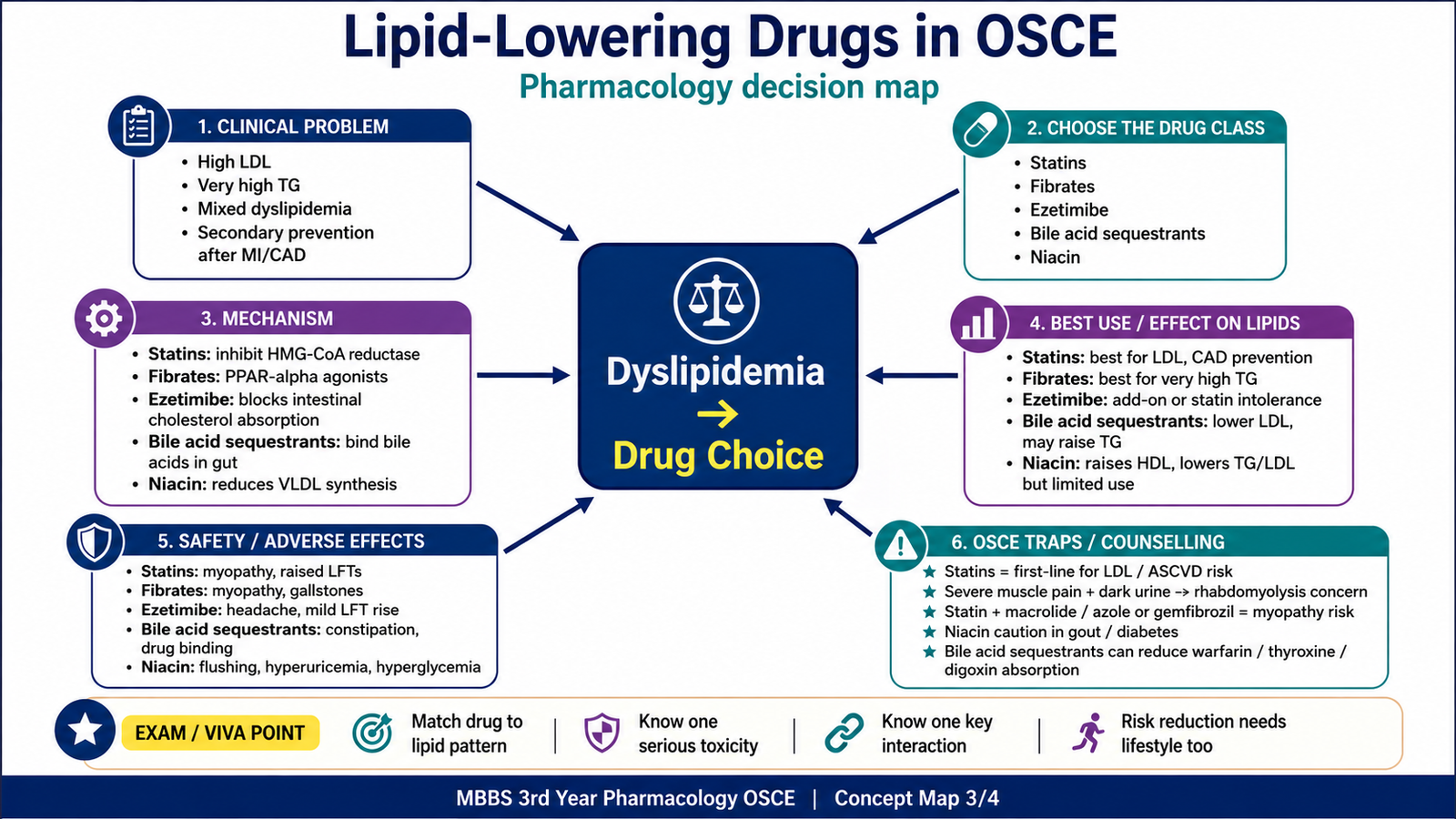
Lipid-lowering drugs reduce atherogenic lipoproteins and therefore lower the chance of plaque progression and cardiovascular events. The five major classes differ in their target, effect on the lipid profile, adverse effects and drug interactions. Statins are the principal drugs for lowering LDL and reducing atherosclerotic cardiovascular risk.
| Class and Examples | Mechanism | Lipid Effect | Important Adverse Effects | Important Interactions or Cautions |
|---|---|---|---|---|
| Statins Atorvastatin, simvastatin, rosuvastatin |
Inhibit HMG-CoA reductase → less hepatic cholesterol → more LDL receptors → increased LDL clearance | Marked ↓ LDL; mild-to-moderate ↓ TG; mild ↑ HDL | Myalgia, myopathy, rare rhabdomyolysis and raised liver enzymes | CYP inhibitors increase levels of susceptible statins; fibrates increase myopathy risk. Avoid in pregnancy and active liver disease. |
| Bile acid sequestrants Cholestyramine, colestipol, colesevelam |
Bind bile acids in the intestine → increased conversion of cholesterol to bile acids → more LDL receptors | ↓ LDL; slight ↑ HDL; TG may rise | Constipation, bloating and reduced absorption of fat-soluble vitamins | Bind many oral drugs and reduce absorption. Avoid in significant hypertriglyceridemia. |
| Ezetimibe | Blocks the intestinal NPC1L1 transporter → reduced cholesterol absorption → increased hepatic LDL receptors | ↓ LDL; mild ↓ TG; mild ↑ HDL | Diarrhea, abdominal discomfort and possible rise in liver enzymes with statins | Bile acid sequestrants reduce its absorption; monitor when combined with drugs affecting the liver. |
| Fibrates Fenofibrate, gemfibrozil |
Activate PPAR-alpha → increased lipoprotein-lipase activity → increased clearance of triglyceride-rich particles | Marked ↓ TG; ↑ HDL; variable LDL effect | Dyspepsia, gallstones, myopathy and raised liver enzymes | Myopathy risk rises with statins, especially gemfibrozil; may enhance the effect of warfarin. |
| Niacin | Reduces adipose lipolysis and hepatic VLDL synthesis → less VLDL and LDL; increases HDL | ↓ TG and LDL; marked ↑ HDL | Flushing, itching, hyperuricemia, hyperglycemia and hepatotoxicity | Alcohol and hepatotoxic drugs increase liver risk; statins may increase myopathy risk. |
Why Important Adverse Effects Occur
Statin-related muscle toxicity becomes more likely when statin blood levels rise because of enzyme inhibition or when another myotoxic drug is added. Bile acid sequestrants remain in the intestine, so their main adverse effects are gastrointestinal and related to binding of vitamins and medicines. Fibrates alter lipid metabolism and the cholesterol content of bile, which explains their association with gallstones. Niacin causes prostaglandin-mediated skin vasodilation, producing flushing, and can worsen glucose and uric-acid control.
Modern agents such as PCSK9 inhibitors and bempedoic acid may be recognized as additional LDL-lowering options, especially in very high-risk or statin-intolerant patients, but the five classes above remain the central undergraduate framework required for this topic.
E. Cardiovascular Risk Stratification and Epidemiology
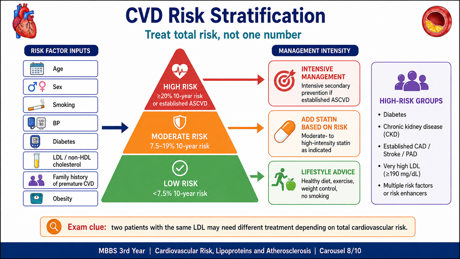
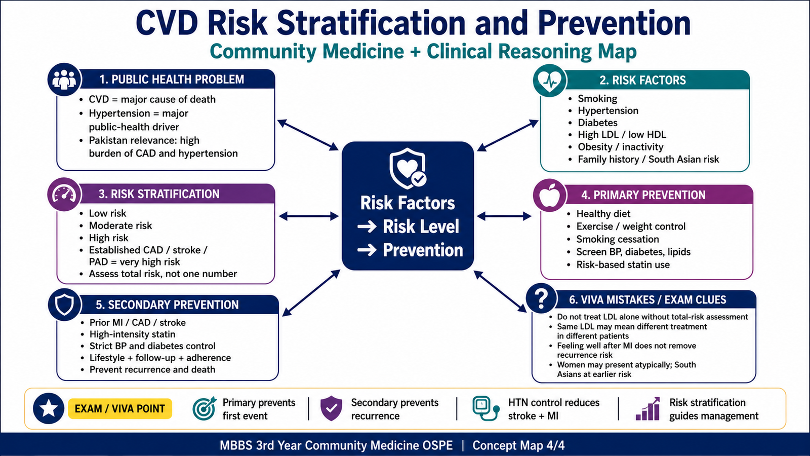
Cardiovascular risk stratification means estimating a person’s overall probability of developing a cardiovascular event over a defined period. It combines several factors rather than considering one laboratory value in isolation. Common factors include age, sex, smoking, blood pressure, diabetes, cholesterol levels, family history and whether atherosclerotic cardiovascular disease is already present.
Risk stratification matters because two people with the same LDL level may not have the same need for treatment. A young person with no other risk factors may initially need lifestyle measures, while an older smoker with hypertension and diabetes has a much higher absolute risk and is more likely to benefit from drug therapy.
Groups with High Cardiovascular Risk
- Established coronary artery disease, stroke or peripheral arterial disease
- Diabetes mellitus or chronic kidney disease
- Very high LDL-C, particularly familial hypercholesterolemia
- Several major risk factors occurring together
- Strong family history of premature coronary disease
Global and Pakistani Public-Health Importance
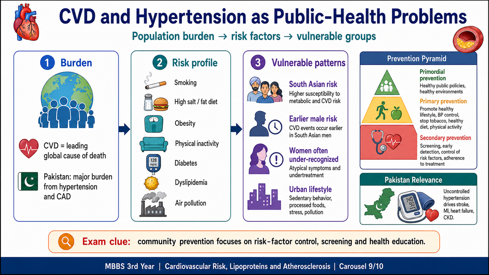
Cardiovascular diseases are among the leading causes of death and disability worldwide, with a particularly heavy burden in low- and middle-income countries. Coronary heart disease and stroke account for a large part of this burden. The public-health effect is increased by the high frequency of smoking, hypertension, diabetes, obesity, unhealthy diet and physical inactivity.
In Pakistan, cardiovascular disease is a major health problem. Hypertension is especially important because it is common, often symptomless and frequently remains undiagnosed or uncontrolled. Uncontrolled blood pressure increases the risk of myocardial infarction, stroke, heart failure, kidney disease and premature death. Population screening, long-term treatment and improved adherence are therefore important public-health priorities.
Cultural, Racial and Gender Differences
Cardiovascular disease does not occur equally in every population. Differences arise from genetics, diet, tobacco exposure, physical activity, access to healthcare, socioeconomic conditions and recognition of symptoms.
- South Asians often develop coronary disease at a younger age and commonly have central obesity, insulin resistance and atherogenic dyslipidemia.
- Men usually develop coronary disease earlier, while risk in women rises after menopause.
- Women may have less typical symptoms and may therefore be under-recognized or diagnosed later.
- Urbanization, sedentary work, high-calorie diets, tobacco, stress and air pollution increase population risk.
- Dietary patterns rich in salt, ghee, trans fats, fried foods and refined carbohydrates can worsen hypertension, obesity and dyslipidemia.
F. Prevention of Coronary Heart Disease
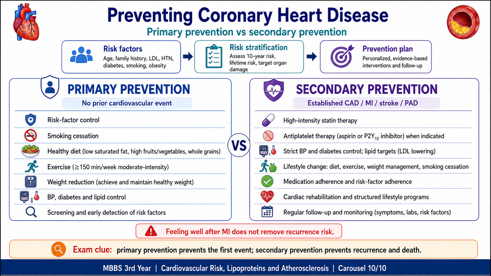
Prevention aims to reduce the risk factors that start or accelerate atherosclerosis. It is divided into primary prevention, which acts before the first cardiovascular event, and secondary prevention, which acts after coronary disease, myocardial infarction, stroke or another atherosclerotic event has already occurred.
| Feature | Primary Prevention | Secondary Prevention |
|---|---|---|
| When used | Before the first MI, stroke or established CAD event | After established CAD, MI, angina, stroke or revascularization |
| Main aim | Prevent the first event | Prevent recurrence, complications and death |
| Core measures | Smoking cessation, healthy diet, physical activity, weight control, BP and diabetes control, and risk-based lipid treatment | Intensive risk-factor control, statin therapy, antiplatelet therapy when indicated, cardiac rehabilitation, adherence and follow-up |
Outpatient Risk-Reduction Strategy
- Stop smoking: this reduces endothelial injury, thrombosis and future cardiovascular risk.
- Control blood pressure: lower pressure reduces mechanical injury to arteries and protects the heart, brain and kidneys.
- Treat dyslipidemia according to total risk: lowering LDL reduces atherogenic particles available for plaque formation.
- Detect and control diabetes: this reduces endothelial dysfunction and associated dyslipidemia.
- Improve diet: reduce trans fats, excess saturated fat, salt and refined sugars; increase healthier food choices.
- Increase physical activity and reduce excess weight: these measures improve blood pressure, insulin sensitivity and the lipid profile.
- Support long-term adherence: cardiovascular risk reduction requires continuous treatment even when the patient feels well.
- Use community measures: health education, tobacco control, screening and improved access to long-term care reduce the population burden.
Integrated Mechanism Flow
↓
Endothelial dysfunction and increased entry of LDL into the intima
↓
LDL modification, monocyte recruitment and macrophage foam-cell formation
↓
Smooth-muscle migration, collagen deposition and fibrous-cap formation
↓
Stable plaque causes chronic stenosis, while vulnerable plaque may rupture
↓
Ischemia, thrombosis, myocardial infarction, stroke or peripheral arterial disease
↓
Risk-factor control, lipid lowering and prevention reduce future events
⭐ AIM High-Yield Review
- Atherosclerosis is a chronic inflammatory disease of the intima of large and medium-sized arteries.
- LDL is the major atherogenic lipoprotein; HDL participates in reverse cholesterol transport.
- HMG-CoA reductase is the rate-limiting enzyme of cholesterol synthesis.
- Major modifiable risks are raised LDL, hypertension, smoking, diabetes, obesity, physical inactivity and unhealthy diet.
- The early lesion is a fatty streak; the mature plaque has a fibrous cap over a necrotic lipid core.
- Foam cells are lipid-filled macrophages produced after uptake of modified LDL.
- A stable plaque usually causes gradual stenosis; a vulnerable plaque has a thin inflamed cap and may rupture suddenly.
- Plaque rupture exposes thrombogenic material and may cause acute coronary thrombosis and myocardial infarction.
- Familial hypercholesterolemia should be suspected with very high LDL, tendon xanthomas and premature coronary disease.
- Statins inhibit HMG-CoA reductase, markedly reduce LDL and are the main drugs for prevention of atherosclerotic cardiovascular events.
- Statin myopathy risk rises with interacting CYP inhibitors and with fibrates, especially gemfibrozil.
- Bile acid sequestrants lower LDL but may raise triglycerides and reduce absorption of other medicines.
- Fibrates mainly lower triglycerides; niacin can cause flushing, hyperuricemia, hyperglycemia and hepatotoxicity.
- Risk stratification uses the patient’s total risk, not one lipid value alone.
- Primary prevention prevents the first event; secondary prevention prevents recurrence and death after established disease.
“`
AIM VIDEO LEARNING
🎥 Hyperlipidemia, Lipoproteins and Atherosclerosis
This video explains lipoproteins, dyslipidemia, atherosclerosis and the main lipid-lowering drugs, including statins and fibrates.
AIM Viewing Focus:
Follow the sequence from abnormal lipoproteins to atherosclerosis, cardiovascular complications and lipid-lowering pharmacology.
“`