
Study tip: This chapter follows the KMU learning outcomes in a simple sequence. First understand how sudden death, hypertrophic cardiomyopathy and chest trauma are connected, then use the final high-yield review for rapid revision.
KMU Curriculum
AIM Learning Cycle
📖 AIM Learning Material
Sudden Cardiac Death, Hypertrophic Cardiomyopathy and Chest Trauma
CVS Module · Forensic Medicine with Medicine and Allied Clinical Integration
Topic Introduction
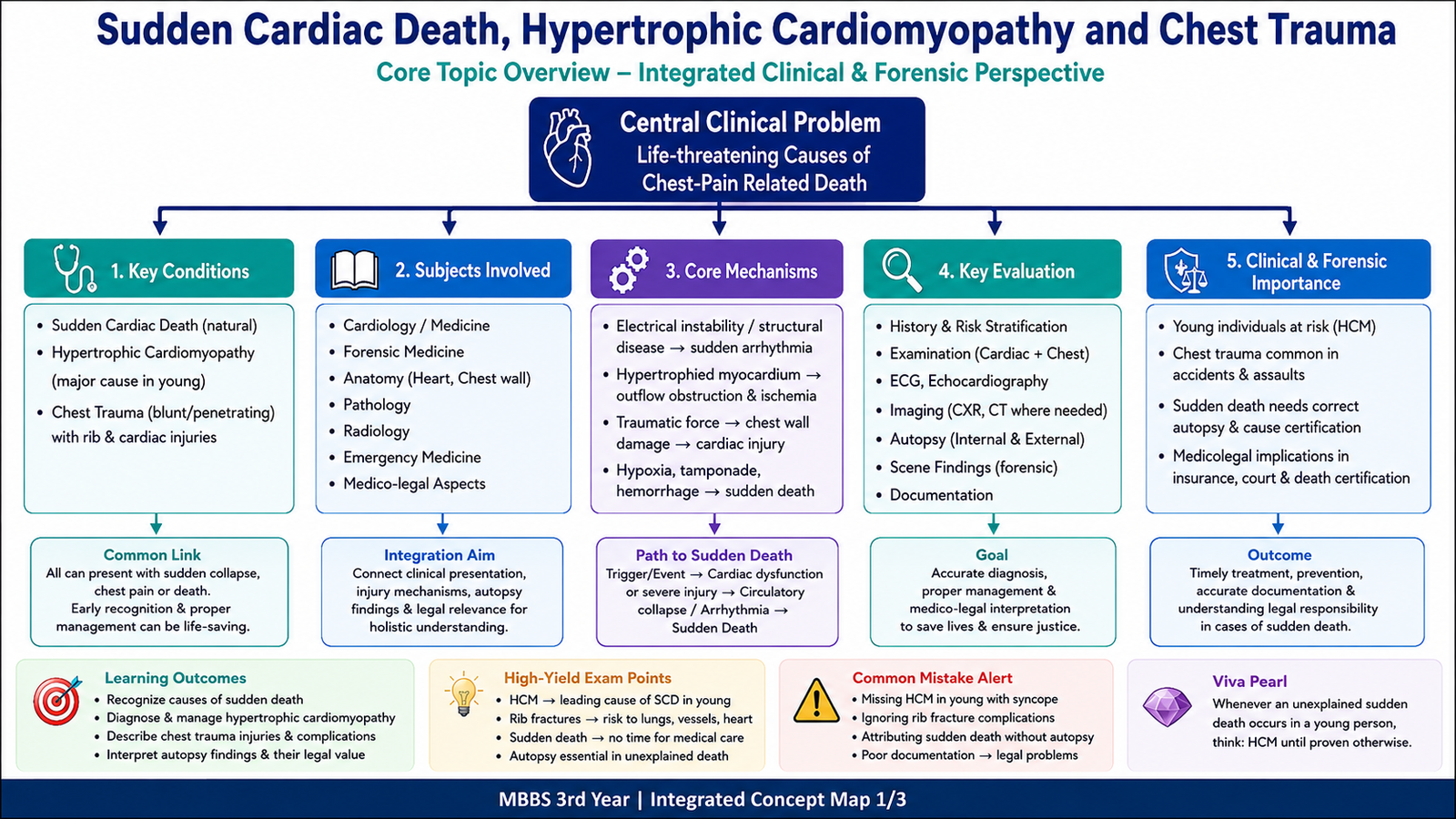
Sudden death is an unexpected death that occurs rapidly in a person who may have appeared healthy. When the heart is responsible, the final event is often a fatal arrhythmia. Hypertrophic cardiomyopathy (HCM) is an important cause in young people because abnormal thickening and disorganization of the myocardium can produce poor filling, outflow obstruction and electrical instability. Chest trauma can also cause sudden death through rib fractures, lung injury, cardiac contusion, rupture, tamponade or commotio cordis. This chapter explains how to recognize HCM, investigate and manage it, assess chest injuries, identify autopsy findings and reach a careful medicolegal conclusion.
A. Sudden Death: Definition, Causes and Forensic Meaning
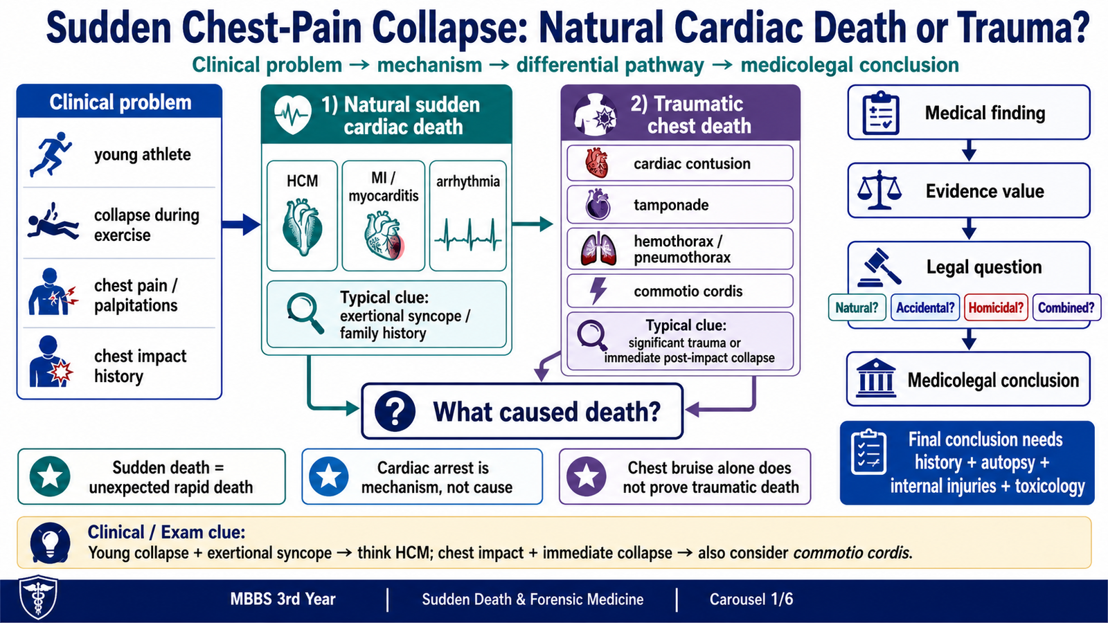
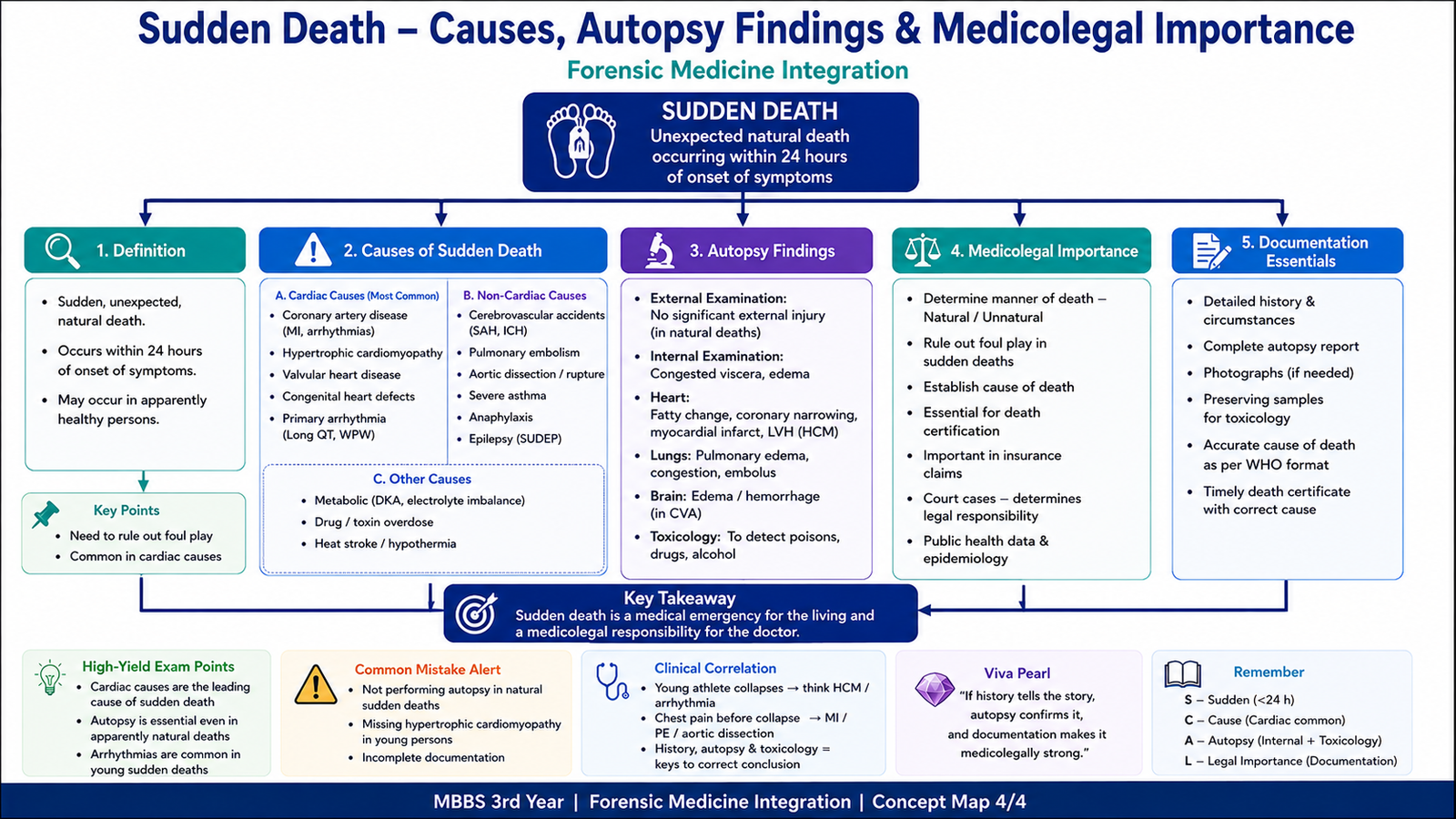
Sudden death is an unexpected natural death occurring abruptly in an apparently healthy person or in someone not known to have a fatal illness. Sudden cardiac death is sudden unexpected death caused by heart disease, most often through ventricular tachycardia or ventricular fibrillation. “Cardiac arrest” describes the final mechanism of dying; it does not identify the disease or injury that caused it.
Important causes
The causes are grouped by the organ system or process responsible. This helps the examiner avoid assuming that every sudden collapse is cardiac.
- Cardiac: coronary artery disease or myocardial infarction, HCM, myocarditis, aortic stenosis, congenital coronary anomalies, arrhythmogenic cardiomyopathy, channelopathies, and aortic dissection or aneurysm rupture.
- Respiratory: pulmonary embolism, severe asthma, pneumothorax and aspiration.
- Central nervous system: subarachnoid hemorrhage, intracerebral hemorrhage and epilepsy-related sudden death.
- Other: poisoning, acute pancreatitis, diabetic emergencies, massive gastrointestinal hemorrhage and adrenal crisis.
Medico-legal importance
Sudden death becomes medicolegally important when it is unexpected, unexplained, suspicious, associated with trauma, or occurs without a clear medical diagnosis. The investigation must decide whether the death was natural, accidental, homicidal, suicidal or undetermined. When trauma is present, the key question is whether it caused death, contributed to death or was merely incidental.
Essential distinction: the cause of death is the disease or injury, the mechanism of death is the physiological disturbance produced by it, and the manner of death is the medicolegal category.
B. Hypertrophic Cardiomyopathy and Sudden Cardiac Death
Hypertrophic cardiomyopathy is a myocardial disease in which the left ventricular myocardium is abnormally thick without another sufficient cause such as long-standing hypertension or aortic stenosis. It is often inherited, so a family history of cardiomyopathy or unexplained sudden death is important.
How HCM develops
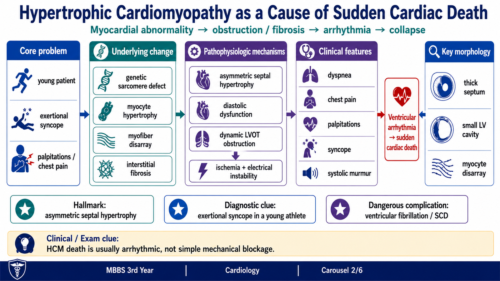
Myocytes become enlarged and arranged in a disorganized pattern called myofibre disarray. The ventricular wall, often the interventricular septum, becomes thick and the ventricular cavity may become small. The thick, stiff ventricle relaxes poorly, so diastolic filling is reduced. In obstructive HCM, systolic anterior motion of the mitral valve narrows the left ventricular outflow tract and may also cause mitral regurgitation.
Abnormal myocardial structure → myocyte hypertrophy and disarray → stiff thick ventricle ± dynamic outflow obstruction → ischemia and fibrosis → ventricular arrhythmia → sudden cardiac death
Why symptoms and sudden death occur
During exercise, the heart rate rises and filling time becomes shorter. A stiff ventricle fills even less effectively, while dynamic obstruction may further reduce forward flow. This can cause exertional breathlessness, chest pain, dizziness or syncope. Myocyte disarray, ischemia and fibrosis also create an electrically unstable myocardium, allowing ventricular tachycardia or ventricular fibrillation to develop. Sudden death therefore usually results from a fatal ventricular arrhythmia rather than complete mechanical blockage of blood flow.
Clinical features
- Exertional dyspnea and reduced exercise tolerance.
- Chest pain or angina-like discomfort due to increased oxygen demand and reduced myocardial perfusion.
- Palpitations caused by atrial or ventricular arrhythmias.
- Dizziness, presyncope or syncope, especially during or after exertion.
- Sudden collapse during sport.
- Family history of HCM or sudden unexplained death.
- Harsh systolic murmur, commonly heard along the left sternal border.
- S4 because atrial contraction forces blood into a stiff ventricle.
In obstructive HCM, the murmur becomes louder when preload falls, such as during standing or the Valsalva manoeuvre. Lower preload makes the ventricular cavity smaller and increases outflow obstruction.
C. Clinical Evaluation, Imaging and Risk Stratification in HCM
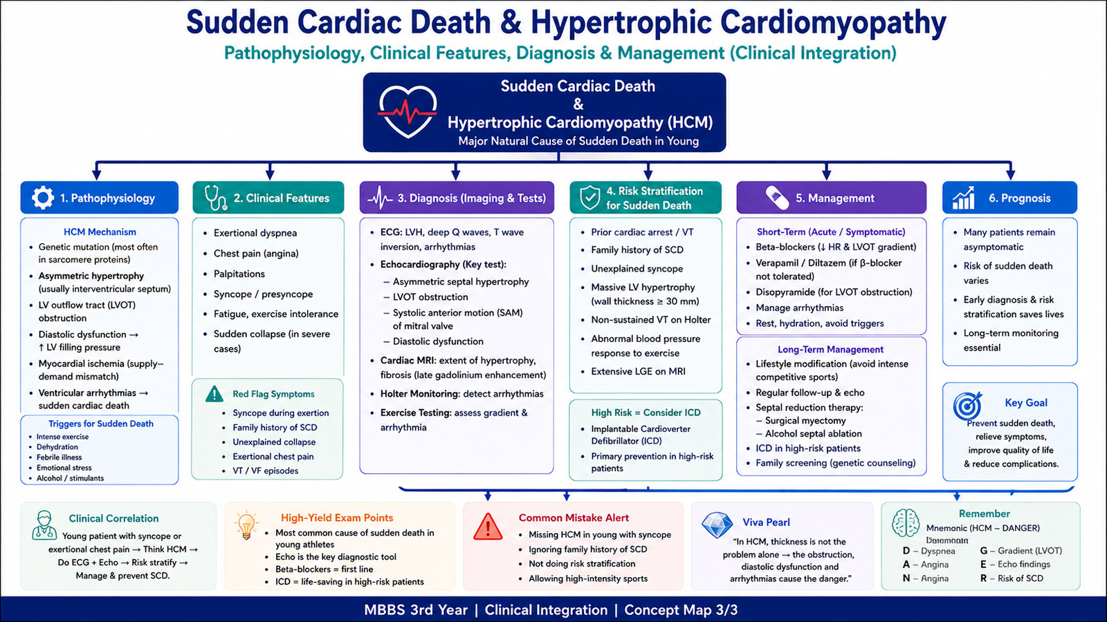
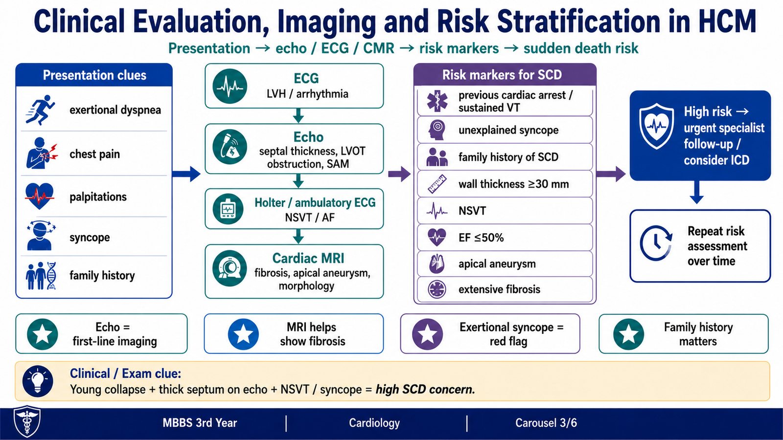
Evaluation has two purposes: to confirm that the patient has HCM and to estimate the risk of sudden cardiac death. The process begins with symptoms and family history, then uses ECG, echocardiography, rhythm monitoring and cardiac MRI to define structure, function and electrical risk.
Clinical assessment
- Ask about exertional dyspnea, chest pain, palpitations, presyncope and syncope.
- Record any previous cardiac arrest or documented ventricular arrhythmia.
- Ask about HCM, cardiomyopathy or premature sudden death in relatives.
- Examine for a systolic murmur and signs of heart failure or arrhythmia.
Investigations and imaging protocol
ECG may show left ventricular hypertrophy, deep Q waves, ST–T changes or arrhythmias. It supports suspicion but cannot define the cardiac anatomy.
Echocardiography is the key initial imaging test. It shows the distribution and degree of hypertrophy, septal thickness, left ventricular cavity size, systolic anterior motion of the mitral valve, mitral regurgitation, diastolic dysfunction, left atrial enlargement and resting or provoked left ventricular outflow tract obstruction.
Ambulatory ECG monitoring looks for intermittent arrhythmias, especially non-sustained ventricular tachycardia. Cardiac MRI is useful when echocardiography is incomplete or when more detail is needed. It can show apical HCM, apical aneurysm, accurate wall thickness and myocardial fibrosis through late gadolinium enhancement.
Risk stratification
Risk stratification identifies patients who may need stronger prevention of sudden cardiac death. Risk is not decided by septal thickness alone; clinical history, rhythm findings, ventricular function and MRI findings must be considered together.
- Previous cardiac arrest or sustained ventricular tachycardia.
- Unexplained syncope suspected to be arrhythmic.
- Family history of premature sudden cardiac death.
- Massive left ventricular hypertrophy, especially a wall thickness of 30 mm or more.
- Non-sustained ventricular tachycardia on ambulatory monitoring.
- Left ventricular systolic dysfunction.
- Left ventricular apical aneurysm.
- Extensive late gadolinium enhancement indicating fibrosis on cardiac MRI.
Red flag: exertional syncope in a patient with possible HCM may represent a transient malignant arrhythmia and requires urgent cardiac assessment.
D. Short-Term and Long-Term Management of HCM
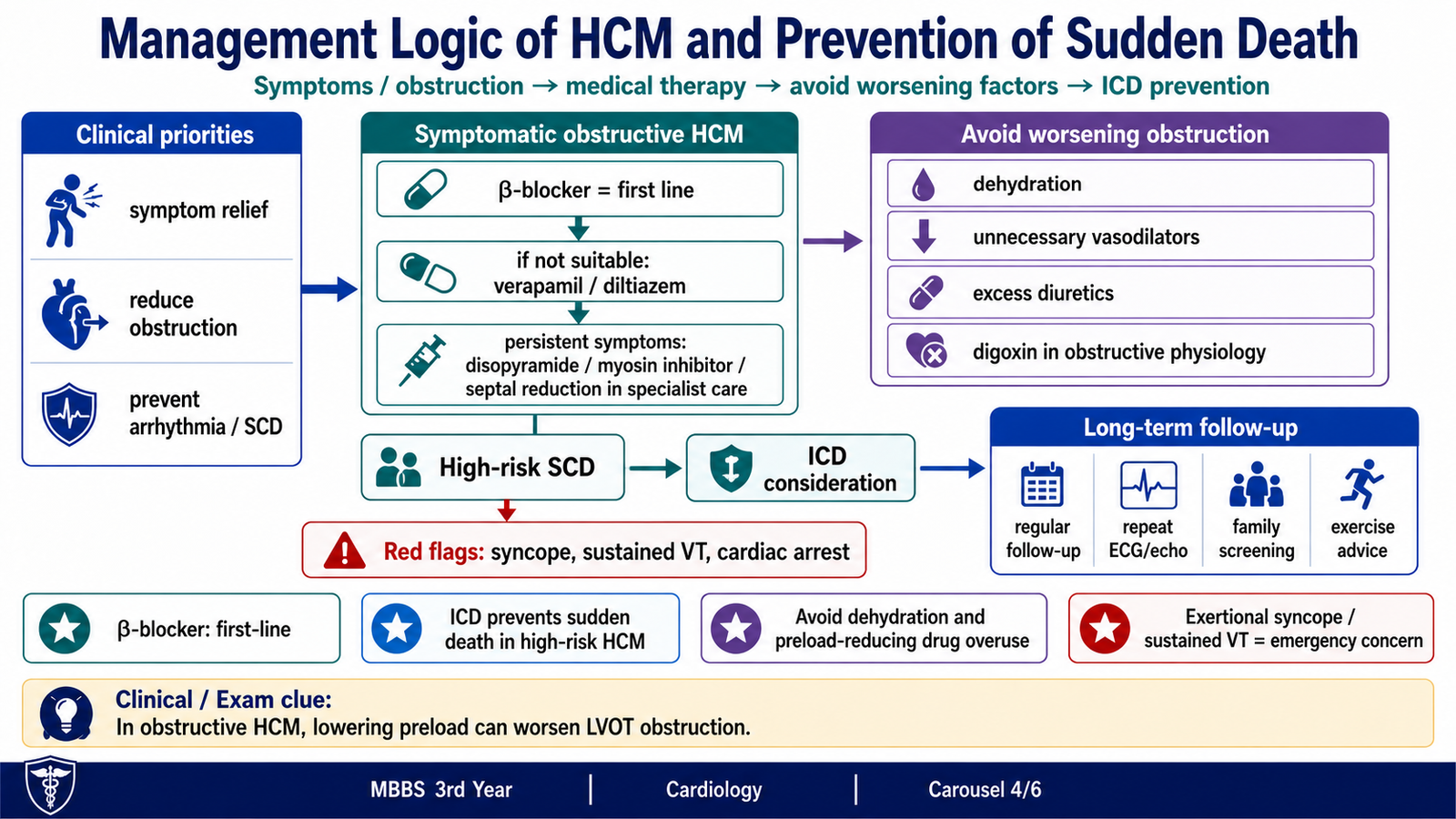
Management aims to relieve symptoms caused by poor filling, obstruction and arrhythmias, while also preventing sudden cardiac death in high-risk patients. The haemodynamic principle is important: in obstructive HCM, reduced ventricular filling or increased contractility can make the outflow obstruction worse.
Short-term approach
A patient with syncope, severe chest pain, hypotension, breathlessness or a ventricular arrhythmia needs urgent assessment and emergency care. Dehydration should be corrected carefully because loss of preload makes the ventricular cavity smaller and can increase obstruction.
- Avoid dehydration and sudden marked reduction in preload.
- Avoid unnecessary vasodilators and excessive diuresis in obstructive physiology.
- Avoid drugs that markedly increase contractility unless specialist-directed.
- Restrict strenuous activity while symptoms are unstable and obtain specialist assessment.
Symptom control
Non-vasodilating beta-blockers are commonly used first because they slow the heart, increase diastolic filling time, reduce myocardial oxygen demand and lessen dynamic obstruction during exertion. If they are unsuitable or ineffective, verapamil or diltiazem may be considered with caution, especially when hypotension or severe obstruction is present. Persistent symptomatic obstructive HCM may require specialist therapies such as disopyramide, a cardiac myosin inhibitor or septal reduction treatment.
Long-term management and prevention
- Regular follow-up with reassessment of symptoms and risk factors.
- Repeat ECG, echocardiography and ambulatory rhythm monitoring as clinically indicated.
- Cardiac MRI when additional structural or fibrosis assessment is required.
- Family screening and genetic counselling where appropriate.
- Individual exercise advice based on symptoms and risk.
- Recognition and treatment of atrial fibrillation and other arrhythmias.
- Implantable cardioverter-defibrillator consideration in patients at high risk.
An implantable cardioverter-defibrillator is particularly important for secondary prevention after survival from cardiac arrest or sustained ventricular arrhythmia. In selected patients without a previous arrest, the decision is based on the combined risk profile.
E. Chest Wall, Rib and Cardiac Injuries
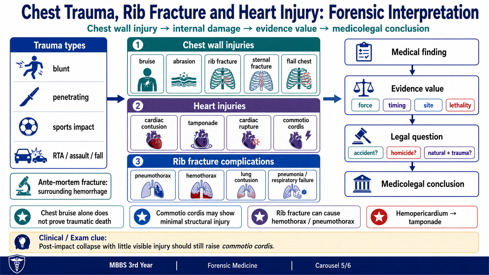
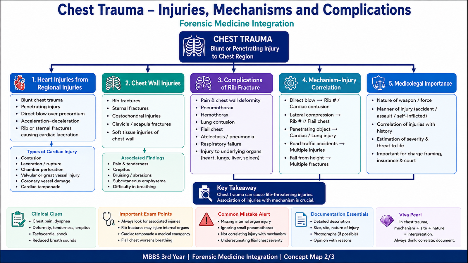
Chest trauma may be blunt, as in road traffic accidents, falls, compression, assault, crush injury or sports impact, or penetrating, as in stab and firearm injuries. The clinical and forensic importance of an external mark depends on the internal damage beneath it. A small bruise may accompany serious internal injury, while a visible bruise alone does not prove that trauma caused death.
Chest wall injuries
Common injuries include abrasions, contusions, lacerations, rib fractures, sternal or clavicular fractures, intercostal vessel injury, soft-tissue hemorrhage and flail chest. Multiple adjacent rib fractures can create a free segment of chest wall. Its paradoxical movement reduces effective ventilation and may contribute to respiratory failure.
Heart injuries caused by regional chest trauma
- Cardiac contusion: blunt force bruises the myocardium and may cause arrhythmia, conduction disturbance or pump failure.
- Cardiac rupture: severe blunt trauma tears a chamber, producing rapid hemorrhage or hemopericardium with tamponade.
- Penetrating cardiac injury: a sharp or firearm wound can puncture the heart and cause massive bleeding or tamponade.
- Hemopericardium and tamponade: blood collects in the pericardial sac, compresses the heart and prevents adequate filling, producing obstructive shock.
- Coronary artery injury: vessel damage can cause myocardial ischemia and arrhythmia.
- Commotio cordis: a sudden precordial blow during a vulnerable phase of the cardiac cycle triggers ventricular fibrillation, often with little or no gross structural injury.
Complications of rib fracture
A fractured rib causes pain and splinting, so the patient breathes less deeply. This promotes atelectasis and pneumonia. Sharp fragments may also injure the pleura, lungs, vessels or upper abdominal organs.
- Severe pain and restricted breathing
- Atelectasis
- Pneumonia
- Pneumothorax
- Hemothorax
- Pulmonary contusion
- Lung laceration
- Surgical emphysema
- Flail chest
- Respiratory failure
- Intercostal vessel injury
- Cardiac or great-vessel injury
- Liver or spleen injury with lower rib fractures
Forensic clue: an ante-mortem rib fracture usually has hemorrhage in the surrounding tissues, showing that circulation was present when the injury occurred.
F. Autopsy Findings and Medico-Legal Conclusion
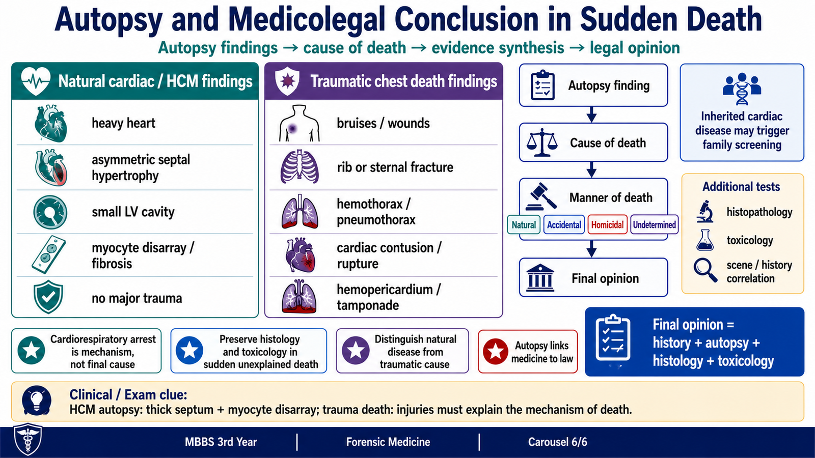
Autopsy connects the medical findings with the legal questions. It aims to identify the cause of death, explain the mechanism, determine the manner of death, document injuries and disease, and decide whether trauma, poisoning or natural disease caused or contributed to death. The opinion should be based on the complete scene history, external and internal examination, histology and toxicology where required.
Autopsy findings in HCM-related sudden death
- Enlarged and heavy heart.
- Thickened left ventricular wall, often with asymmetric septal hypertrophy.
- Small left ventricular cavity.
- Possible endocardial thickening in the outflow tract.
- Myocardial fibrosis.
- Microscopic myocyte hypertrophy, myofibre disarray and interstitial fibrosis.
- No major coronary obstruction in many young patients.
These findings support HCM as a natural cause of sudden cardiac death, but they must be interpreted with the circumstances and exclusion of other causes.
Other possible sudden cardiac death findings
Depending on the cause, autopsy may reveal coronary atherosclerosis or thrombosis, acute or healed myocardial infarction, myocarditis, valvular disease, aortic dissection, congenital coronary abnormalities or another cardiomyopathy. Some primary electrical disorders may leave no specific gross structural lesion, making the history, toxicology and specialist testing important.
Findings in chest trauma death
- External abrasions, bruises, lacerations or patterned injuries.
- Soft-tissue hemorrhage and rib, sternal or clavicular fractures.
- Pneumothorax, hemothorax, pulmonary contusion or lung laceration.
- Cardiac contusion, chamber rupture or penetrating cardiac wound.
- Hemopericardium and cardiac tamponade.
- Injury to coronary arteries or great vessels.
- Minimal structural injury in commotio cordis, requiring careful correlation with a witnessed precordial blow and sudden collapse.
Reaching the medicolegal opinion
The examiner should link each finding to its evidentiary value. The presence, severity, timing and anatomical consequences of an injury must be assessed. A chest bruise alone is not enough to certify traumatic death, and the presence of natural heart disease does not automatically exclude trauma. The final opinion may be natural, traumatic, combined or undetermined when the available evidence is insufficient.
Careful interpretation: when gross findings do not fully explain death, histology and toxicology may reveal hidden disease or poisoning. Negative findings must also be interpreted cautiously because some arrhythmic deaths leave little structural evidence.
Integrated Mechanism Flow
AIM High-Yield Review
- ⭐ Sudden death is an unexpected natural death occurring abruptly; sudden cardiac death is caused by heart disease, usually through a fatal arrhythmia.
- “Cardiac arrest” is a mechanism, not an adequate underlying cause of death.
- Young athlete with exertional syncope, palpitations or collapse and a family history of sudden death should raise suspicion of HCM.
- HCM shows myocardial hypertrophy, myofibre disarray, a stiff ventricle and sometimes dynamic left ventricular outflow obstruction.
- Sudden death in HCM usually results from ventricular tachycardia or ventricular fibrillation.
- The HCM murmur becomes louder when preload falls, such as during standing or Valsalva.
- Echocardiography is the main initial imaging test; cardiac MRI better defines difficult morphology and myocardial fibrosis.
- Important HCM risk markers include previous arrest or sustained VT, unexplained syncope, family history, massive LV hypertrophy, NSVT, LV dysfunction, apical aneurysm and extensive MRI fibrosis.
- Beta-blockers reduce heart rate, improve filling and lessen dynamic obstruction; dehydration, excessive diuresis and unnecessary vasodilation may worsen obstructive HCM.
- Commotio cordis is ventricular fibrillation after a precordial blow and may leave little or no gross structural injury.
- Rib fractures may cause pneumothorax, hemothorax, pulmonary contusion, flail chest, respiratory failure and injury to vessels or upper abdominal organs.
- Hemothorax is blood in the pleural cavity; cardiac tamponade is blood or fluid in the pericardial sac compressing the heart.
- Ante-mortem rib fractures usually show surrounding soft-tissue hemorrhage.
- A chest bruise alone does not prove traumatic death; internal injury and the complete circumstances must support the mechanism.
- Autopsy in sudden death should distinguish cause, mechanism and manner of death and may require histology and toxicology.
🎥 AIM Video Learning
Watch this video to strengthen your understanding of hypertrophic cardiomyopathy, risk assessment and sudden cardiac death.
AIM focus: Relate myocardial hypertrophy and fibrosis to ventricular arrhythmia, risk stratification and prevention of sudden cardiac death.