
Study tip: This chapter follows the KMU learning outcomes in a simple sequence. First understand the explanations and cause-and-effect links, then use the AIM High-Yield Review for quick revision.
📖 AIM Learning Material
Topic 8 — Venous Disorders, Vascular Tumors and Cardiac Poisons
CVS Module | Pathology, Forensic Medicine & Toxicology, and PRIME
Topic Introduction
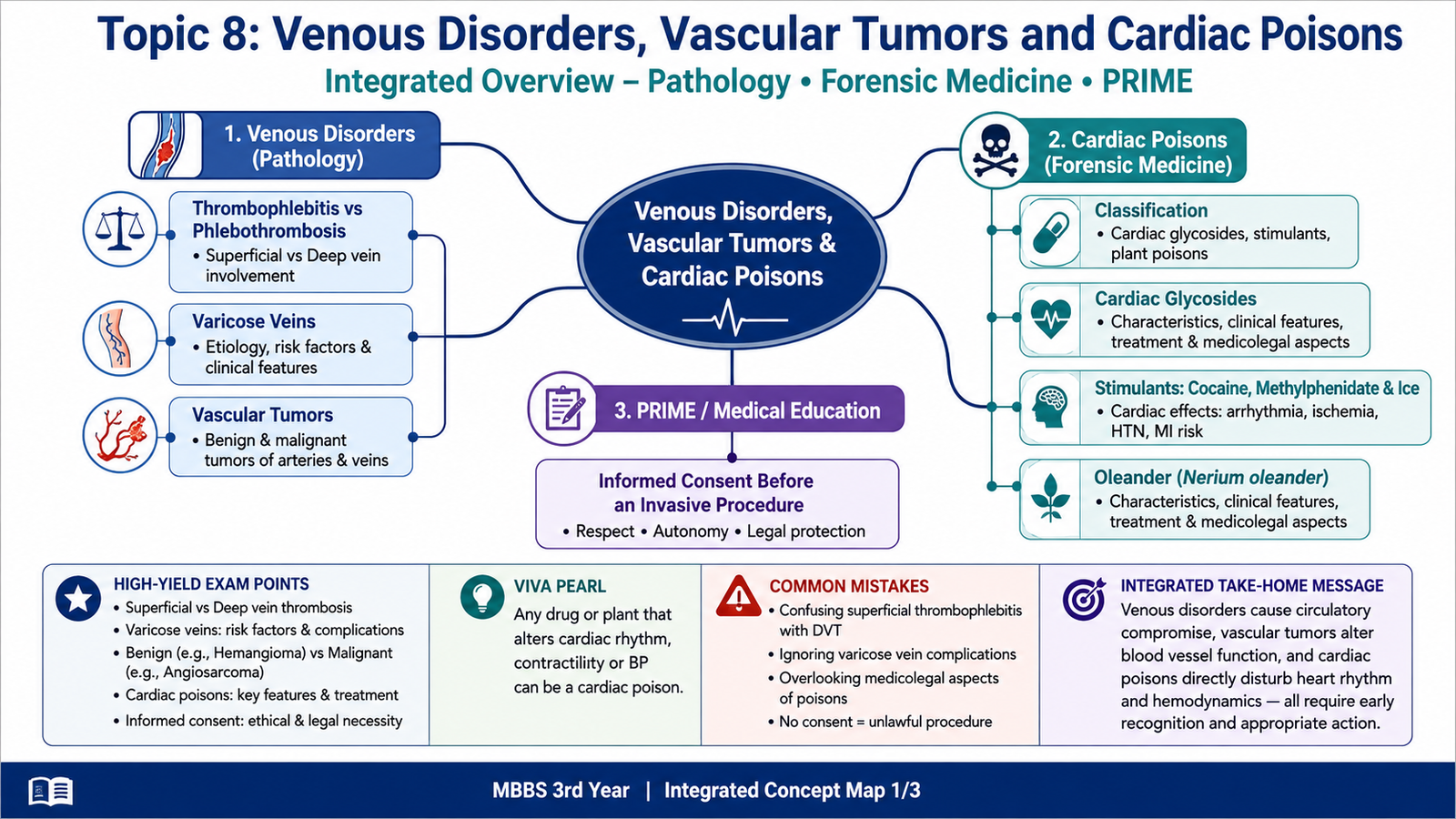
This topic brings together three important areas. First, it explains common venous disorders, including thrombophlebitis, phlebothrombosis and varicose veins. Second, it introduces benign and malignant tumors arising from blood vessels. Third, it describes cardiac poisons, especially cardiac glycosides, Oleander and stimulant drugs such as methylphenidate, cocaine and Ice. The central idea is simple: venous disease mainly disturbs blood flow, vascular tumors represent abnormal growth of vessel-forming tissues, and cardiac poisons disturb heart rate, conduction, contractility or coronary blood flow. The chapter also explains informed consent before an invasive procedure.
A. Thrombophlebitis and Phlebothrombosis
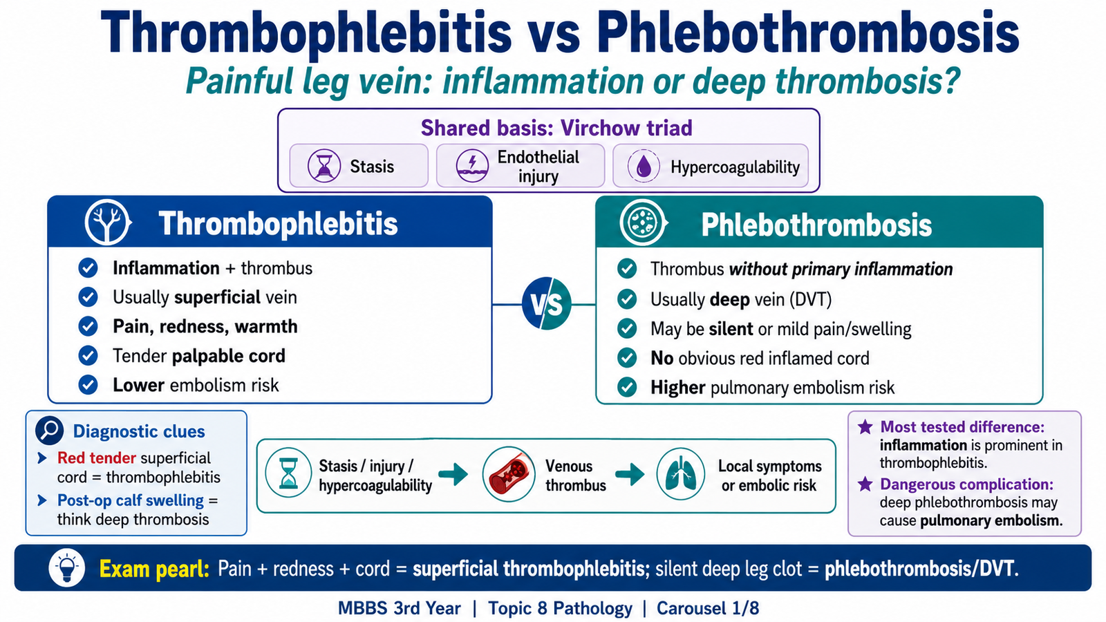
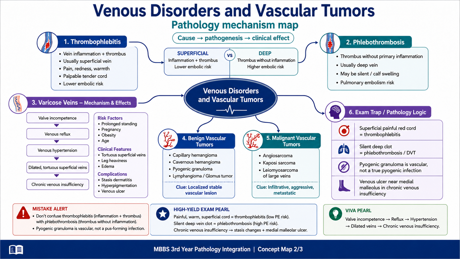
Both conditions involve a thrombus inside a vein, but they differ in the role of inflammation. Thrombophlebitis is inflammation of a vein associated with thrombus formation. It commonly affects superficial veins and therefore usually produces obvious local inflammation. Phlebothrombosis is venous thrombosis without marked primary inflammation. It commonly affects deep veins and may remain clinically silent. “`
How venous thrombosis develops
Venous thrombi usually develop through Virchow’s triad. Stasis slows blood flow, endothelial injury exposes a thrombogenic surface, and hypercoagulability increases the tendency of blood to clot. In thrombophlebitis, inflammation of the vein wall is prominent and helps anchor the thrombus. In phlebothrombosis, stasis and hypercoagulability are often more important.
Stasis, endothelial injury or hypercoagulability → venous thrombus → obstruction of venous flow → swelling and pain → possible embolization if the thrombus is in a deep vein
| Feature | Thrombophlebitis | Phlebothrombosis |
|---|---|---|
| Basic process | Vein inflammation with thrombosis | Thrombosis without marked primary inflammation |
| Common site | Usually superficial veins | Usually deep leg veins |
| Local signs | Pain, redness, warmth and a tender cord | May cause swelling and pain, but can be silent |
| Thrombus attachment | Often firmly attached to inflamed wall | May extend within a deep vein |
| Embolism risk | Usually lower when confined to superficial veins | Higher because deep venous thrombi may cause pulmonary embolism |
A painful, red, warm, cord-like superficial vein strongly suggests thrombophlebitis. Deep phlebothrombosis may present with calf swelling, pain and tenderness, but the absence of obvious inflammation does not exclude it. Duplex ultrasonography is useful when deep venous thrombosis is suspected. The major danger of deep phlebothrombosis is pulmonary embolism.
B. Varicose Veins
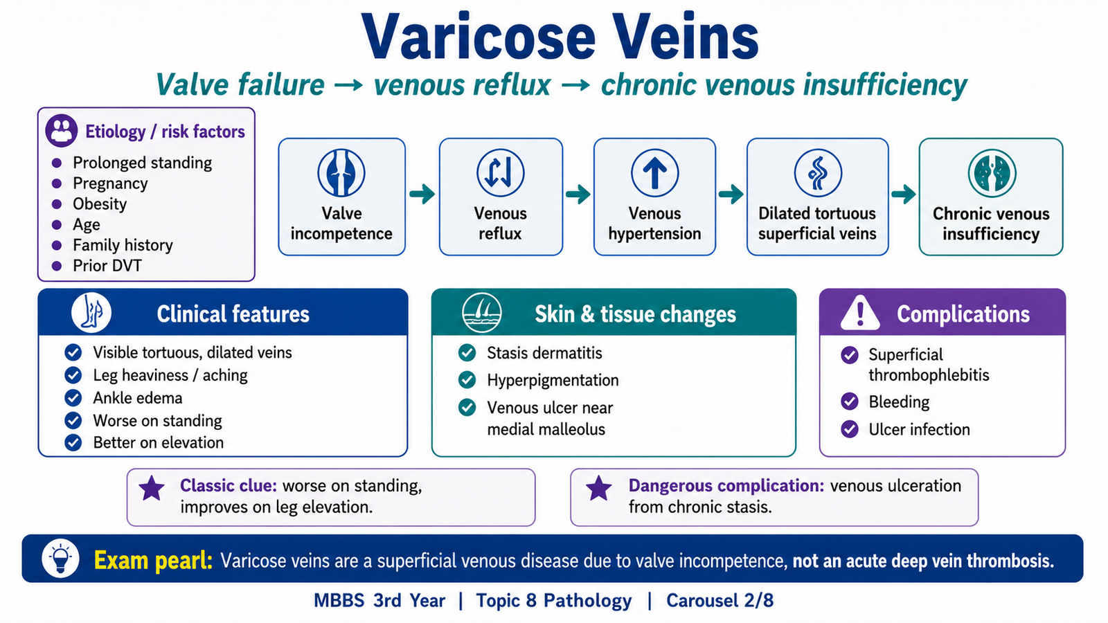
Varicose veins are abnormally dilated, elongated and tortuous superficial veins. The basic problem is failure of venous valves. When valves do not close properly, blood falls backward under gravity, especially during prolonged standing. This reflux raises venous pressure, stretches the vein and separates the valve cusps even more. The result is a self-perpetuating cycle of valve failure and venous dilatation. “`
Etiology and risk factors
Anything that weakens the venous wall, damages valves or increases venous pressure can promote varicose veins.
- Prolonged standing and increasing age
- Pregnancy, obesity and increased intra-abdominal pressure
- Family tendency or weak connective-tissue support
- Previous deep venous thrombosis causing secondary obstruction and valve damage
- Congenital venous weakness or arteriovenous fistula
Valve incompetence → downward reflux → venous hypertension → superficial vein dilatation → further valve separation → chronic venous congestion
Morphology and clinical features
Grossly, the affected superficial veins are elongated, dilated and tortuous. Their walls may become irregularly thickened or thinned, and valves become deformed or incompetent. Microscopically, the wall may show changes in smooth muscle and elastic tissue, fibrosis and, in severe chronic stasis, inflammation in surrounding tissues.
| Clinical feature | Why it occurs |
|---|---|
| Visible tortuous veins | Superficial veins dilate under persistently raised venous pressure |
| Heaviness and aching | Venous congestion increases during standing |
| Ankle edema | Raised hydrostatic pressure pushes fluid into tissues |
| Stasis dermatitis and itching | Chronic venous stasis injures the skin |
| Brown pigmentation | Red cells escape and break down, leaving hemosiderin |
| Venous ulcer | Long-standing congestion causes poor oxygenation and tissue nutrition, commonly near the medial malleolus |
Complications include superficial thrombophlebitis, chronic venous insufficiency, stasis dermatitis, venous ulceration, bleeding from a ruptured superficial varix and secondary infection of an ulcer. Symptoms usually worsen during prolonged standing and improve with leg elevation because elevation lowers venous pressure.
C. Benign and Malignant Vascular Tumors
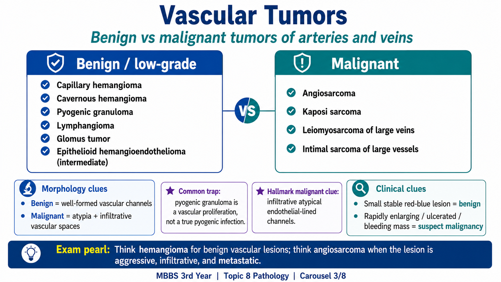
Vascular tumors arise from endothelial cells, vascular supporting tissues or smooth muscle in the walls of larger vessels. Their behaviour ranges from harmless localized lesions to aggressive malignant tumors. Students should know the major examples and the morphological clues that separate benign from malignant growth. “`
| Group | Important examples | General behaviour |
|---|---|---|
| Benign | Capillary hemangioma, cavernous hemangioma, pyogenic granuloma, lymphangioma, glomus tumor, vascular malformations and rare benign smooth-muscle tumors of vessel wall | Usually localized and well differentiated |
| Intermediate | Epithelioid hemangioendothelioma | Behaviour lies between clearly benign and highly malignant lesions |
| Malignant | Angiosarcoma, Kaposi sarcoma, leiomyosarcoma of large veins and rare intimal sarcoma of large vessels | Infiltrative growth, tissue destruction and potential metastasis |
Important morphological patterns
Hemangioma usually appears as a red-blue lesion and contains blood-filled vascular channels lined by bland endothelial cells. Capillary hemangioma contains small capillary-sized vessels, whereas cavernous hemangioma contains large dilated vascular spaces. Pyogenic granuloma is a rapidly growing red lesion that bleeds easily, often after trauma or irritation. Despite its name, it is neither a true pus-forming infection nor a true granuloma; it is a lobular capillary vascular proliferation. Glomus tumor is a small, very painful benign tumor, often beneath a fingernail. It arises from structures involved in temperature regulation. Angiosarcoma is malignant and infiltrative. Microscopically, it forms irregular vascular channels lined by atypical endothelial cells. It may ulcerate, bleed and metastasize. Kaposi sarcoma is associated with HHV-8 and typically shows spindle cells, slit-like vascular spaces and extravasated red blood cells. Leiomyosarcoma may arise from the smooth muscle of a large vein, particularly the inferior vena cava. A small, stable and well-circumscribed lesion favours benign behaviour. Rapid enlargement, infiltration, ulceration, repeated bleeding and marked cellular atypia raise concern for malignancy. Histopathology confirms the diagnosis when malignancy is suspected.
D. Classification of Cardiac Poisons
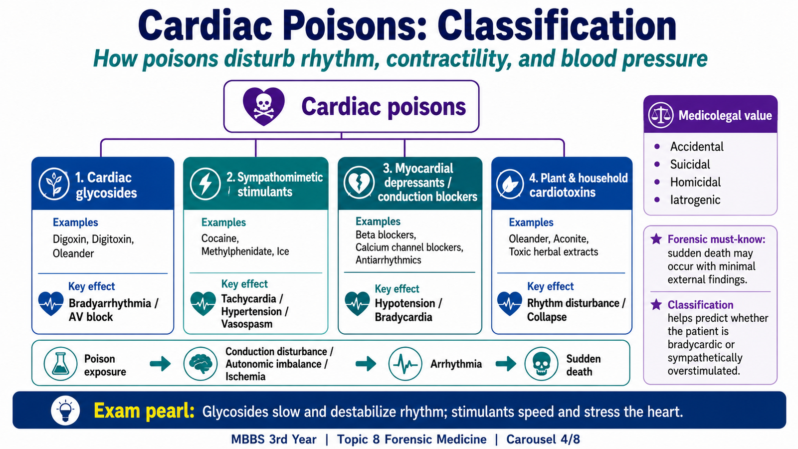
Cardiac poisons are substances that produce serious illness or death by disturbing cardiac rhythm, conduction, contractility, blood pressure, electrolyte balance or coronary blood flow. A practical classification is based on the main way in which the substance harms the heart. “`
| Class | Examples | Main cardiac disturbance |
|---|---|---|
| Cardiac glycosides | Digoxin, digitoxin, foxglove, Oleander and yellow Oleander | Abnormal conduction and many types of arrhythmia |
| Sympathomimetic stimulants | Methylphenidate, cocaine and methamphetamine or Ice | Tachycardia, hypertension, vasoconstriction, ischemia and arrhythmia |
| Myocardial depressants and conduction blockers | Substances that suppress myocardial activity or block ion-channel-dependent conduction | Bradycardia, conduction block, hypotension and reduced contractility |
| Electrolyte and metabolic poisons | Poisons producing major potassium, calcium, acid-base or metabolic disturbance | Electrical instability and rhythm disturbance |
| Plant and household cardiotoxins | Oleander and other cardiotoxic plant exposures | Often glycoside-like rhythm and conduction abnormalities |
Different poisons may produce similar final effects, such as arrhythmia, hypotension, myocardial ischemia or sudden cardiac death. For this reason, the history of exposure, clinical pattern, electrocardiogram and relevant laboratory findings must be interpreted together.
E. Cardiac Glycosides and Oleander Poisoning
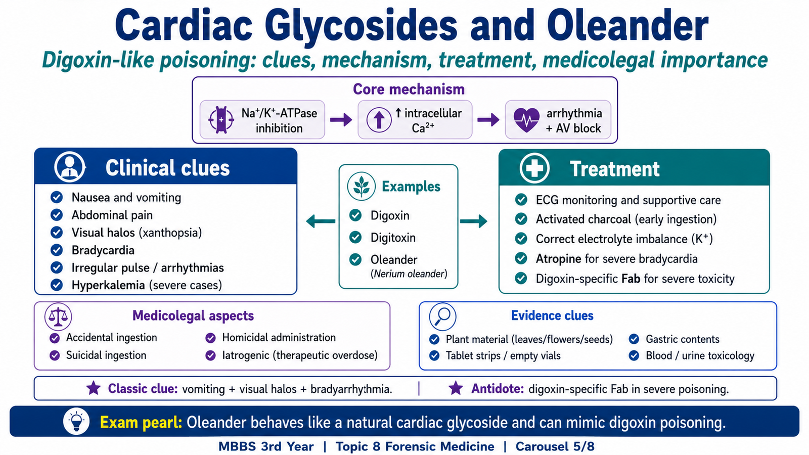
Cardiac glycosides include therapeutic drugs such as digoxin and naturally occurring glycosides found in foxglove and Oleander. Their toxicity is important because the same mechanism that increases cardiac contractility can also make the conducting system electrically unstable. “`
Mechanism
Cardiac glycosides inhibit the sodium-potassium ATPase pump. Intracellular sodium rises, reducing the activity of the sodium-calcium exchanger. Calcium then accumulates inside cardiac cells, which increases contractility. Toxic concentrations also increase vagal activity and disturb automaticity and conduction. The result may be bradycardia, atrioventricular block or many different atrial and ventricular arrhythmias. Severe acute poisoning may be associated with hyperkalemia because potassium cannot enter cells normally.
Na⁺/K⁺-ATPase inhibition → increased intracellular Na⁺ → reduced Na⁺/Ca²⁺ exchange → increased intracellular Ca²⁺ → stronger contraction but electrical instability and arrhythmias
Clinical signs and symptoms
Gastrointestinal symptoms often appear early because glycosides affect excitable tissues and the autonomic nervous system. Neurological and visual symptoms may follow, while cardiac toxicity is the major danger.
- Nausea, vomiting, abdominal discomfort and loss of appetite
- Weakness, dizziness, confusion and visual disturbance
- Bradycardia, irregular pulse, atrioventricular block or tachyarrhythmia
- Hypotension and circulatory collapse in severe poisoning
- Hyperkalemia in severe acute toxicity
Treatment principles
Management begins with airway, breathing and circulation, followed by continuous cardiac monitoring. The poison should be stopped, electrolyte and acid-base abnormalities corrected, and clinically important arrhythmias treated. Activated charcoal may be considered after a recent significant ingestion when the airway is protected. Digoxin-specific antibody fragments are the specific antidote for severe cardiac glycoside poisoning.
Oleander
Oleander is an ornamental plant containing cardiac glycosides in its leaves, flowers, stems and seeds. Ingestion may be accidental, suicidal or related to traditional herbal use. Its poisoning resembles digoxin toxicity: vomiting and abdominal symptoms are followed by bradycardia, conduction block, ventricular arrhythmias, hypotension and possible hyperkalemia. Treatment follows the same supportive and antidotal principles used for severe cardiac glycoside poisoning.
Medico-legal importance
The circumstances of exposure must be documented carefully because poisoning may be accidental, suicidal or, rarely, homicidal. Relevant plant material, medicine containers, vomitus or gastric contents, blood and urine may assist investigation when collected according to local procedure. Findings should be interpreted cautiously because clinical features and arrhythmias are not specific to a single poison.
F. Cardiac Effects of Methylphenidate, Cocaine and Ice
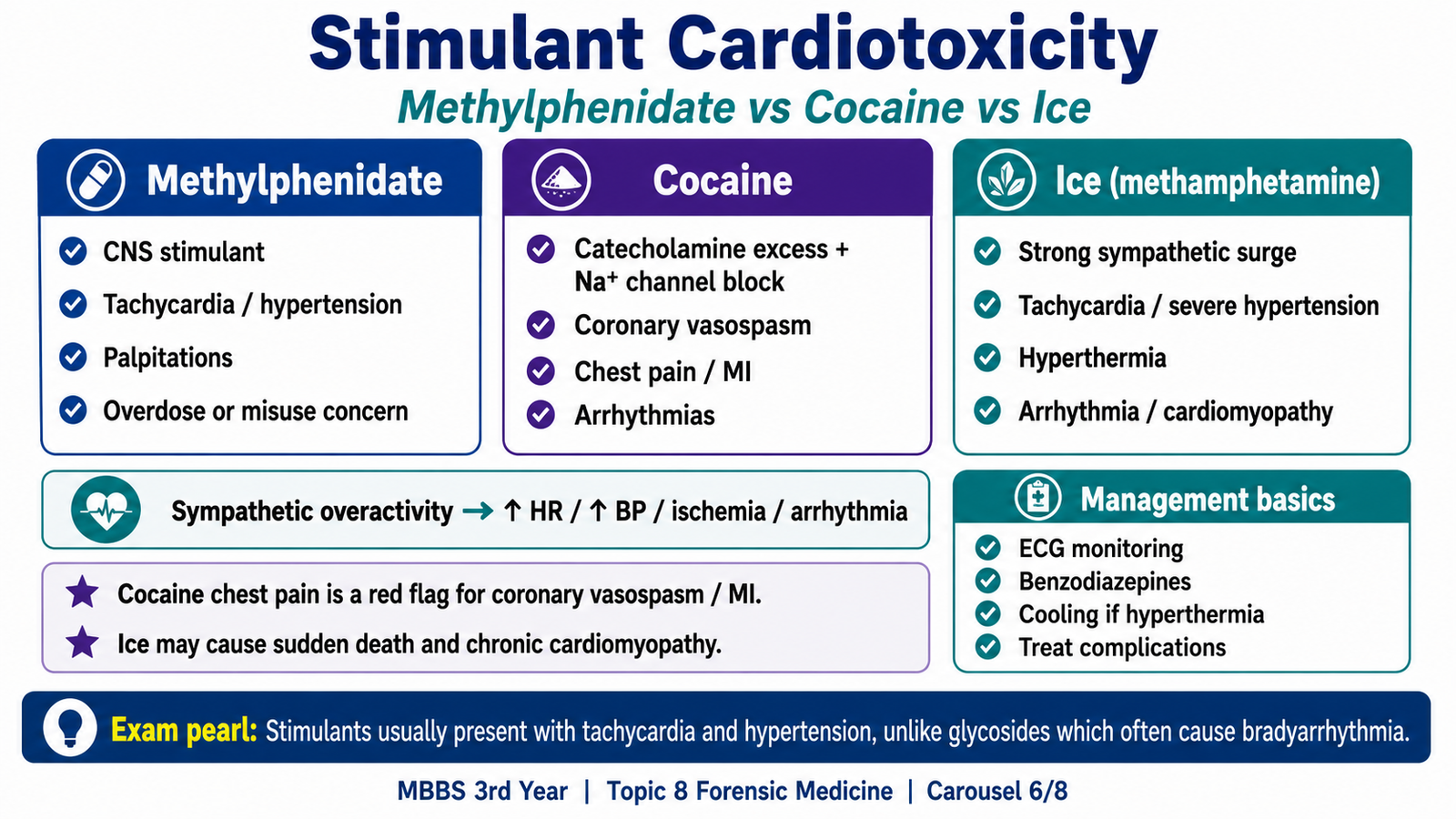
These drugs increase sympathetic activity. Excess sympathetic stimulation raises heart rate and blood pressure, increases myocardial oxygen demand and causes vasoconstriction. The combination of increased demand and reduced coronary blood supply can produce chest pain, ischemia, infarction, arrhythmia or sudden death. “`
Methylphenidate
Methylphenidate is a central nervous system stimulant. By increasing catecholaminergic activity, it may cause palpitations, tachycardia and increased blood pressure. Serious cardiotoxic effects are more likely with excessive doses, misuse or underlying cardiac disease. Possible complications include arrhythmia, marked hypertension, chest pain and, rarely, serious cardiovascular events.
Cocaine
Cocaine blocks reuptake of catecholamines and produces intense sympathetic stimulation. It also causes coronary vasoconstriction and can disturb cardiac ion channels. Therefore, it simultaneously increases oxygen demand, reduces coronary supply and increases electrical instability. Cardiac effects include severe hypertension, tachycardia, coronary spasm, myocardial ischemia or infarction, arrhythmias, aortic stress and sudden cardiac death.
Ice
Ice refers to crystalline methamphetamine. It causes prolonged release of catecholamines and intense sympathetic stimulation. Acute effects include tachycardia, hypertension, chest pain, vasospasm and arrhythmias. Repeated use may produce persistent hypertension, myocardial injury and cardiomyopathy, increasing the risk of heart failure and sudden death.
Stimulant exposure → excess catecholamine effect → tachycardia and hypertension + coronary vasoconstriction → oxygen supply-demand mismatch → ischemia, arrhythmia or sudden death
From a medico-legal perspective, the exact substance, route, timing, dose if known, co-ingestants and circumstances of use should be documented. Toxicology results must be interpreted together with the history, examination and postmortem findings because detection of a stimulant does not by itself prove that it caused death.
G. Initial Management and Forensic Handling of Cardiac Poisoning
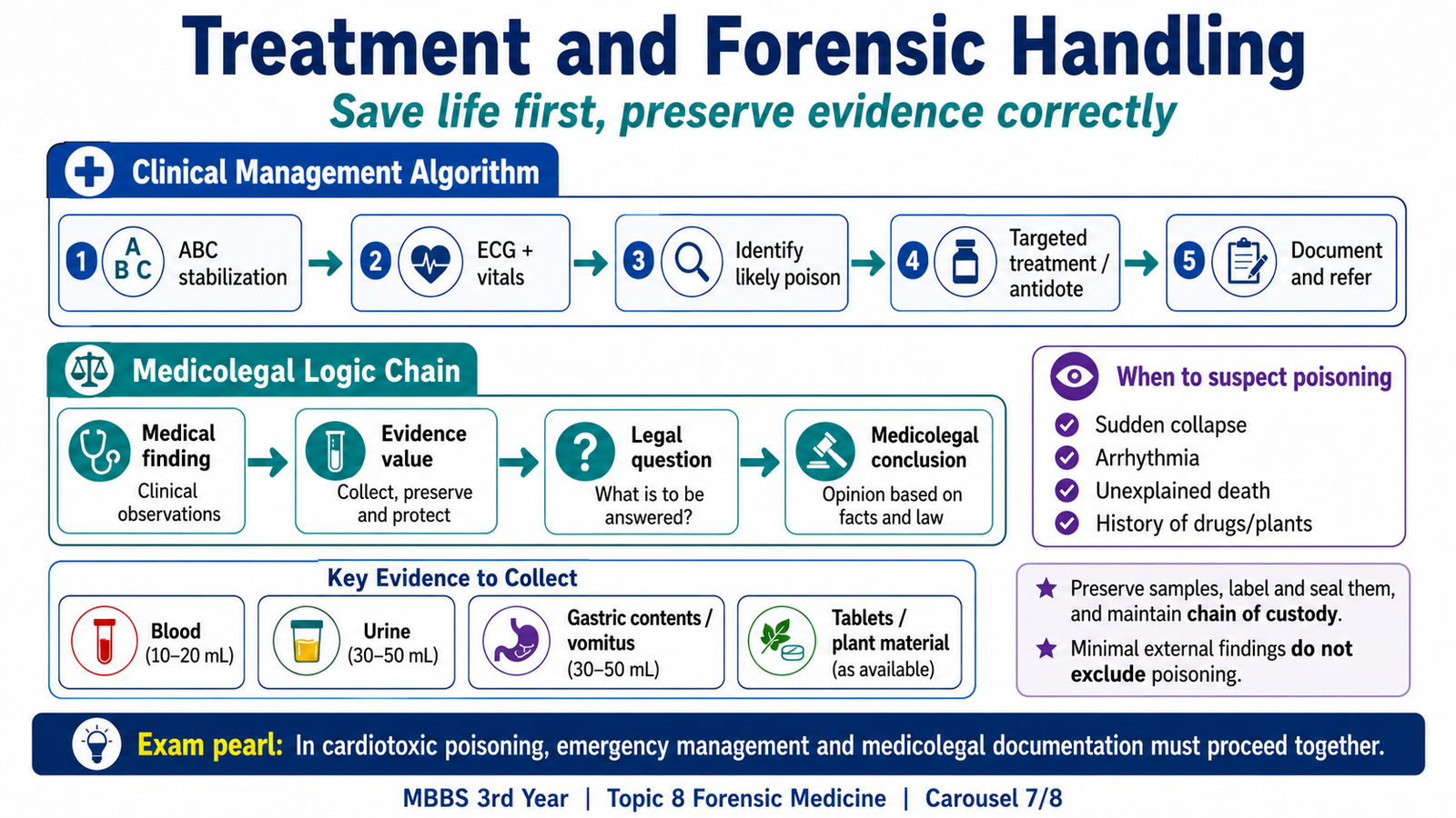
Cardiac poisoning is managed by treating the patient’s immediate physiological danger while also preserving reliable information about the exposure. The clinical priority is stabilization. The forensic priority is accurate documentation and appropriate preservation of relevant evidence. “`
Clinical management sequence
- Assess airway, breathing and circulation.
- Record vital signs and begin continuous ECG monitoring.
- Identify the suspected poison, route, amount and time of exposure when possible.
- Stop further exposure and consider gastrointestinal decontamination only when appropriate and safe.
- Check and correct electrolyte, glucose and acid-base abnormalities.
- Treat hypotension, bradycardia, conduction block or arrhythmia according to the toxicological pattern.
- Use a specific antidote when indicated, such as digoxin-specific antibody fragments in severe glycoside poisoning.
Forensic documentation and evidence
Record the patient’s condition, statements, treatment, timing and changes in clinical status. Where relevant, preserve medicine strips or bottles, plant material, powders, tablets or containers. Biological samples may include blood, urine, vomitus or gastric contents according to the case and local procedure. Each item should be labelled and handled in a way that maintains identity and chain of custody. The toxicology result must not be interpreted in isolation. A detected substance may represent therapeutic use, misuse, contamination or a level unrelated to death. The final opinion should integrate the circumstances, clinical course, ECG findings, pathology and toxicology.
H. Informed Consent Before an Invasive Procedure
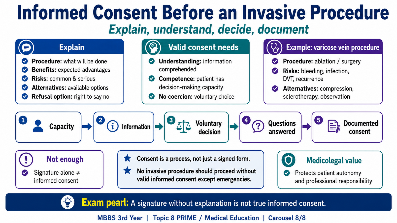
Informed consent is a voluntary decision made by a patient who has received and understood relevant information about a proposed procedure. It is more than obtaining a signature. The purpose is to respect patient autonomy and allow the patient to make a meaningful choice. “`
Essential elements
- The patient has decision-making capacity.
- The nature and purpose of the procedure are explained in understandable language.
- Expected benefits and important risks are discussed.
- Reasonable alternatives, including no procedure where relevant, are explained.
- The patient has an opportunity to ask questions.
- The decision is voluntary and free from coercion.
- Consent is documented before the procedure.
Before an invasive procedure for varicose veins, the clinician should explain what will be done, why it is recommended, expected improvement, possible complications such as bleeding, infection, thrombosis, recurrence or nerve injury where relevant, and reasonable alternatives such as conservative measures or another procedure. The explanation should match the patient’s level of understanding. Consent may be invalid when essential information is withheld, the patient lacks capacity without an appropriate substitute decision process, or the apparent agreement is obtained through pressure or deception. Good consent protects the patient’s right to choose and supports professional accountability.
Integrated Mechanism Flow
Venous pathway: Valve failure or venous stasis → raised venous pressure or thrombus formation → impaired venous return → swelling, pain and chronic skin change → ulceration or embolic complication.
Poisoning pathway: Toxic effect on pumps, ion channels, catecholamines or coronary vessels → abnormal conduction, contractility or perfusion → arrhythmia, ischemia or circulatory collapse → urgent stabilization, targeted treatment and medico-legal documentation.
AIM High-Yield Review
- Thrombophlebitis combines venous inflammation with thrombosis and commonly causes a painful, red, warm superficial cord.
- Phlebothrombosis commonly affects deep veins, may be clinically silent and carries a greater risk of pulmonary embolism.
- Virchow’s triad consists of stasis, endothelial injury and hypercoagulability.
- Varicose veins are dilated, elongated and tortuous superficial veins caused mainly by valve incompetence and chronic venous hypertension.
- Brown skin pigmentation in chronic venous disease results from red-cell breakdown and hemosiderin deposition.
- Venous ulcers commonly develop near the medial malleolus because chronic congestion impairs tissue oxygenation and nutrition.
- Hemangiomas contain blood-filled vascular channels; capillary types have small vessels and cavernous types have large dilated spaces.
- Pyogenic granuloma is a lobular capillary proliferation, not a true pyogenic infection or true granuloma.
- Angiosarcoma is an infiltrative malignant endothelial tumor with atypical vascular channels.
- Cardiac glycosides inhibit Na⁺/K⁺-ATPase and may cause bradycardia, conduction block, many arrhythmias and hyperkalemia in severe acute poisoning.
- Oleander poisoning produces a digoxin-like clinical picture because the plant contains cardiac glycosides.
- Cocaine and Ice increase sympathetic activity, causing tachycardia, hypertension, vasoconstriction, ischemia and arrhythmia.
- Digoxin-specific antibody fragments are the specific antidote for severe cardiac glycoside poisoning.
- Toxicology findings must be interpreted with history, clinical findings, pathology and circumstances; detection alone does not prove cause of death.
- Valid informed consent requires capacity, adequate information, understanding, voluntariness and documentation.
Study tip: This chapter follows the KMU learning outcomes in a simple sequence. First understand the explanations and cause-and-effect links, then use the AIM High-Yield Review for quick revision.
🎥 AIM Video Learning
Watch these focused videos after reading the learning material to reinforce venous thrombosis, vascular tumors and cardiac glycoside poisoning.