
Study tip: This chapter follows the KMU learning outcomes in a simple sequence. First understand how thrombosis develops and causes embolism, then connect pulmonary embolism with its diagnosis, risk assessment, management and pulmonary hypertension. Use the final high-yield review only after reading the explanations.
📖 AIM Learning Material
Topic 12 — Thrombosis, Embolism, Pulmonary Embolism and Pulmonary Hypertension
CVS Module · Pathology and Medicine & Allied Clinical Integration
Topic Introduction
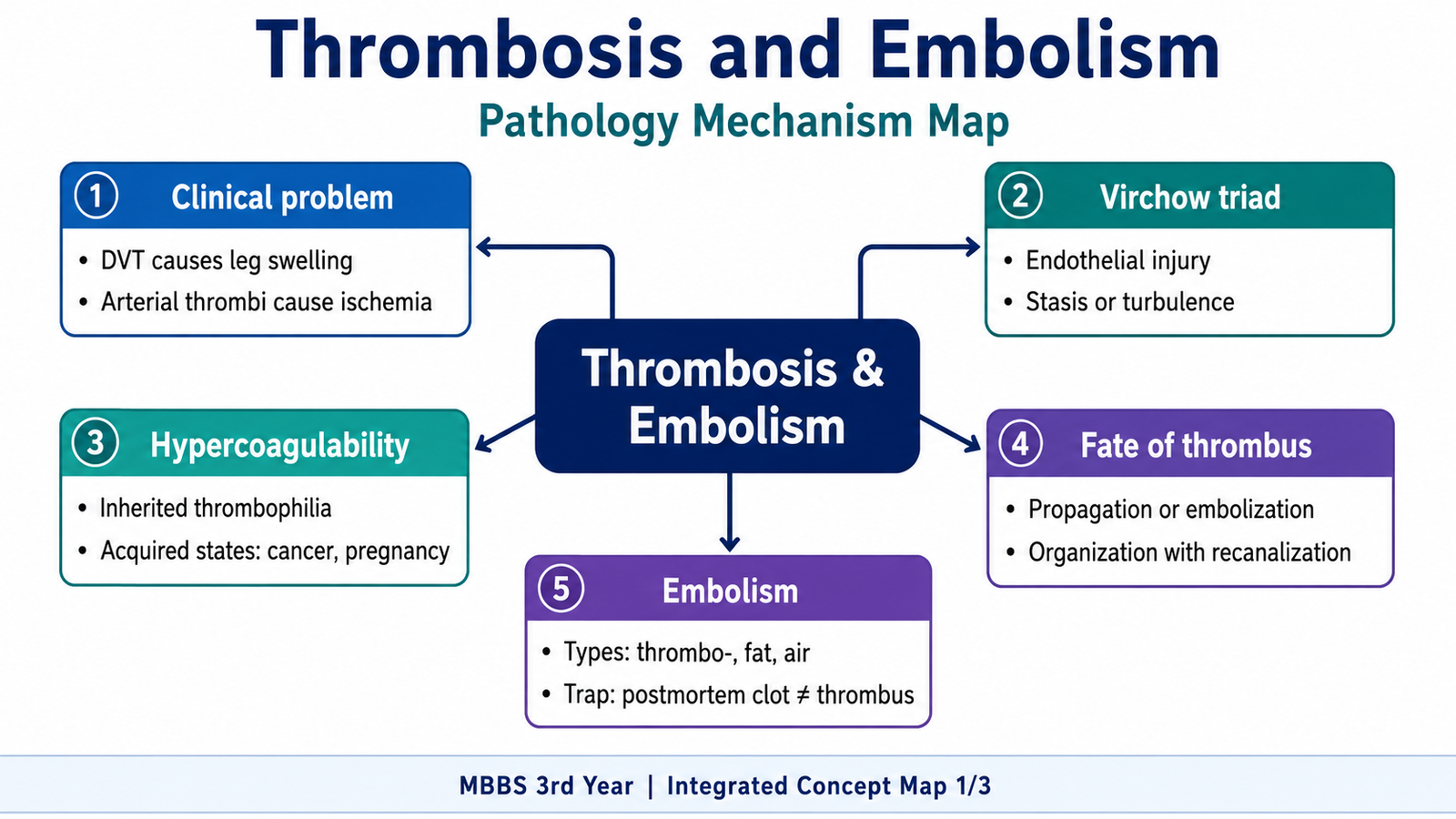
Thrombosis is the abnormal formation of a blood clot inside the cardiovascular system during life. A thrombus may obstruct blood flow at its site of formation or detach and travel as an embolus. When a venous thrombus reaches the pulmonary arteries, it produces pulmonary embolism, which may range from a small silent obstruction to acute right-heart failure, shock and sudden death. Repeated pulmonary emboli or long-standing cardiac disease may also increase pressure in the pulmonary circulation and produce pulmonary hypertension. This chapter explains why thrombi form, the important hypercoagulable states, the types of embolism, and the pathology, clinical features, diagnosis, risk stratification and broad management of pulmonary embolism and cardiac causes of pulmonary hypertension.
A. Why Thrombosis Develops and Who Is at Risk
Thrombosis is the pathological formation of a solid mass from blood constituents within an intact vessel or cardiac chamber during life. It differs from normal hemostasis, which is a controlled response that stops bleeding after injury. Pathological thrombosis occurs excessively or at an inappropriate site, so it may block circulation or become the source of an embolus.
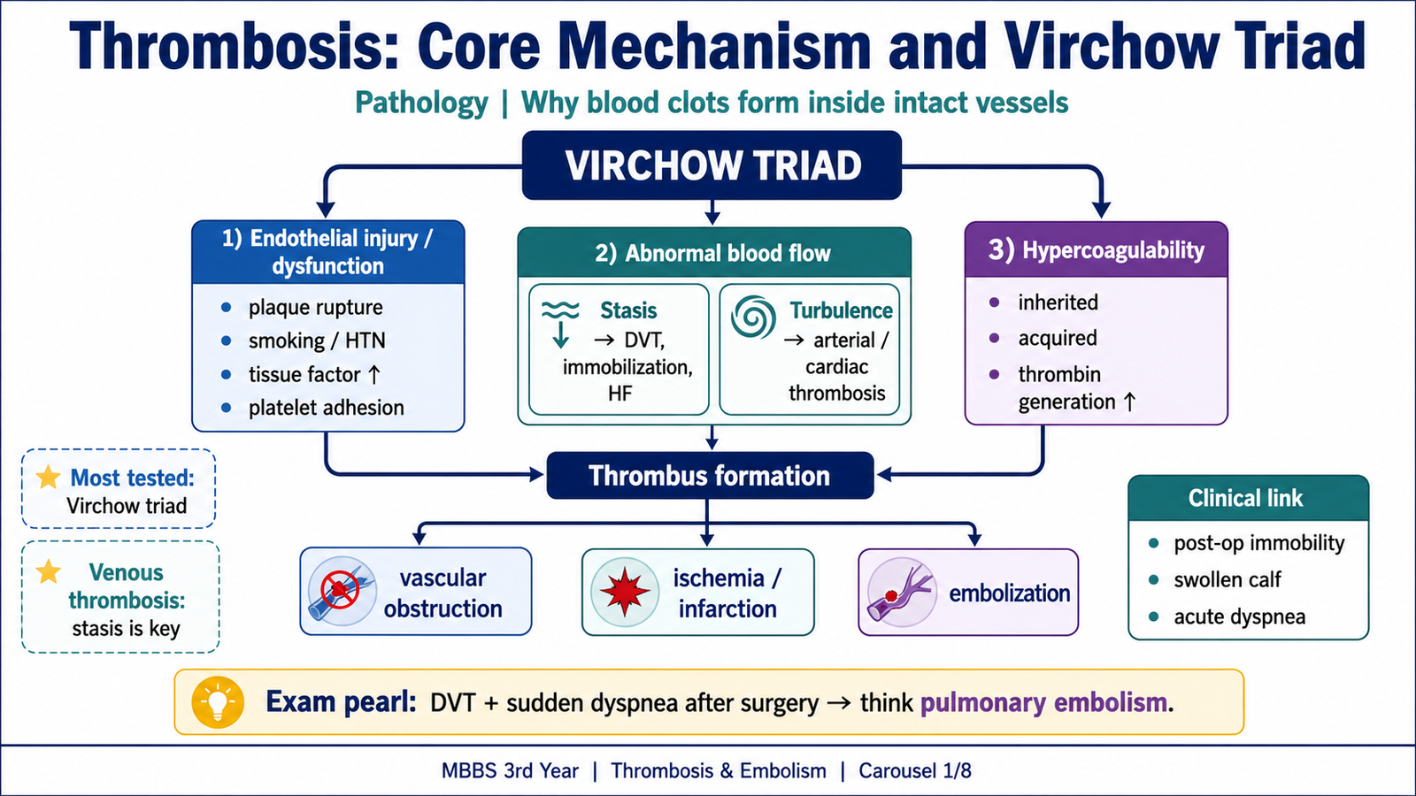
Virchow triad
The three major pathogenetic factors are endothelial injury, abnormal blood flow and hypercoagulability. They often act together.
- Endothelial injury or dysfunction: Normal endothelium prevents platelet adhesion, produces prostacyclin and nitric oxide, expresses anticoagulant molecules and supports fibrinolysis. Injury exposes collagen and von Willebrand factor, activates platelets, increases tissue factor and reduces anticoagulant and fibrinolytic activity. This is especially important in arterial and cardiac thrombosis, such as thrombosis over a disrupted atherosclerotic plaque or damaged heart valve.
- Abnormal blood flow: Stasis is most important in venous thrombosis. It prevents dilution and clearance of activated clotting factors, delays the arrival of anticoagulants and brings platelets into contact with the endothelium. Prolonged immobilization, heart failure, paralysis, pregnancy and varicose veins are important settings. Turbulence is more important in arteries and cardiac chambers because it injures endothelium and creates local pockets of stasis. It occurs around atherosclerotic plaques, aneurysms, abnormal valves and dilated chambers. In atrial fibrillation, ineffective atrial contraction causes stasis, especially in the left atrial appendage.
- Hypercoagulability: This means an increased tendency of blood to clot. It may be inherited or acquired and is particularly important in recurrent venous thrombosis, thrombosis at a young age or thrombosis at an unusual site.
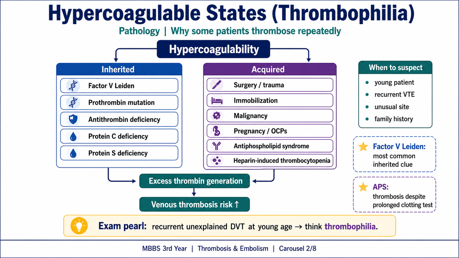
Inherited hypercoagulable states
Inherited thrombophilias disturb the normal balance between coagulation and anticoagulant mechanisms.
- Factor V Leiden: factor V becomes resistant to inactivation by activated protein C, so thrombin generation continues for longer.
- Prothrombin gene mutation: increased prothrombin levels promote excess thrombin formation.
- Antithrombin deficiency: reduced inhibition of thrombin and activated coagulation factors.
- Protein C deficiency: reduced inactivation of factors Va and VIIIa.
- Protein S deficiency: reduced cofactor support for activated protein C.
- Less common causes include increased factor VIII, defects in fibrinolysis and homozygous homocystinuria.
Acquired hypercoagulable states
Acquired states are more common and frequently combine with stasis or endothelial injury.
- Major surgery, trauma and prolonged immobilization
- Malignancy, particularly some adenocarcinomas
- Pregnancy, puerperium, oral contraceptives and estrogen therapy
- Obesity, increasing age and smoking
- Antiphospholipid antibody syndrome
- Heparin-induced thrombocytopenia
- Nephrotic syndrome, myeloproliferative neoplasms and severe heart failure
- Previous venous thromboembolism
In malignancy, tumour-derived tissue factor and other procoagulant mediators may cause recurrent thrombosis. Migratory superficial thrombophlebitis associated with an underlying malignancy is called Trousseau syndrome. Antiphospholipid antibody syndrome can cause venous or arterial thrombosis, recurrent pregnancy loss and thrombocytopenia even though phospholipid-dependent clotting tests may be prolonged. In heparin-induced thrombocytopenia, antibodies against heparin–platelet factor 4 complexes activate platelets; therefore, the fall in platelet count is accompanied by a dangerous risk of thrombosis.
A hypercoagulable state should be suspected when thrombosis is recurrent, unprovoked, occurs at a young age or unusual site, or is associated with a strong family history or repeated pregnancy-related events.
B. Morphology, Fate and Consequences of a Thrombus
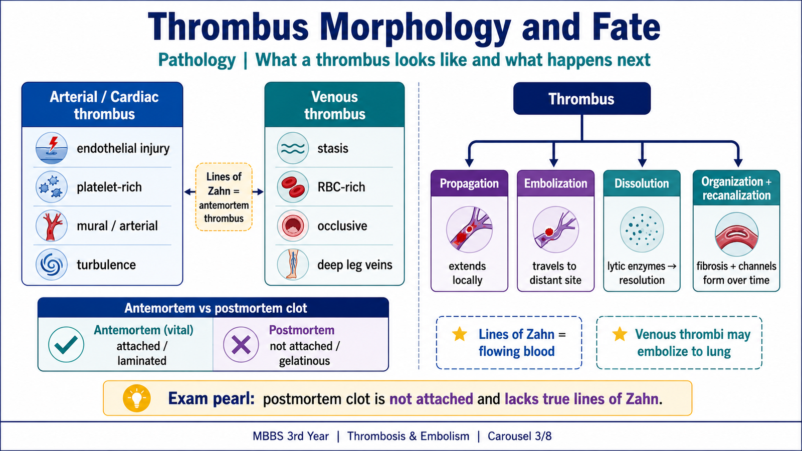
The appearance of a thrombus reflects its site and the conditions under which it formed. Arterial and cardiac thrombi usually arise where there is endothelial injury or turbulence. They are relatively rich in platelets and fibrin and may appear pale. Cardiac thrombi may form over an infarcted ventricular wall, inside a dilated chamber, in an atrium during atrial fibrillation or on damaged valves. A thrombus attached to the wall of a cardiac chamber or large vessel is called a mural thrombus.
Venous thrombi develop mainly because of stasis and hypercoagulability. They are usually dark red, rich in red cells, occlusive and may form a long cast of the affected vein. Clinically important deep-vein thrombi commonly occur in the popliteal, femoral and iliac veins. A calf-vein thrombus may remain localized or extend proximally; proximal extension increases the risk of pulmonary embolism.
Lines of Zahn and antemortem thrombosis
Thrombi formed in flowing blood may show alternating pale layers of platelets and fibrin and darker layers rich in red cells. These laminations are called lines of Zahn. They support formation during life. An antemortem thrombus is attached to the vessel wall, firm, friable and often laminated. A postmortem clot is usually unattached, smooth and gelatinous, with a dark dependent portion and a yellow upper portion.
Fate of a thrombus
- Propagation: further platelets and fibrin enlarge the thrombus. Venous thrombi tend to extend toward the heart.
- Embolization: part or all of the thrombus detaches and travels elsewhere.
- Dissolution: recent thrombi may be removed by fibrinolysis.
- Organization and recanalization: endothelial cells, smooth muscle cells and fibroblasts grow into an older thrombus. Fibrous tissue replaces it, and small channels may form through it to partly restore blood flow.
The clinical effect depends on the vessel involved. Thrombosis may cause venous congestion and edema, tissue ischemia, infarction, pulmonary or systemic embolism, chronic venous insufficiency, pulmonary hypertension or sudden death.
C. Embolism and Its Major Types
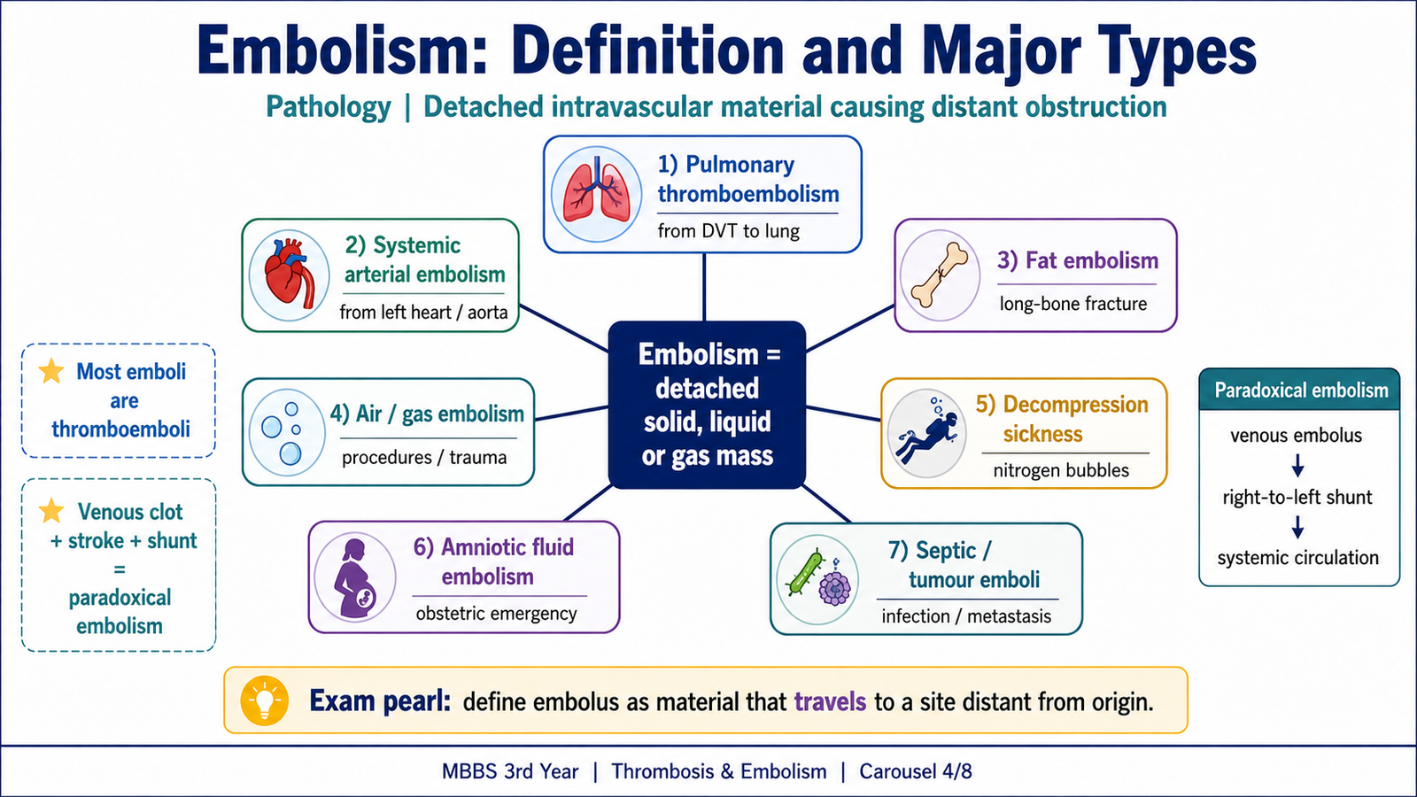
An embolus is a detached solid, liquid or gaseous intravascular mass carried by blood to a site distant from its origin. The resulting vascular obstruction is called embolism. Most emboli are detached thrombi and are therefore called thromboemboli.
- Pulmonary thromboembolism: usually originates from deep veins of the lower limb and travels through the inferior vena cava, right atrium and right ventricle into the pulmonary arterial circulation.
- Systemic arterial thromboembolism: commonly originates from left ventricular mural thrombi after myocardial infarction, left atrial thrombi in atrial fibrillation, diseased valves, aortic aneurysms or atherosclerotic plaques. It may lodge in the lower limbs, brain, intestines, kidneys or spleen.
- Fat embolism: fat globules may enter blood after long-bone fracture, severe soft-tissue trauma or bone-marrow injury. Fat embolism syndrome usually appears after a delay with respiratory distress, neurological symptoms, thrombocytopenia, anemia and a petechial rash.
- Air or gas embolism: air may enter during surgery, obstetric procedures, chest injury or central venous catheterization. A large volume can obstruct circulation and cause acute collapse.
- Decompression sickness: rapid reduction in atmospheric pressure causes dissolved nitrogen to form bubbles. Acute disease may cause joint and muscle pain, respiratory distress and neurological symptoms; chronic disease may cause ischemic bone necrosis.
- Amniotic-fluid embolism: amniotic fluid and fetal material enter the maternal circulation during labour or soon after delivery, producing sudden dyspnea, cyanosis, shock, seizures and disseminated intravascular coagulation.
- Septic embolism: infected thrombotic material causes vascular occlusion, infarction and secondary abscess formation.
- Tumour and cholesterol emboli: tumour cells may spread through blood, while cholesterol crystals from ulcerated plaques may obstruct small arteries.
Paradoxical embolism
A paradoxical embolus is a venous embolus that enters the systemic arterial circulation through a right-to-left cardiac or vascular communication. A patent foramen ovale, a septal defect with right-to-left flow or a pulmonary arteriovenous malformation may provide the pathway. Thus, a venous thrombus may rarely cause a stroke or another systemic arterial event.
D. Pulmonary Embolism: Cause, Pathogenesis, Morphology and Clinical Features
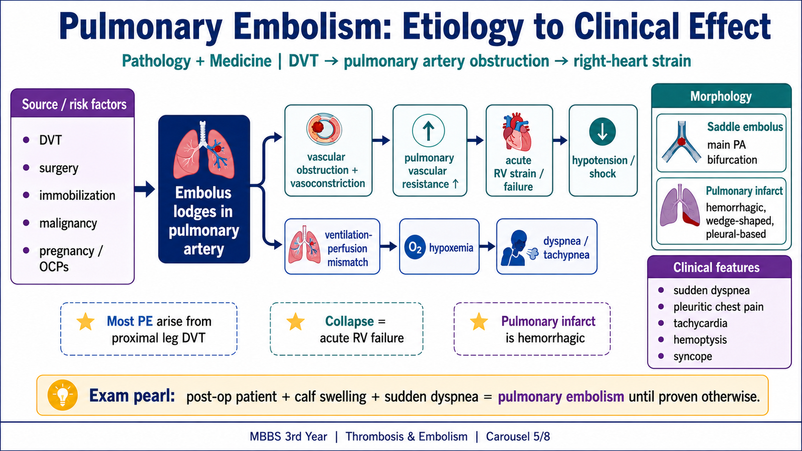
Most pulmonary emboli arise from deep-vein thromboses in the popliteal, femoral, iliac or pelvic veins. Less common sources include upper-limb veins, the right side of the heart and some abdominal veins. The risk factors are the same processes described by Virchow triad: immobility and heart failure produce stasis; trauma, fractures, surgery and venous catheters injure endothelium; malignancy, pregnancy, estrogen exposure and inherited thrombophilias increase coagulability.
How pulmonary embolism causes disease
After detaching, a venous thrombus passes through the right side of the heart and lodges in the pulmonary arterial circulation. The effect depends on the size and number of emboli, the degree and speed of arterial obstruction, vasoconstriction and the patient’s cardiopulmonary reserve.
- Mechanical obstruction: reduced pulmonary vascular cross-sectional area raises pulmonary vascular resistance and right-ventricular afterload.
- Pulmonary vasoconstriction: platelet and endothelial mediators further increase pulmonary arterial pressure.
- Acute right-ventricular strain: the thin-walled right ventricle is adapted to a low-resistance circulation. A sudden pressure rise causes dilatation, increased wall tension, ischemia and acute right-heart failure. Septal shift toward the left ventricle reduces left-ventricular filling, so cardiac output falls and hypotension or obstructive shock may develop.
- Ventilation-perfusion mismatch: an embolized area may remain ventilated but receive little blood flow. This increases physiological dead space and produces dyspnea, tachypnea, hypoxemia and often respiratory alkalosis.
Morphology
A large embolus may straddle the bifurcation of the main pulmonary artery and is called a saddle embolus. It can produce severe dyspnea, acute right-heart failure, cardiovascular collapse and sudden death. Smaller emboli lodge in peripheral arteries and may be asymptomatic or cause pulmonary hemorrhage, infarction and pleuritic pain.
The lung has dual blood supply from pulmonary and bronchial arteries, so embolic obstruction does not always cause infarction. Infarction is more likely when cardiopulmonary reserve or bronchial perfusion is poor, such as in left-sided heart failure or chronic lung disease.
A pulmonary infarct is classically hemorrhagic, wedge-shaped, peripheral and pleural-based. Its apex points toward the obstructed vessel, while its base lies against the pleura. Microscopically, early changes include alveolar hemorrhage, coagulative necrosis of alveolar walls and neutrophils at the margins. Later, macrophages remove debris, hemosiderin may accumulate and a fibrous scar forms.
Clinical features
Pulmonary embolism has a wide clinical range, from no symptoms to sudden death. The presentation depends on the degree of obstruction and the patient’s reserve.
- Sudden unexplained dyspnea, tachypnea and tachycardia
- Pleuritic chest pain, cough or hemoptysis, especially with peripheral infarction
- Hypoxemia, anxiety, syncope or low-grade fever
- Signs of deep-vein thrombosis, such as unilateral calf swelling and tenderness
- Raised jugular venous pressure, right-ventricular heave, loud pulmonary component of the second heart sound and hypotension in severe disease
Small emboli may be silent. Pulmonary infarction commonly causes pleuritic pain and hemoptysis. A large high-risk embolus may cause acute right-ventricular failure, shock or sudden death. Recurrent emboli may gradually obstruct the pulmonary vascular bed and contribute to chronic pulmonary hypertension.
E. Diagnostic Workup of Suspected Pulmonary Embolism
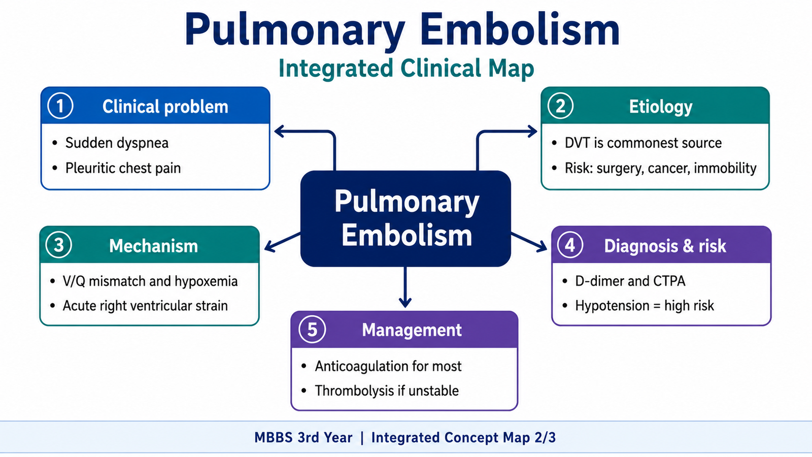
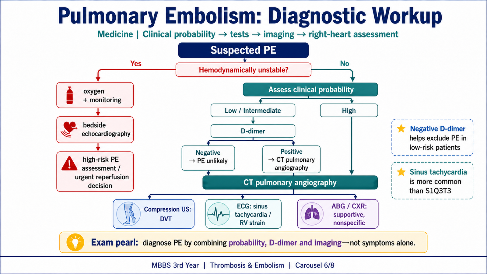
No single symptom or routine test confirms pulmonary embolism in every patient. Diagnosis begins by assessing hemodynamic stability and estimating clinical probability from symptoms, signs and risk factors. Testing is then selected according to that probability.
Initial assessment
First assess airway, breathing, circulation, oxygen saturation, blood pressure, pulse and evidence of shock. Look for features of deep-vein thrombosis and for alternative causes of chest pain or dyspnea. Hypotension, syncope, severe hypoxemia or signs of acute right-heart strain indicate a high-risk presentation.
Clinical probability and D-dimer
Clinical prediction tools organize findings such as previous venous thromboembolism, recent surgery or immobilization, malignancy, tachycardia, hemoptysis and signs of deep-vein thrombosis. In a patient with low or intermediate clinical probability, a negative D-dimer helps exclude pulmonary embolism because D-dimer reflects breakdown of cross-linked fibrin. A positive result is not specific and may occur after surgery, infection, inflammation, trauma, pregnancy, malignancy and with increasing age.
Imaging and supportive tests
- CT pulmonary angiography: the main imaging test in many stable patients. It directly shows a filling defect in the pulmonary arteries and may reveal right-ventricular enlargement or another diagnosis.
- Ventilation-perfusion scan: useful when CT contrast is unsuitable or CT pulmonary angiography cannot be performed. A perfusion defect with preserved ventilation suggests embolic obstruction.
- Compression ultrasonography: demonstrates deep-vein thrombosis and is particularly useful when leg signs are present or chest imaging is difficult.
- Echocardiography: may show right-ventricular dilatation, reduced right-ventricular function, septal flattening or elevated pulmonary pressure. It is especially useful in an unstable patient, but a normal study does not exclude a small embolus.
- ECG: often shows sinus tachycardia. Right-heart strain patterns may occur but are not sufficiently sensitive or specific to confirm the diagnosis.
- Chest radiograph: may be normal or show nonspecific changes. Its main value is identifying alternative diagnoses and helping interpret a ventilation-perfusion scan.
- Arterial blood gases: may show hypoxemia and respiratory alkalosis, but normal values do not exclude pulmonary embolism.
- Cardiac biomarkers: troponin and natriuretic peptides may rise because of right-ventricular strain and are more useful for risk assessment than for confirming the diagnosis.
F. Risk Stratification and Management of Pulmonary Embolism
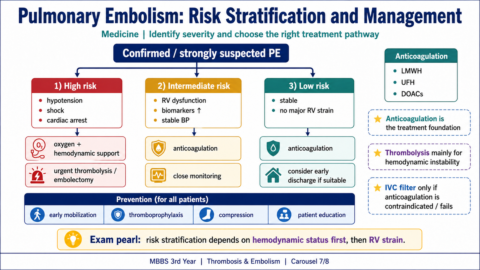
Risk stratification estimates the chance of early deterioration or death and determines how urgently circulation must be restored. The most important distinction is whether the patient is hemodynamically unstable.
Risk categories
- High-risk pulmonary embolism: persistent hypotension, obstructive shock or cardiac arrest. This indicates severe right-ventricular failure and requires urgent reperfusion assessment.
- Intermediate-risk pulmonary embolism: normal blood pressure but evidence of right-ventricular dysfunction and/or myocardial injury. These patients require close monitoring because they may deteriorate.
- Low-risk pulmonary embolism: stable circulation without significant right-ventricular dysfunction or major clinical risk features.
Immediate support
Initial treatment supports oxygenation and circulation while preventing further embolization. Give oxygen when hypoxemia is present. In shock, use cautious circulatory support because excessive fluid may worsen right-ventricular dilatation. Vasopressor support may be required when hypotension persists. Severe respiratory failure may require ventilatory support, but positive-pressure ventilation can further reduce venous return and must be used carefully.
Anticoagulation
Anticoagulation is the main treatment for most patients. It does not directly remove the existing embolus; instead, it prevents extension of the current thrombus and formation of new thrombi while the body’s fibrinolytic system gradually clears the obstruction. Options include unfractionated heparin, low-molecular-weight heparin, fondaparinux, direct oral anticoagulants and vitamin K antagonists. The choice depends on hemodynamic stability, renal function, bleeding risk, pregnancy, cancer and the possible need for an urgent procedure.
Unfractionated heparin is useful when rapid reversal or a procedure may be needed because its effect can be adjusted quickly. A falling platelet count with new thrombosis should raise concern for heparin-induced thrombocytopenia.
Reperfusion and other interventions
Systemic thrombolytic therapy accelerates fibrin breakdown and may rapidly reduce pulmonary obstruction. It is mainly considered in high-risk pulmonary embolism with shock or persistent hypotension when bleeding risk is acceptable. Its major danger is serious bleeding, including intracranial hemorrhage.
Catheter-directed therapy or surgical embolectomy may be considered when thrombolysis is contraindicated, has failed or a severe embolus requires another route of reperfusion. An inferior vena cava filter may be considered when anticoagulation is absolutely contraindicated or when recurrent embolism occurs despite appropriate anticoagulation. It prevents some emboli from reaching the lungs but does not treat the existing thrombus and may itself increase deep-vein thrombosis risk.
Duration and prevention
The duration of anticoagulation depends on whether the event was provoked by a temporary factor, remains unprovoked, is recurrent or is associated with a persistent risk such as malignancy or thrombophilia. Prevention focuses on early mobilization after illness or surgery, mechanical methods where appropriate and anticoagulant prophylaxis in patients whose thrombotic risk outweighs bleeding risk.
G. Pulmonary Hypertension Due to Cardiac Disease
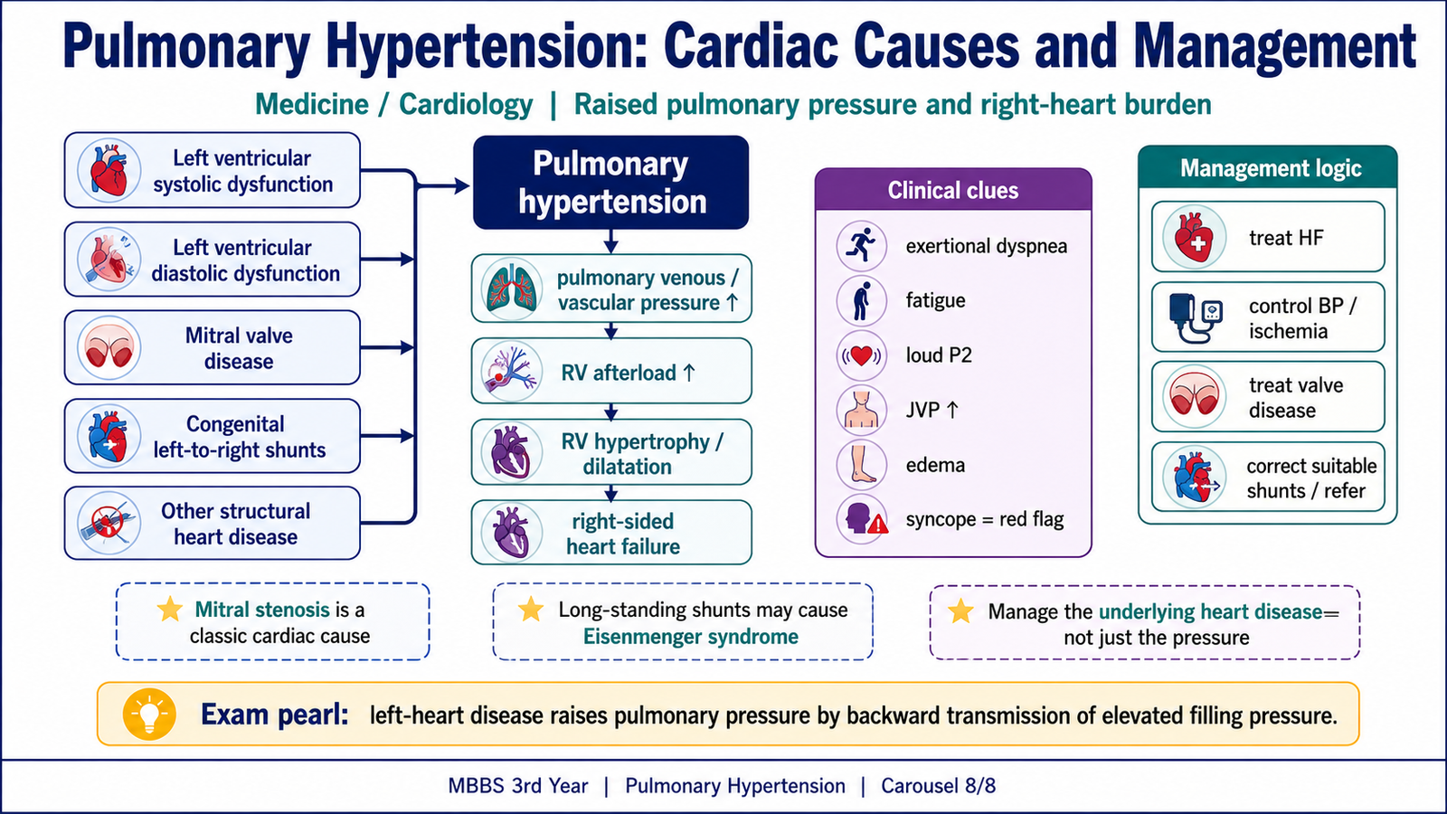
Pulmonary hypertension means abnormally increased pressure within the pulmonary circulation. Sustained elevation increases right-ventricular workload. The right ventricle first hypertrophies to overcome the pressure, but progressive disease may cause dilatation, tricuspid regurgitation and right-heart failure.
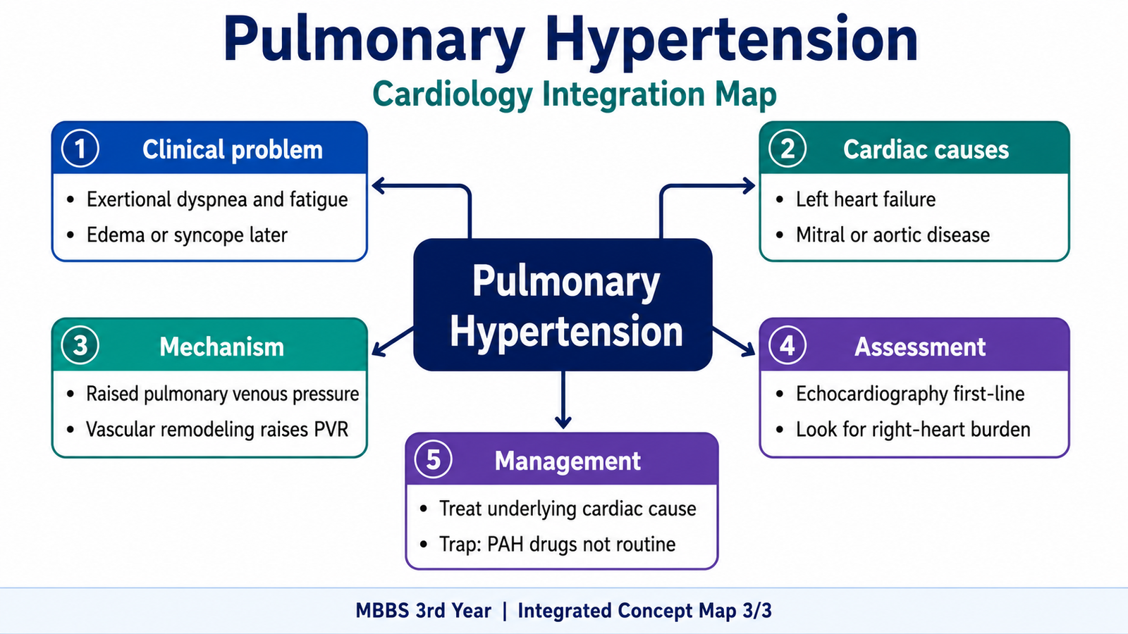
Patients may develop exertional dyspnea and fatigue because the right ventricle cannot adequately increase output during exercise. Later features include chest discomfort, syncope, a loud pulmonary component of the second heart sound, raised jugular venous pressure, peripheral edema and other signs of right-heart failure.
Cardiac causes
The most important cardiac mechanism is transmission of raised left-sided filling pressure backward into the pulmonary veins and capillaries. Chronic venous pressure may then cause pulmonary arterial vasoconstriction and vascular remodeling, which further increase pulmonary pressure.
- Left-ventricular systolic dysfunction: poor ventricular contraction raises left-ventricular end-diastolic and left atrial pressure. This pressure is transmitted to the lungs.
- Left-ventricular diastolic dysfunction: a stiff ventricle fills poorly, so filling pressure rises even when ejection fraction is preserved.
- Mitral stenosis: obstruction to flow from left atrium to left ventricle markedly raises left atrial and pulmonary venous pressure.
- Mitral regurgitation: backward systolic flow into the left atrium raises left atrial pressure and may cause chronic pulmonary vascular changes.
- Aortic valve disease: aortic stenosis or regurgitation may eventually cause left-ventricular dysfunction and raised left-sided filling pressure.
- Congenital left-to-right shunts: increased pulmonary blood flow may cause progressive pulmonary vascular remodeling. Advanced disease may reverse the shunt and produce cyanosis.
- Other contributors include left atrial obstruction and chronic cardiomyopathy.
Diagnostic approach and management logic
Assessment includes history and examination for left-sided heart disease and right-heart failure, ECG and chest radiography, and echocardiography to evaluate chamber size, ventricular function, valve disease, shunts and estimated pulmonary pressure. Further tests are selected according to the suspected cause.
Management is directed mainly at the underlying cardiac disorder. This may include treating heart failure, controlling volume overload, managing valve disease and correcting a significant shunt when appropriate. Oxygen is used when hypoxemia is present. Pulmonary vasodilator drugs used for primary pulmonary arterial hypertension are not routinely appropriate for pulmonary hypertension caused by left-heart disease because they do not correct the raised left-sided filling pressure and may worsen pulmonary congestion.
Integrated Mechanism Flow
→ deep-vein thrombus formation and propagation
→ detachment and passage through the right heart
→ pulmonary arterial obstruction and vasoconstriction
→ increased right-ventricular afterload plus ventilation-perfusion mismatch
→ hypoxemia, acute right-ventricular strain and reduced left-ventricular filling
→ dyspnea, hypotension, obstructive shock or recurrent-embolism-related pulmonary hypertension
Important Comparison
| Feature | Arterial/Cardiac Thrombus | Venous Thrombus | Postmortem Clot |
|---|---|---|---|
| Main mechanism | Endothelial injury and turbulence | Stasis and hypercoagulability | Blood coagulation after death |
| Composition/appearance | Platelet and fibrin rich; relatively pale | Red-cell rich; dark red and often occlusive | Gelatinous with dependent red and upper yellow portions |
| Attachment | Attached to vessel or chamber wall | Attached to vein wall | Not attached |
| Lines of Zahn | May be present | May be present | Absent |
| Main embolic destination | Systemic organs | Pulmonary arteries | No embolic consequence |
⭐ AIM High-Yield Review
- Virchow triad consists of endothelial injury, abnormal blood flow and hypercoagulability.
- Stasis is the major contributor to venous thrombosis; endothelial injury and turbulence are especially important in arterial and cardiac thrombosis.
- Factor V Leiden causes resistance to activated protein C.
- Antiphospholipid antibody syndrome may cause recurrent thrombosis despite prolonged phospholipid-dependent clotting tests.
- Heparin-induced thrombocytopenia causes a fall in platelets with a paradoxical risk of severe thrombosis.
- Lines of Zahn support thrombus formation in flowing blood during life.
- Most pulmonary emboli arise from proximal deep veins of the lower limb.
- A pulmonary infarct is typically hemorrhagic, wedge-shaped, peripheral and pleural-based.
- Massive pulmonary embolism causes collapse mainly through acute right-ventricular failure and reduced left-ventricular filling.
- A negative D-dimer can help exclude pulmonary embolism in a patient with low or intermediate clinical probability; a positive result does not confirm it.
- CT pulmonary angiography directly demonstrates pulmonary arterial filling defects in many stable patients.
- Anticoagulation prevents extension and new thrombosis; it does not instantly remove the existing embolus.
- Thrombolysis is mainly considered for high-risk pulmonary embolism with shock or persistent hypotension when bleeding risk is acceptable.
- Cardiac pulmonary hypertension commonly results from backward transmission of raised left-sided filling pressure.
- Treatment of cardiac pulmonary hypertension focuses on the underlying left-heart or shunt lesion rather than routine use of pulmonary arterial vasodilators.
This Ninja Nerd lecture is the best single-video match because it covers venous thrombosis, DVT, pulmonary embolism, diagnosis, management, complications and chronic thromboembolic pulmonary hypertension. ([YouTube][1]) “`html
Venous Thromboembolism: DVT and Pulmonary Embolism
This video explains venous thrombosis, pulmonary embolism, clinical features, diagnostic assessment, management and important complications.