
Study tip: This chapter follows the KMU learning outcomes in a logical sequence. First understand how pressure and valve movement create heart sounds and murmurs, then connect these principles with valve disease, rheumatic fever and endocarditis. Use the final high-yield review after completing the explanations.
📖 AIM Learning Material · CVS
Cardiac Cycle, Murmurs, Valvular Disease, Rheumatic Heart Disease and Endocarditis
Understand normal valve movement first, then use it to explain murmurs, the four major left-sided valve lesions, rheumatic valve damage and infective or sterile vegetations.
Topic Introduction
The heart pumps blood through a repeating cardiac cycle in which pressure changes open and close the valves. Normal valve closure produces heart sounds, while abnormal or turbulent blood flow produces murmurs. Valve disease may obstruct forward flow, called stenosis, or allow backward flow, called regurgitation. This chapter explains how these changes produce characteristic murmurs and clinical features. It then connects group A streptococcal infection with acute rheumatic fever and chronic rheumatic valve deformity, and explains infective endocarditis and the main non-infected vegetations. By following cause, structural change and functional effect, the topic becomes easier to understand rather than memorize.
A. Cardiac Cycle, Heart Sounds and Murmur Formation
The cardiac cycle is the sequence of pressure, volume and valve changes occurring during one heartbeat. A valve opens or closes passively according to the pressure difference across it.
The essential rule is:
Blood flows from a chamber with higher pressure toward a chamber with lower pressure. Valves open when the pressure behind them exceeds the pressure ahead of them.
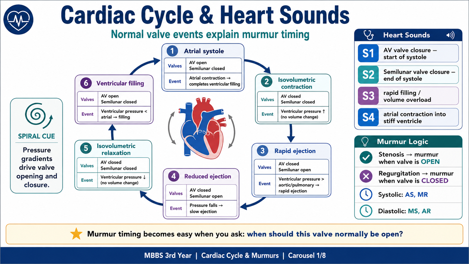
Phase 1: Ventricular filling
During ventricular diastole:
- Ventricular pressure falls below atrial pressure.
- Mitral and tricuspid valves open.
- Aortic and pulmonary valves remain closed.
- Blood enters the ventricles.
Most ventricular filling occurs passively during early diastole.
Rapid filling
As the atrioventricular valves open, blood rapidly enters the relaxed ventricles. Rapid filling may produce an S3 heart sound.
Diastasis
The pressure difference between atria and ventricles decreases, so ventricular filling slows.
Atrial systole
Atrial contraction supplies the final portion of ventricular filling. This contribution becomes particularly important when the ventricle is stiff or the heart rate is increased.
Atrial contraction against a poorly compliant ventricle may produce an S4 heart sound.
Phase 2: Isovolumetric ventricular contraction
The QRS complex is followed by ventricular contraction.
As ventricular pressure rises above atrial pressure:
- Mitral and tricuspid valves close.
- Aortic and pulmonary valves are still closed.
- All four valves are closed.
- Ventricular pressure rises rapidly without a change in ventricular volume.
Closure of the mitral and tricuspid valves produces S1, marking the beginning of systole.
Phase 3: Ventricular ejection
When ventricular pressure exceeds aortic and pulmonary arterial pressure:
- Aortic and pulmonary valves open.
- Blood is ejected into the great vessels.
- Ventricular volume falls.
Ejection is initially rapid and later becomes slower as ventricular contraction declines.
Phase 4: Isovolumetric ventricular relaxation
Following ventricular repolarization:
- Ventricular contraction ends.
- Aortic and pulmonary arterial pressure becomes greater than ventricular pressure.
- Aortic and pulmonary valves close, producing S2.
- All four valves are temporarily closed.
- Ventricular pressure falls without a change in volume.
When ventricular pressure finally becomes lower than atrial pressure, the atrioventricular valves reopen and a new cycle begins.
Cardiac-cycle summary
| Phase | Mitral/tricuspid valves | Aortic/pulmonary valves | Main event |
|---|---|---|---|
| Atrial systole | Open | Closed | Final ventricular filling |
| Isovolumetric contraction | Closed | Closed | S1; pressure rises |
| Rapid ejection | Closed | Open | Maximum forward flow |
| Reduced ejection | Closed | Open | Slower forward flow |
| Isovolumetric relaxation | Closed | Closed | S2; pressure falls |
| Rapid filling | Open | Closed | Possible S3 |
| Diastasis | Open | Closed | Slow passive filling |
Physiology of the heart sounds
First heart sound — S1
S1 results mainly from closure and sudden tensing of the mitral and tricuspid valve apparatus at the beginning of ventricular systole.
S1 is:
- Loud when the mitral leaflets remain mobile but are widely separated before closure, as in early mitral stenosis.
- Soft in severe mitral regurgitation because the valve does not close effectively.
- Soft in heavily calcified mitral stenosis because the leaflets have lost mobility.
Second heart sound — S2
S2 is produced by closure of the aortic and pulmonary valves at the end of systole.
It has two components:
- A2: aortic valve closure
- P2: pulmonary valve closure
During inspiration, increased right-sided venous return prolongs right ventricular ejection and delays P2, producing physiological splitting of S2.
Third heart sound — S3
S3 occurs during rapid early ventricular filling.
It may be physiological in:
- Children
- Young adults
- Pregnancy
In an older adult, it commonly suggests:
- Volume overload
- Ventricular dilatation
- Mitral regurgitation
- Heart failure
Fourth heart sound — S4
S4 occurs when atrial contraction forces blood into a stiff, poorly compliant ventricle.
It may occur in:
- Aortic stenosis
- Systemic hypertension
- Ventricular hypertrophy
S4 is absent in atrial fibrillation because effective atrial contraction is absent.
How a murmur is generated
A murmur is a prolonged sound caused by turbulent blood flow. Turbulence may result from:
- Flow through a narrowed valve
- Backward flow through an incompetent valve
- Increased blood flow across a normal valve
- Reduced blood viscosity, such as in severe anemia
- Flow through an abnormal communication
The most important physiological rule is:
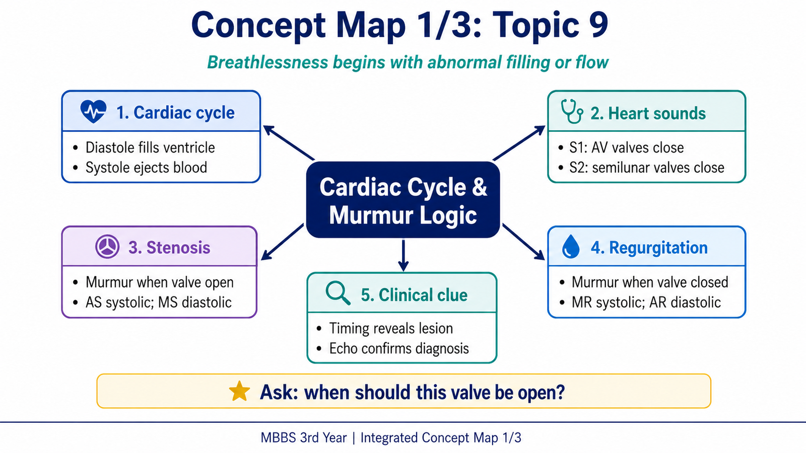
A stenotic valve produces a murmur while that valve should normally be open. A regurgitant valve produces a murmur while that valve should normally be closed.
Therefore:
- Aortic stenosis → systolic murmur because the aortic valve is open during systole.
- Mitral stenosis → diastolic murmur because the mitral valve is open during diastole.
- Mitral regurgitation → systolic murmur because the mitral valve should be closed during systole.
- Aortic regurgitation → diastolic murmur because the aortic valve should be closed during diastole.
B. Systematic Interpretation of Cardiac Murmurs
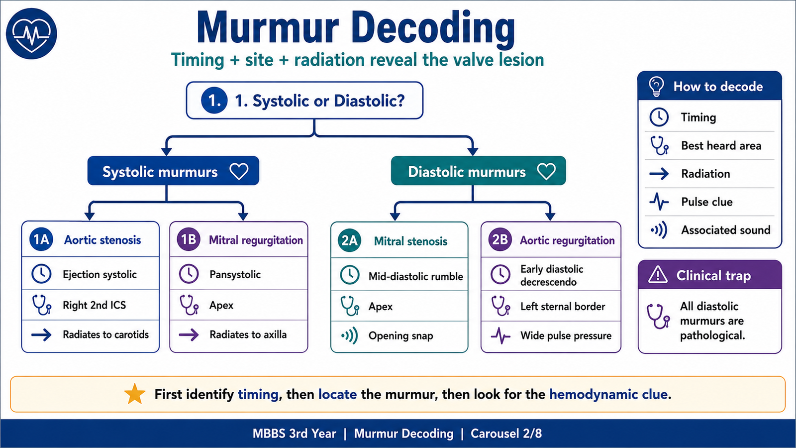
A murmur should not be memorized as an isolated sound. It should be decoded through five questions:
- Is it systolic or diastolic?
- Where is it loudest?
- Where does it radiate?
- What is its character and shape?
- Which pressure gradient produces it?
Systolic murmurs
Ejection systolic murmur
An ejection systolic murmur begins after S1, increases in intensity and then decreases before S2.
It occurs when blood passes through an obstructed ventricular outflow tract.
The classical example is aortic stenosis.
Pansystolic murmur
A pansystolic murmur begins with S1 and continues at a relatively uniform intensity until S2.
It occurs when a persistent pressure difference exists throughout systole.
The classical example is mitral regurgitation, because left ventricular pressure remains higher than left atrial pressure throughout ventricular systole.
Diastolic murmurs
Diastolic murmurs are almost always pathological.
Early diastolic murmur
An early diastolic murmur begins immediately after S2 and gradually decreases in intensity.
The classical example is aortic regurgitation, as aortic pressure is initially much higher than left ventricular pressure.
Mid-diastolic murmur
A mid-diastolic murmur occurs when blood flows through a narrowed atrioventricular valve during ventricular filling.
The classical example is mitral stenosis.
Four-valve-lesion murmur map
| Lesion | Timing and character | Best heard | Typical radiation or associated sound |
| Aortic stenosis | Crescendo–decrescendo ejection systolic | Right second intercostal space | Radiates to carotids |
| Aortic regurgitation | High-pitched early diastolic decrescendo | Left sternal border | Best with patient leaning forward |
| Mitral stenosis | Low-pitched mid-diastolic rumble | Apex | Opening snap; little radiation |
| Mitral regurgitation | Blowing pansystolic | Apex | Radiates toward axilla |
Using bedside maneuvers
- Left-sided murmurs are generally better heard during expiration.
- Right-sided murmurs commonly become louder during inspiration.
- Sustained handgrip increases systemic vascular resistance and usually makes mitral and aortic regurgitation more prominent.
- Reduced venous return during standing or the strain phase of Valsalva decreases the intensity of most murmurs.
These maneuvers support—but do not replace—correct timing, site and radiation.
Diagnostic conclusion
Once a pathological murmur is suspected, echocardiography is used to:
- Identify abnormal valve anatomy
- Demonstrate stenosis or regurgitation
- Assess chamber enlargement
- Estimate pressure gradients
- Evaluate ventricular response
- Detect vegetations or other complications
History and physical findings should be interpreted together with echocardiography rather than in isolation.
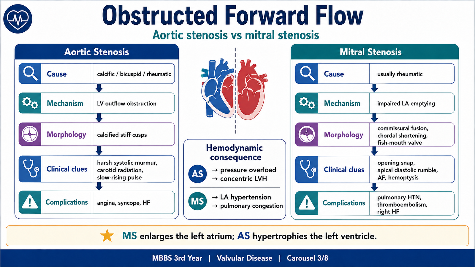
C. Valvular Stenosis: Aortic Stenosis and Mitral Stenosis
Stenosis means failure of a valve to open completely. The narrowed orifice obstructs forward flow and creates a pressure load upstream from the valve.
However, aortic and mitral stenosis affect different chambers and different phases of the cardiac cycle.
Aortic stenosis
Core concept
Aortic stenosis is narrowing of the aortic valve opening, obstructing left ventricular ejection during systole.
Etiology
Important causes include:
- Calcific degeneration of a normal tricuspid aortic valve
- Usually occurs later in life.
- Progressive calcification reduces cusp mobility.
- Congenital bicuspid aortic valve
- The valve has two functional cusps instead of three.
- Abnormal mechanical stress causes earlier calcification and stenosis.
- Rheumatic aortic stenosis
- Commissural fusion and fibrosis occur.
- It commonly accompanies rheumatic mitral valve disease.
Pathogenesis
The stenotic valve creates resistance to left ventricular outflow:
Aortic narrowing → increased left ventricular systolic pressure → chronic pressure overload → concentric left ventricular hypertrophy → reduced ventricular compliance → increased filling pressure → pulmonary congestion and breathlessness
Concentric hypertrophy initially maintains cardiac output but increases myocardial oxygen demand and may reduce coronary perfusion.
Morphology
Calcific aortic stenosis:
- Nodular calcific deposits develop in the cusps.
- Cusp mobility becomes restricted.
- Commissures are generally not fused in ordinary degenerative calcific disease.
Rheumatic aortic stenosis:
- Commissural fusion is present.
- Cusps become thickened and fibrotic.
- Calcification may develop later.
Clinical features explained by pathology
Ejection systolic murmur: Blood accelerates through the narrowed valve during systole, producing a harsh crescendo–decrescendo murmur.
Radiation to the carotids: The turbulent jet travels from the aortic valve into the ascending aorta and neck vessels.
Slow-rising, low-volume pulse: Obstruction causes delayed and reduced systolic ejection.
Narrow pulse pressure: Systolic pressure may be reduced because of impaired forward output.
S4: The atrium contracts against a hypertrophied, stiff left ventricle.
Classical symptomatic triad:
- Exertional angina
- Exertional syncope
- Exertional dyspnea or heart failure
Angina may occur even without coronary artery obstruction because the hypertrophied myocardium has increased oxygen demand.
Syncope commonly occurs during exercise when the fixed obstruction prevents an adequate rise in cardiac output.
Important complications
- Left ventricular failure
- Myocardial ischemia
- Arrhythmia
- Sudden cardiac death
- Infective endocarditis
- Progressive ventricular dysfunction
Mitral stenosis
Core concept
Mitral stenosis is narrowing of the mitral valve orifice, obstructing blood flow from the left atrium to the left ventricle during diastole.
Etiology
The most important cause worldwide is chronic rheumatic heart disease.
Less common causes include:
- Congenital mitral stenosis
- Severe mitral annular calcification
- Rare obstructive masses
Pathogenesis
Mitral narrowing → impaired left atrial emptying → increased left atrial pressure → left atrial enlargement → pulmonary venous hypertension → pulmonary edema and breathlessness → reactive pulmonary arterial hypertension → right ventricular hypertrophy and right-sided failure
Left atrial enlargement also promotes:
- Atrial fibrillation
- Blood stasis
- Mural thrombus formation
- Systemic embolism
The left ventricle is generally not hypertrophied by isolated mitral stenosis because it receives less blood rather than being exposed to increased pressure or volume.
Gross morphology
Chronic rheumatic mitral stenosis produces:
- Diffuse leaflet thickening
- Commissural fusion
- Shortening and fusion of chordae tendineae
- Calcification in advanced disease
- A narrowed “fish-mouth” or “buttonhole” valve opening
Clinical features explained by pathology
Progressive exertional dyspnea: Raised left atrial and pulmonary venous pressures cause pulmonary congestion.
Orthopnea and paroxysmal nocturnal dyspnea: Venous return increases when lying flat, worsening pulmonary congestion.
Hemoptysis: Pulmonary venous hypertension may rupture small bronchial veins or capillaries.
Palpitations: Left atrial enlargement predisposes to atrial fibrillation.
Embolic stroke: A thrombus may form in the enlarged left atrium or atrial appendage and embolize.
Hoarseness: A markedly enlarged left atrium may compress the left recurrent laryngeal nerve—Ortner syndrome.
Loud S1: Mobile stenotic mitral leaflets close forcefully.
Opening snap: A still-mobile but stenotic mitral valve suddenly tenses when it opens after S2.
Low-pitched mid-diastolic rumble: Turbulent flow occurs across the narrowed valve during ventricular filling.
The shorter the interval between A2 and the opening snap, the greater the left atrial pressure and generally the more severe the stenosis.
Presystolic accentuation of the murmur depends on atrial contraction and disappears in atrial fibrillation.
Important complications
- Atrial fibrillation
- Left atrial thrombosis
- Systemic embolism
- Pulmonary edema
- Pulmonary hypertension
- Right ventricular failure
- Infective endocarditis
Pressure-overload comparison
| Feature | Aortic stenosis | Mitral stenosis |
| Obstruction occurs during | Systole | Diastole |
| Primary upstream chamber | Left ventricle | Left atrium |
| Initial adaptation | Concentric LV hypertrophy | LA dilatation |
| Main pulmonary effect | Late pulmonary congestion | Early pulmonary venous hypertension |
| Murmur | Ejection systolic | Mid-diastolic |
| Major rhythm complication | Ventricular arrhythmia | Atrial fibrillation |
D. Valvular Regurgitation: Aortic and Mitral Regurgitation
Regurgitation means failure of a valve to close completely. Blood moves backward while the valve should be closed.
Unlike stenosis, regurgitation creates a volume load.
Aortic regurgitation
Core concept
Aortic regurgitation is backward flow of blood from the aorta into the left ventricle during diastole.
Etiology
The cause may involve either the valve cusps or the aortic root.
Valve cusp disease:
- Rheumatic valve disease
- Infective endocarditis
- Congenital bicuspid valve
- Degenerative cusp disease
- Trauma
Aortic root disease:
- Long-standing systemic hypertension
- Aortic dissection
- Connective-tissue disorders causing aortic dilatation
- Inflammatory aortitis
- Classically, syphilitic aortitis
Pathogenesis of chronic aortic regurgitation
Aortic incompetence → diastolic return of blood to the left ventricle → increased left ventricular end-diastolic volume → ventricular dilatation and eccentric hypertrophy → increased stroke volume → wide pulse pressure → eventual systolic dysfunction and heart failure
The left ventricle initially accommodates the regurgitant volume by dilating. This compensation may preserve forward output for years.
Pathogenesis of acute aortic regurgitation
In acute regurgitation, the left ventricle has no time to dilate.
Sudden regurgitation → rapid rise in left ventricular diastolic pressure → early mitral valve closure and pulmonary venous congestion → pulmonary edema, hypotension and cardiogenic shock
Acute severe aortic regurgitation may occur in infective endocarditis or aortic dissection and is an emergency.
Clinical features
Early diastolic decrescendo murmur: The pressure difference between the aorta and left ventricle is greatest immediately after aortic valve closure and then decreases.
Wide pulse pressure: Systolic pressure rises because of increased stroke volume, while diastolic pressure falls as blood returns into the ventricle.
Bounding or collapsing pulse: The arterial pulse rises rapidly and falls rapidly.
Displaced, hyperdynamic apex: Chronic volume overload enlarges the left ventricle.
Austin Flint murmur: A low-pitched apical mid-diastolic murmur may occur when the aortic regurgitant jet functionally interferes with mitral valve opening. It does not represent true mitral stenosis.
Complications
- Progressive left ventricular dilatation
- Left ventricular failure
- Pulmonary edema
- Arrhythmia
- Infective endocarditis
- Sudden deterioration in acute severe disease
Mitral regurgitation
Core concept
Mitral regurgitation is backward flow from the left ventricle into the left atrium during systole.
Etiology
Mitral regurgitation may result from disease of any component of the mitral valve apparatus:
- Valve leaflets
- Chordae tendineae
- Papillary muscles
- Mitral annulus
- Left ventricular wall
Important causes include:
- Rheumatic heart disease
- Mitral valve prolapse
- Infective endocarditis
- Papillary muscle dysfunction after myocardial ischemia
- Papillary muscle rupture after myocardial infarction
- Chordal rupture
- Left ventricular dilatation
- Dilated cardiomyopathy
Pathogenesis of chronic mitral regurgitation
Mitral incompetence → systolic blood flow into the left atrium → left atrial volume overload and dilatation → increased ventricular filling during the next diastole → left ventricular volume overload → eccentric left ventricular hypertrophy → eventual pulmonary hypertension and heart failure
A dilated left atrium can initially accommodate the regurgitant volume without a major pressure rise. Therefore, chronic disease may remain compensated for some time.
Pathogenesis of acute mitral regurgitation
Acute valve perforation, chordal rupture or papillary muscle rupture causes blood to enter a normal-sized, noncompliant left atrium.
This produces:
- Sudden rise in left atrial pressure
- Severe pulmonary edema
- Reduced forward cardiac output
- Hypotension or cardiogenic shock
The murmur may occasionally be shorter or less impressive than expected when ventricular and atrial pressures equalize rapidly.
Clinical features
Pansystolic murmur: Left ventricular pressure exceeds left atrial pressure throughout systole.
Best heard at the apex: The mitral valve is anatomically closest to the apical area.
Radiation toward the axilla: The regurgitant jet is commonly directed posterolaterally.
Soft S1: The valve fails to close completely.
S3: Rapid filling of a volume-overloaded ventricle may produce an S3.
Displaced hyperdynamic apex: Chronic left ventricular volume overload causes eccentric enlargement.
Complications
- Left atrial enlargement
- Atrial fibrillation
- Pulmonary hypertension
- Heart failure
- Systemic embolism in atrial fibrillation
- Infective endocarditis
Volume-overload comparison
| Feature | Aortic regurgitation | Mitral regurgitation |
| Backward flow | Aorta → LV | LV → LA |
| Timing | Diastole | Systole |
| Main chamber overloaded | LV | LA and LV |
| Murmur | Early diastolic decrescendo | Pansystolic |
| Pulse pressure | Wide | Usually not characteristically wide |
| Acute severe presentation | Pulmonary edema and shock | Pulmonary edema and shock |
E. Acute Rheumatic Fever: Etiology, Pathogenesis and Diagnosis
Etiology
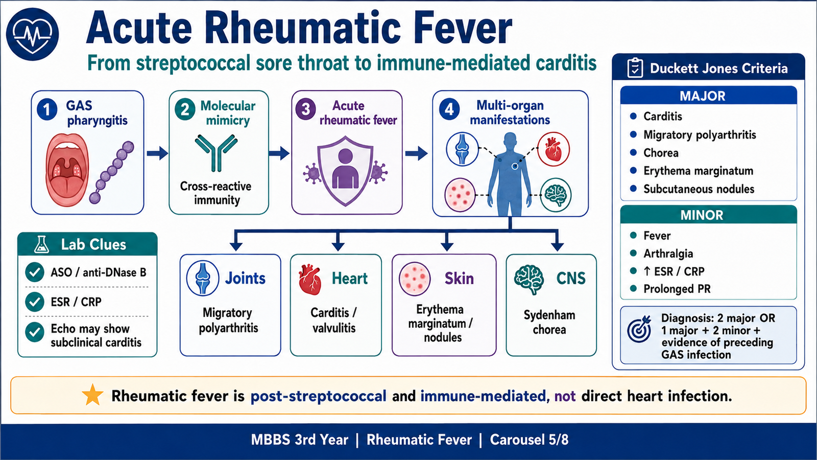
Acute rheumatic fever is a delayed, non-suppurative inflammatory complication associated classically with inadequately treated pharyngeal infection caused by rheumatogenic strains of group A beta-hemolytic Streptococcus—Streptococcus pyogenes.
It most commonly affects school-aged children.
The original throat infection may have resolved before rheumatic manifestations appear. Current WHO guidance describes rheumatic fever as an autoimmune inflammatory reaction following GAS pharyngitis and notes a possible association with some superficial skin infections, but the classical undergraduate and examination association remains untreated GAS pharyngitis.
It is not direct bacterial invasion
The organism does not usually infect the heart valves during acute rheumatic fever.
Instead:
GAS infection → immune response against streptococcal antigens → cross-reaction with human tissues → inflammation of heart, joints, skin and central nervous system
Immunological pathogenesis
Certain streptococcal antigens, particularly components associated with M protein, resemble host antigens.
This molecular mimicry produces:
- Cross-reactive antibodies
- Activation of T lymphocytes
- Inflammation of cardiac tissue
- Injury to connective tissue and valvular endothelium
The process resembles an antibody-mediated or type II hypersensitivity mechanism, with an important T-cell contribution.
Why the manifestations differ
The same immune process targets different tissues:
- Synovial and periarticular tissues → migratory arthritis
- Endocardium, myocardium and pericardium → carditis
- Basal ganglia → Sydenham chorea
- Skin → erythema marginatum
- Subcutaneous connective tissue → nodules
Duckett Jones criteria
No single laboratory test confirms acute rheumatic fever. Diagnosis is clinical and uses the revised Jones criteria together with evidence of a preceding GAS infection.
For an initial episode, the usual diagnostic pattern is:
- Two major manifestations, or
- One major plus two minor manifestations
In most patients, evidence of a preceding GAS infection is also required.
Major manifestations
A useful memory aid is J♥NES:
- J — Joints: migratory polyarthritis
- ♥ — Carditis, including subclinical echocardiographic carditis
- N — Subcutaneous nodules
- E — Erythema marginatum
- S — Sydenham chorea
In moderate- or high-risk populations, joint criteria are broadened and may include monoarthritis or polyarthralgia after excluding alternative causes.
Minor manifestations
- Fever
- Arthralgia not already counted as a major manifestation
- Raised ESR and/or CRP
- Prolonged PR interval on ECG, unless carditis is already counted as a major criterion
Risk-adjusted criteria use broader joint manifestations and a lower ESR threshold in moderate- and high-risk populations. A joint manifestation must not be counted simultaneously as both major and minor. Similarly, prolonged PR interval should not be counted as a minor criterion when carditis has already been used as a major manifestation.
Evidence of preceding GAS infection
Evidence includes:
- Positive throat culture
- Positive rapid antigen test
- Elevated or rising antistreptococcal antibody titre
- Recent scarlet fever
Common antibody tests include:
- Antistreptolysin O titre
- Anti-DNase B titre
A negative throat culture does not exclude rheumatic fever because the pharyngeal infection may have resolved before immune manifestations develop.
Important diagnostic exceptions
Sydenham chorea and indolent carditis may appear late. In appropriate settings, they may support a diagnosis even when complete Jones criteria or laboratory evidence of recent GAS infection are no longer demonstrable.
For recurrent rheumatic fever, accepted combinations include:
- Two major manifestations
- One major plus two minor manifestations
- Three minor manifestations after excluding more likely diagnoses
Routine echocardiography with Doppler is recommended in suspected or confirmed cases because carditis may be subclinical and produce no obvious murmur.
Laboratory and investigation findings
Evidence of inflammation
- Raised ESR
- Raised CRP
- Leukocytosis
These indicate inflammation but are not specific for rheumatic fever.
Evidence of preceding streptococcal infection
- Raised or rising ASO titre
- Raised anti-DNase B
- Positive throat culture or rapid antigen test
An ASO titre proves recent streptococcal exposure; it does not by itself prove active rheumatic carditis.
Cardiac investigations
ECG:
- Prolonged PR interval may occur.
- Tachycardia disproportionate to fever may suggest carditis.
Echocardiography:
- Mitral or aortic regurgitation
- Valvular thickening
- Subclinical valvulitis
- Ventricular dysfunction
- Pericardial effusion in some patients
Basic clinical reasoning
A child with fever and one painful swollen joint may have septic arthritis, but rheumatic arthritis typically:
- Involves large joints
- Migrates from one joint to another
- Responds rapidly to anti-inflammatory treatment
- Does not usually cause permanent joint deformity
The cardiac lesion, however, may leave permanent damage.
F. Rheumatic Carditis and Chronic Rheumatic Heart Disease
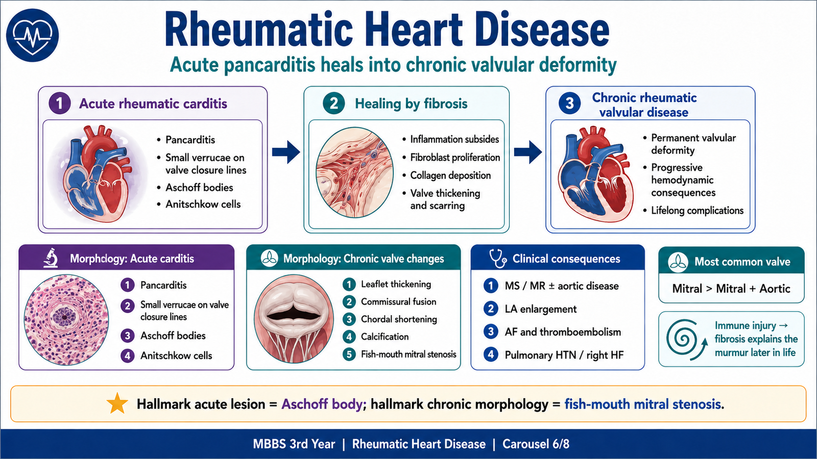
Acute rheumatic fever may cause inflammation throughout the heart—pancarditis. Repeated or severe attacks result in permanent valvular scarring.
The pathological sequence is:
Immune injury → fibrinoid tissue damage and inflammation → healing by fibrosis → leaflet and chordal deformity → stenosis and/or regurgitation → chamber enlargement and heart failure
Acute rheumatic heart disease
Endocardial changes
The valves become edematous and inflamed.
Small, sterile vegetations called verrucae may appear along the lines of valve closure.
These vegetations are:
- Small
- Warty
- Sterile
- Firmly attached
- Usually non-destructive
The mitral valve is most often involved, followed by combined mitral and aortic involvement.
Acute rheumatic valvulitis commonly produces mitral regurgitation because swollen leaflets cannot close properly.
Myocardial changes
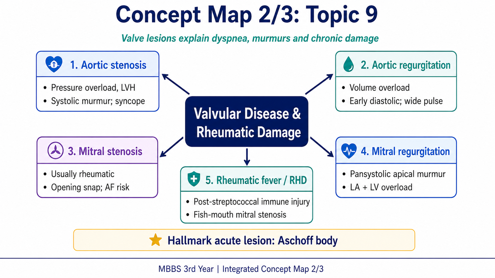
The characteristic lesion is the Aschoff body.
An Aschoff body contains:
- Central fibrinoid necrosis
- Surrounding T lymphocytes
- Plasma cells
- Activated macrophages
The characteristic macrophages are called Anitschkow cells. Their nuclei have central, wavy chromatin and are sometimes described as “caterpillar cells.”
Some fuse to form multinucleated Aschoff giant cells.
Myocarditis may reduce myocardial contractility and contribute to cardiac failure.
Pericardial changes
The pericardium may develop fibrinous inflammation, producing a rough “bread-and-butter” appearance.
Fibrinous pericarditis may cause:
- Pleuritic chest pain
- Pericardial friction rub
- Small pericardial effusion
It usually heals without producing constrictive pericarditis.
Chronic rheumatic heart disease
Chronic disease represents healing and organization of previous valvular inflammation.
Gross valvular morphology
- Leaflet thickening
- Fibrosis
- Commissural fusion
- Shortening and fusion of chordae tendineae
- Calcification in advanced disease
- Fish-mouth mitral stenosis
The mitral valve is affected most commonly.
Patterns include:
- Isolated mitral stenosis
- Mixed mitral stenosis and regurgitation
- Combined mitral and aortic valve disease
- Less commonly, isolated aortic disease
The tricuspid valve may be involved, but pulmonary valve involvement is unusual.
Microscopic morphology
- Dense collagenous fibrosis
- Neovascularization of valve leaflets
- Chronic inflammatory cells in some lesions
- Calcification in advanced disease
Active Aschoff bodies are generally absent from old, inactive chronic scars.
MacCallum plaques
Irregular subendocardial fibrotic thickening may occur in the posterior wall of the left atrium.
These plaques develop due to repeated impact of regurgitant jets on the atrial endocardium.
How morphology produces the clinical picture
Commissural fusion → narrowed mitral orifice → mitral stenosis → left atrial hypertension and enlargement → atrial fibrillation and pulmonary hypertension
Leaflet retraction or chordal shortening → failure of valve closure → regurgitation → atrial and ventricular volume overload
Major complications of chronic rheumatic heart disease
- Mitral stenosis
- Mitral or aortic regurgitation
- Atrial fibrillation
- Left atrial mural thrombus
- Systemic thromboembolism
- Pulmonary hypertension
- Right-sided heart failure
- Infective endocarditis
- Progressive cardiac failure
Key disease sequence: Untreated GAS pharyngitis → acute rheumatic fever → chronic mitral scarring → mitral stenosis → left atrial enlargement → atrial fibrillation and pulmonary congestion.
G. Infective Endocarditis
Definition
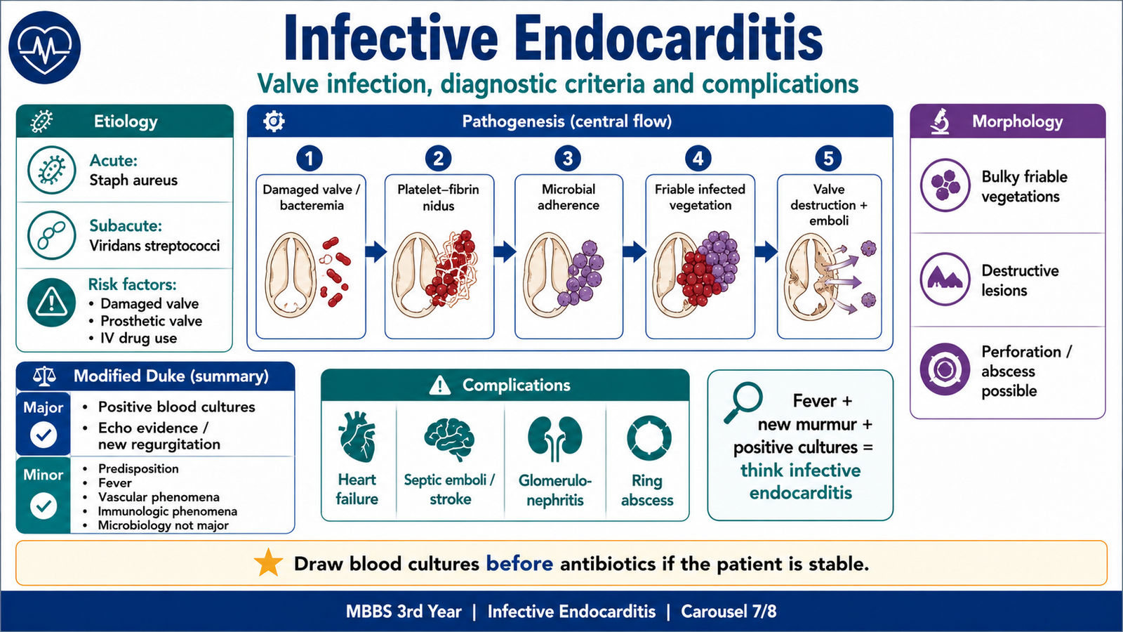
Infective endocarditis is microbial infection of the endocardial surface of the heart, usually involving a heart valve.
The lesion consists of infected vegetations containing:
- Fibrin
- Platelets
- Inflammatory material
- Microorganisms
- Necrotic tissue
Unlike rheumatic verrucae, infective vegetations are often bulky, friable and destructive.
Predisposing conditions
Important risk factors include:
- Previously damaged rheumatic valves
- Congenital heart disease
- Prosthetic valves
- Previous infective endocarditis
- Cardiac devices
- Intravenous drug use
- Poor dentition
- Invasive procedures associated with bacteremia
- Long-term intravenous access
- Hemodialysis
A structurally abnormal valve develops irregular blood flow and endothelial injury, making it easier for circulating organisms to attach.
Etiology and clinical patterns
Acute infective endocarditis
Acute endocarditis is caused by highly virulent organisms that can infect even previously normal valves.
A major organism is:
- Staphylococcus aureus
It commonly produces:
- Rapid onset
- High fever
- Marked toxicity
- Rapid valve destruction
- Abscess formation
- Acute regurgitation
- Early embolic complications
Subacute infective endocarditis
Subacute disease is caused by less virulent organisms and usually occurs on an already abnormal valve.
A classical organism is:
- Viridans-group streptococci following transient oral bacteremia
It commonly produces:
- Low-grade fever
- Fatigue
- Weight loss
- Anemia
- Changing murmur
- Splenomegaly
- Symptoms developing over weeks
Other important associations include:
- Enterococci after genitourinary or gastrointestinal sources
- Coagulase-negative staphylococci in prosthetic valve infection
- Staphylococcus aureus in intravenous drug use
- Streptococcus gallolyticus bacteremia associated with colonic pathology
- HACEK organisms causing relatively indolent disease
Pathogenesis
Step 1: Endothelial injury
Turbulent flow across a damaged valve injures the endocardium.
Step 2: Formation of a sterile platelet-fibrin deposit
Platelets and fibrin accumulate at the damaged area, creating a non-infected thrombotic surface.
Step 3: Transient bacteremia
Organisms enter the bloodstream through:
- Oral infection or manipulation
- Skin infection
- Intravenous injection
- Intravascular devices
- Gastrointestinal or genitourinary sources
Step 4: Microbial adherence
Circulating organisms adhere to the platelet-fibrin deposit.
Step 5: Vegetation growth
Further deposition of fibrin and platelets protects organisms from host defenses, allowing rapid multiplication.
Step 6: Local destruction and systemic spread
The vegetation may:
- Destroy valve leaflets
- Perforate the valve
- Rupture chordae
- Extend into the valve ring
- Form an abscess
- Fragment and embolize
Virulent Staphylococcus aureus may directly invade relatively normal endocardium without requiring major pre-existing valve damage.
Morphology
Gross appearance
Infective vegetations are typically:
- Bulky
- Friable
- Irregular
- Destructive
- Located on valve leaflets
The mitral and aortic valves are most commonly affected in left-sided disease.
Tricuspid valve infection is particularly associated with intravenous drug use and may produce septic pulmonary emboli.
Severe infection may produce:
- Leaflet perforation
- Chordal rupture
- Ring abscess
- Myocardial abscess
- Prosthetic valve dehiscence
Microscopic appearance
Vegetations contain:
- Fibrin
- Platelets
- Numerous microorganisms
- Necrotic debris
- Variable inflammatory cells
Older or partially treated lesions may show organization and fibrosis at their base.
Clinical features explained by pathology
Fever
Persistent bacteremia and inflammatory cytokine release produce fever and constitutional symptoms.
New or changing murmur
Valve destruction creates new regurgitation or worsens a pre-existing lesion.
Petechiae and splinter hemorrhages
Small emboli or vascular injury affect skin, nail beds and mucosal surfaces.
Janeway lesions
These are painless erythematous or hemorrhagic lesions of the palms and soles, classically associated with septic microemboli.
Osler nodes
These are painful nodules on fingers or toes, classically related to immune-complex phenomena.
Roth spots
Retinal hemorrhages with pale centers may occur.
Renal findings
Patients may develop:
- Hematuria due to immune-complex glomerulonephritis
- Renal infarction due to embolism
- Renal abscess due to septic emboli
Neurological findings
Embolization may produce:
- Ischemic stroke
- Brain abscess
- Intracranial hemorrhage
- Infected arterial, traditionally called mycotic, aneurysm
Diagnostic approach
Infective endocarditis should be suspected in a patient with:
- Persistent unexplained fever
- A new or changing murmur
- Positive blood cultures
- Predisposing valve disease
- Embolic or immunological manifestations
Blood cultures
In a hemodynamically stable patient:
- Obtain multiple blood-culture sets, commonly three, before starting antibiotics.
- Samples should not all be collected from an existing intravenous line.
- Prior antibiotic exposure may produce culture-negative endocarditis.
Antibiotics should not be delayed when the patient is septic or unstable, but cultures should be obtained first whenever safely possible.
Echocardiography
Echocardiography may demonstrate:
- Vegetation
- Valve perforation
- Abscess
- New regurgitation
- Prosthetic valve dehiscence
Transthoracic echocardiography is commonly the initial investigation.
Transesophageal echocardiography is more sensitive, particularly when:
- Initial transthoracic imaging is negative but suspicion remains high
- A prosthetic valve is present
- An abscess or device infection is suspected
Modified Duke diagnostic framework
For undergraduate examination purposes, the Modified Duke framework organizes findings into major and minor criteria.
Major criteria
- Positive blood cultures
- Typical organisms from appropriate cultures
- Persistently positive cultures
- Evidence of endocardial involvement
- Echocardiographic vegetation
- Abscess
- New prosthetic-valve dehiscence
- New valvular regurgitation
Minor criteria
- Predisposing cardiac lesion or intravenous drug use
- Fever of at least 38°C
- Vascular phenomena
- Immunological phenomena
- Microbiological evidence not fulfilling a major criterion
Clinical combinations traditionally used
Definite infective endocarditis:
- Two major criteria, or
- One major plus three minor criteria, or
- Five minor criteria
Possible infective endocarditis:
- One major plus one minor criterion, or
- Three minor criteria
The 2023 Duke-ISCVID update retains the overall diagnostic logic but expands accepted microbiological and imaging evidence, including selected molecular tests, cardiac CT and metabolic imaging in appropriate settings. At 3rd-year level, students should first master the classic major/minor framework.
Complications
Cardiac complications
- Acute valve regurgitation
- Heart failure
- Valve perforation
- Chordal rupture
- Ring abscess
- Conduction disturbance due to extension near the conduction system
- Prosthetic valve dehiscence
Embolic complications
Left-sided infective endocarditis may embolize to:
- Brain
- Kidneys
- Spleen
- Intestines
- Limbs
- Coronary arteries
Right-sided infective endocarditis produces septic pulmonary emboli.
Immunological complications
- Glomerulonephritis
- Osler nodes
- Roth spots
- Positive rheumatoid factor in prolonged disease
Metastatic infection
- Brain abscess
- Vertebral osteomyelitis
- Septic arthritis
- Splenic abscess
Basic management logic
Management requires:
- Prompt blood cultures
- Echocardiography
- Prolonged targeted intravenous antimicrobial therapy
- Identification and control of the bacteremia source
- Early specialist involvement
Urgent cardiac-surgical assessment is particularly important when there is:
- Severe valve dysfunction causing heart failure
- Uncontrolled infection or abscess
- Prosthetic valve dehiscence
- Recurrent embolization
- Persistent bacteremia despite appropriate treatment
Infective endocarditis is uncommon but potentially fatal, and modern diagnostic pathways combine clinical assessment, microbiology and multimodality imaging.
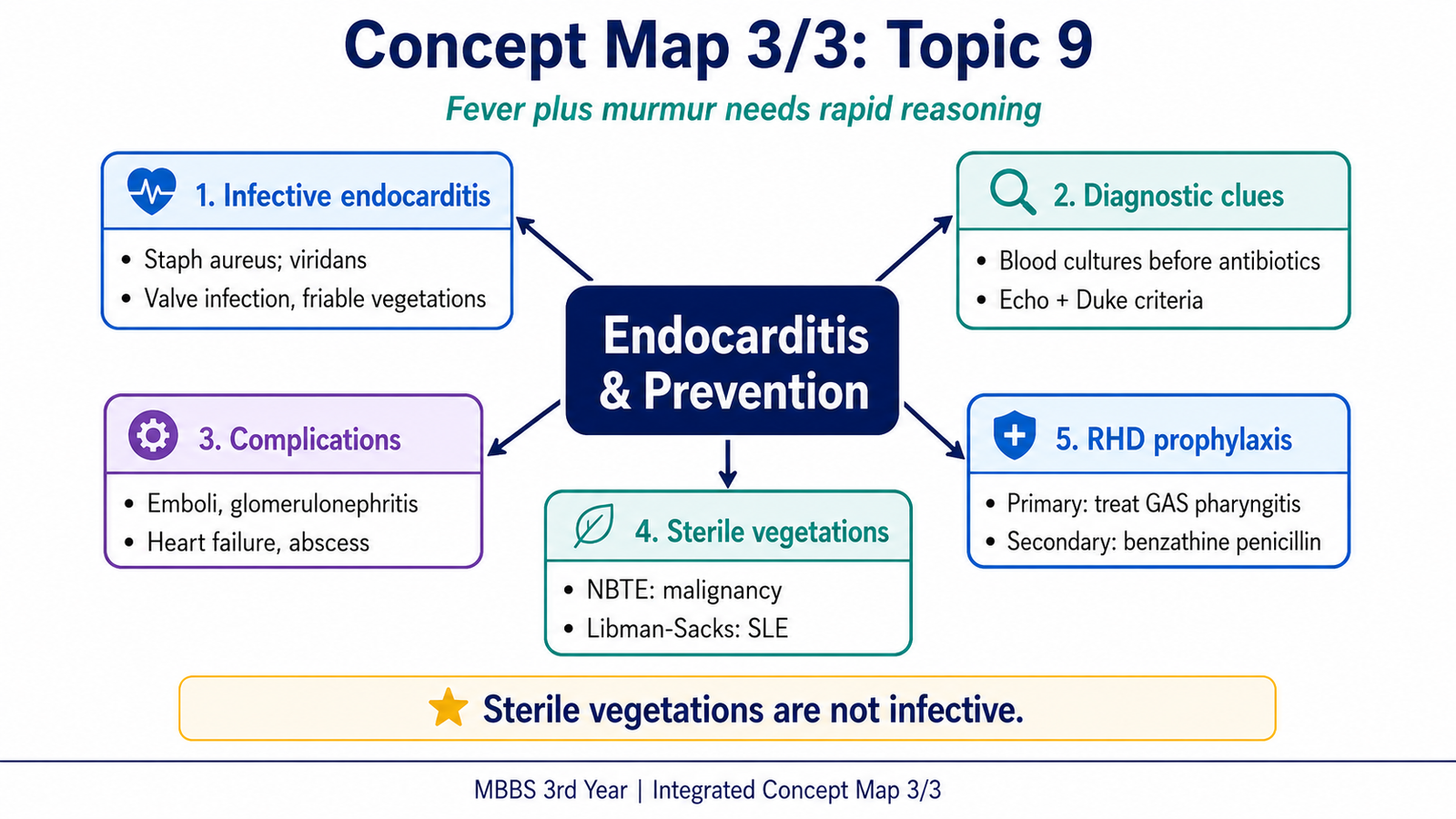
H. Non-Infected Vegetations and Rheumatic Fever Prophylaxis
Not every vegetation is infected. The location, size, inflammatory response and clinical setting help identify its type.
Rheumatic vegetations
Acute rheumatic fever produces small sterile verrucae:
- Located along the lines of valve closure
- Associated with valvular inflammation
- Usually firmly attached
- Usually non-destructive
- Most often involving the mitral valve
Their main long-term consequence is healing by fibrosis and permanent valve deformity.
Nonbacterial thrombotic endocarditis
Nonbacterial thrombotic endocarditis, also called marantic endocarditis, consists of sterile platelet-fibrin vegetations.
It is associated with hypercoagulable states, including:
- Advanced malignancy, especially mucin-producing adenocarcinoma
- Severe wasting illness
- Disseminated intravascular coagulation
- Other marked prothrombotic states
Morphology
- Small to medium sterile vegetations
- Located along valve closure lines
- Minimal underlying inflammation
- Little valve destruction
- Loosely attached and prone to embolization
Clinical significance
The major danger is systemic embolism rather than valve failure.
A patient with advanced malignancy and multiple cerebral or systemic infarcts but no infection should raise suspicion of nonbacterial thrombotic endocarditis.
Libman–Sacks endocarditis
Libman–Sacks endocarditis is associated mainly with:
- Systemic lupus erythematosus
- Antiphospholipid antibody syndrome
Morphology
Sterile vegetations may occur:
- On either surface of a valve leaflet
- On both surfaces simultaneously
- On chordae tendineae
- On adjacent mural endocardium
Unlike bland nonbacterial thrombotic endocarditis, there may be:
- Fibrinoid necrosis
- Inflammation
- Hematoxylin bodies in some lesions
- Subsequent scarring and deformity
The mitral valve is frequently involved.
Vegetation comparison
| Feature | Rheumatic | Infective | Nonbacterial thrombotic | Libman–Sacks |
| Infection | No | Yes | No | No |
| Typical setting | Acute rheumatic fever | Bacteremia with valve/device risk | Malignancy or hypercoagulability | SLE/antiphospholipid syndrome |
| Size | Small | Bulky | Small–medium | Variable |
| Attachment | Firm | Friable | Often loose | Variable |
| Valve destruction | Minimal acutely | Common | Minimal | May scar |
| Location | Closure lines | Commonly flow side of valve | Closure lines | Either or both valve surfaces |
| Major danger | Chronic fibrosis | Valve destruction and septic emboli | Systemic emboli | Emboli and valve dysfunction |
Primary prophylaxis of rheumatic fever
Primary prophylaxis means preventing the first attack of acute rheumatic fever.
This requires:
- Recognizing possible GAS pharyngitis
- Confirming infection when testing is available and appropriate
- Treating confirmed GAS infection adequately
- Improving adherence to the complete antibiotic course
- Reducing transmission through hand hygiene and respiratory etiquette
Adequate antibiotic treatment of confirmed GAS pharyngitis prevents the first attack of rheumatic fever. Penicillin is the standard first-line drug; an appropriate alternative is selected when true penicillin allergy is present. The exact regimen should follow the locally adopted guideline.
Secondary prophylaxis
Secondary prophylaxis means preventing recurrent attacks in a patient who has already experienced rheumatic fever or has established rheumatic heart disease.
Each recurrent episode may add further valve damage.
Long-term penicillin prophylaxis is used to prevent recurrent GAS infection and further rheumatic attacks. Intramuscular benzathine penicillin is generally preferred because regular administration improves protection. An appropriate oral alternative may be used when intramuscular therapy is unsuitable or penicillin cannot be given. The exact regimen and duration depend on locally adopted guidance.
Duration is individualized according to:
- Previous carditis
- Presence of residual valve disease
- Age
- Time since the last attack
- Risk of GAS exposure
- Local disease burden
Secondary prophylaxis is continued longer when previous carditis or residual valve disease is present. Duration is individualized according to age, time since the last attack, continuing exposure risk and the severity of established valve disease.
Public-health logic
Rheumatic heart disease is largely preventable.
Effective control requires:
- Early access to care for sore throat
- Reliable diagnostic pathways
- Adequate antibiotic supply
- Patient registries
- Recall systems for regular injections
- Education about adherence
- Reduction of overcrowding and barriers to healthcare
WHO identifies primary prevention, long-term secondary prophylaxis and early detection of rheumatic heart disease as central control strategies.
Important distinction: rheumatic prophylaxis versus endocarditis prophylaxis
Long-term penicillin in rheumatic heart disease is given to prevent recurrent GAS infection and recurrent rheumatic fever.
It does not automatically mean the patient requires antibiotic prophylaxis before every dental procedure.
Routine infective-endocarditis prophylaxis is not recommended solely because a patient has native rheumatic valve disease. It is reserved for selected highest-risk cardiac conditions, such as certain patients with prosthetic valves or previous infective endocarditis, according to the applicable guideline.
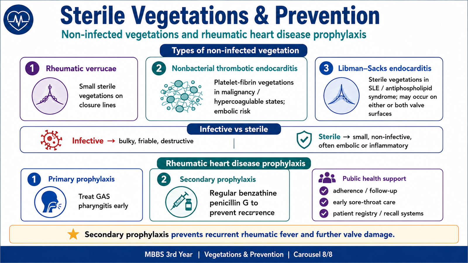
⭐ AIM High-Yield Review
- Heart valves open and close passively according to pressure gradients.
- S1 is produced mainly by mitral and tricuspid valve closure; S2 by aortic and pulmonary valve closure.
- A stenotic valve produces a murmur while it should be open; a regurgitant valve produces a murmur while it should be closed.
- Aortic stenosis causes left ventricular pressure overload, concentric hypertrophy and a systolic murmur radiating to the carotids.
- Mitral stenosis most often follows rheumatic heart disease and causes left atrial enlargement, pulmonary hypertension and atrial fibrillation.
- Aortic regurgitation produces a diastolic decrescendo murmur, left ventricular volume overload and a wide pulse pressure.
- Mitral regurgitation produces a pansystolic apical murmur radiating toward the axilla and causes left atrial and left ventricular volume overload.
- Acute rheumatic fever is an immune-mediated complication of preceding group A streptococcal infection, not direct bacterial invasion of the heart.
- Jones criteria require major and minor manifestations together with evidence of preceding streptococcal infection in most patients.
- Acute rheumatic carditis may show Aschoff bodies and small sterile verrucae; chronic disease produces fibrosis, commissural fusion and chordal shortening.
- Infective endocarditis produces bulky, friable, destructive vegetations and is diagnosed by integrating blood cultures, echocardiography and clinical criteria.
- Nonbacterial thrombotic endocarditis is associated with hypercoagulability and mainly causes systemic emboli.
- Libman–Sacks vegetations may occur on either surface of valve leaflets and are associated with SLE or antiphospholipid syndrome.
- Primary prophylaxis prevents the first rheumatic attack by treating GAS pharyngitis; secondary prophylaxis prevents recurrent attacks and further valve damage.
This Ninja Nerd lecture covers **aortic and mitral stenosis/regurgitation, murmurs, rheumatic fever, complications, and diagnostic evaluation**, making it a strong match for the main topic. ([YouTube][1])
🎥 AIM Video Learning — Valvular Heart Disease and Murmurs
“`
Watch this video after reading the chapter to strengthen your understanding of mitral and aortic stenosis and regurgitation, rheumatic valve disease, heart murmurs, complications and diagnostic evaluation.
“`
AIM Study Tip: Relate the timing of each murmur to whether the affected valve should normally be open or closed during systole or diastole.