
Study tip: This chapter follows the KMU learning outcomes in a logical sequence. First understand how coronary thrombosis causes myocardial injury and then connect that mechanism with diagnosis and antithrombotic treatment. Use the final high-yield review only after reading the explanations.
Acute Coronary Syndrome, Myocardial Infarction and Antithrombotic Therapy
CVS Module — Understand the ACS spectrum, myocardial infarction, clinical assessment, complications, and the rational use of antiplatelet, anticoagulant and thrombolytic drugs.
Topic Introduction
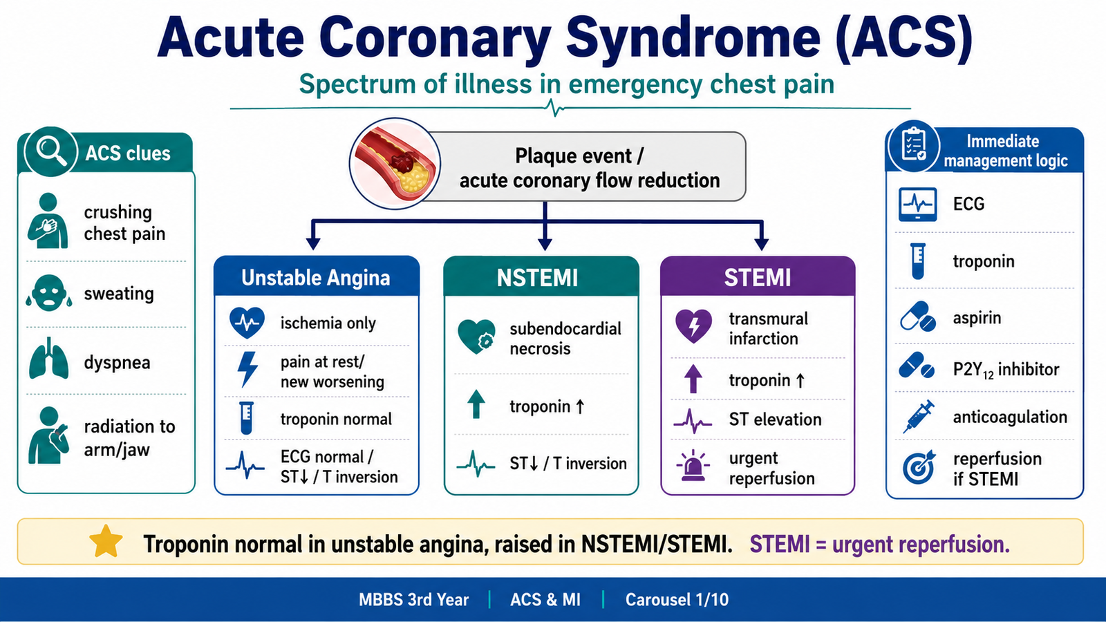
Acute coronary syndrome (ACS) is a group of emergencies caused by a sudden fall in coronary blood flow. It includes unstable angina, NSTEMI and STEMI. The usual event is disruption of an atherosclerotic plaque, followed by platelet activation and formation of a thrombus. When ischemia is prolonged, myocardial cells die and myocardial infarction develops. This chapter explains the disease process, the clinical features and diagnosis of myocardial infarction, risk assessment, immediate management and major complications. It also explains why antiplatelet drugs, anticoagulants and thrombolytic drugs are used, how they work, their important adverse effects, monitoring requirements and key comparisons.
A. Acute Coronary Syndrome and Coronary Thrombosis
Acute coronary syndrome means a group of emergency clinical conditions caused by acute myocardial ischemia, usually due to sudden reduction of blood flow in a coronary artery.
The ACS spectrum includes:
| ACS Type | Main Pathology | ECG | Troponin | Basic Meaning |
|---|---|---|---|---|
| Unstable angina | Ischemia without necrosis | ST depression/T inversion or normal | Normal | Threatened infarction |
| NSTEMI | Partial-thickness myocardial necrosis | ST depression/T inversion or nonspecific | Raised | Infarction without ST elevation |
| STEMI | Usually transmural infarction | ST elevation/new LBBB pattern | Raised | Complete or near-complete occlusion |
⭐ Common KMU trap: unstable angina and NSTEMI may look clinically similar, but troponin is normal in unstable angina and raised in NSTEMI.
ACS is not simply “chest pain.” It is a time-dependent myocardial injury pathway. The longer the coronary artery remains blocked, the more myocardium dies.
B. What Is Happening in the Coronary Artery?
Most ACS cases begin with an atherosclerotic plaque in a coronary artery.
The usual sequence is:
- Long-standing endothelial injury due to risk factors such as smoking, hypertension, diabetes, dyslipidemia, and age.
- Lipid accumulation and inflammation inside the arterial wall.
- Formation of an atheromatous plaque with a lipid core and fibrous cap.
- Plaque rupture or erosion.
- Exposure of thrombogenic material to blood.
- Platelet adhesion, activation, and aggregation.
- Coagulation cascade activation.
- Fibrin-rich thrombus formation.
- Partial or complete coronary occlusion.
⭐ Most tested mechanism: ACS is commonly due to rupture of an unstable atherosclerotic plaque with superadded thrombosis.
A stable plaque causes predictable exertional angina. An unstable plaque can rupture suddenly and cause ACS even at rest.

B. Myocardial Infarction: Pathophysiology and Morphology
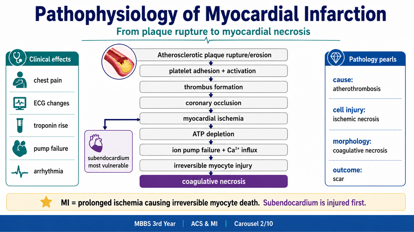
Myocardial infarction means myocardial cell death due to prolonged ischemia.
The core pathology is:
Coronary occlusion → oxygen deprivation → ATP depletion → failure of ion pumps → calcium overload → mitochondrial damage → irreversible myocyte injury → necrosis.
Early ischemia may be reversible. If ischemia persists, irreversible injury begins. The subendocardial region is usually most vulnerable because it has the highest oxygen demand and relatively poorest perfusion during systole.
Cellular injury sequence
When oxygen supply falls:
- Oxidative phosphorylation decreases.
- ATP falls.
- Na⁺/K⁺ ATPase fails.
- Cells swell.
- Anaerobic glycolysis increases.
- Lactic acid accumulates.
- Contractility decreases.
- Calcium enters cells.
- Membrane and mitochondrial injury become irreversible.
⭐ Basic science bridge: normal myocardium depends heavily on aerobic metabolism; therefore, loss of oxygen rapidly causes ATP failure and contractile dysfunction.
D. Morphology of Myocardial Infarction
Pathology explains why the clinical picture changes over time.
Gross changes
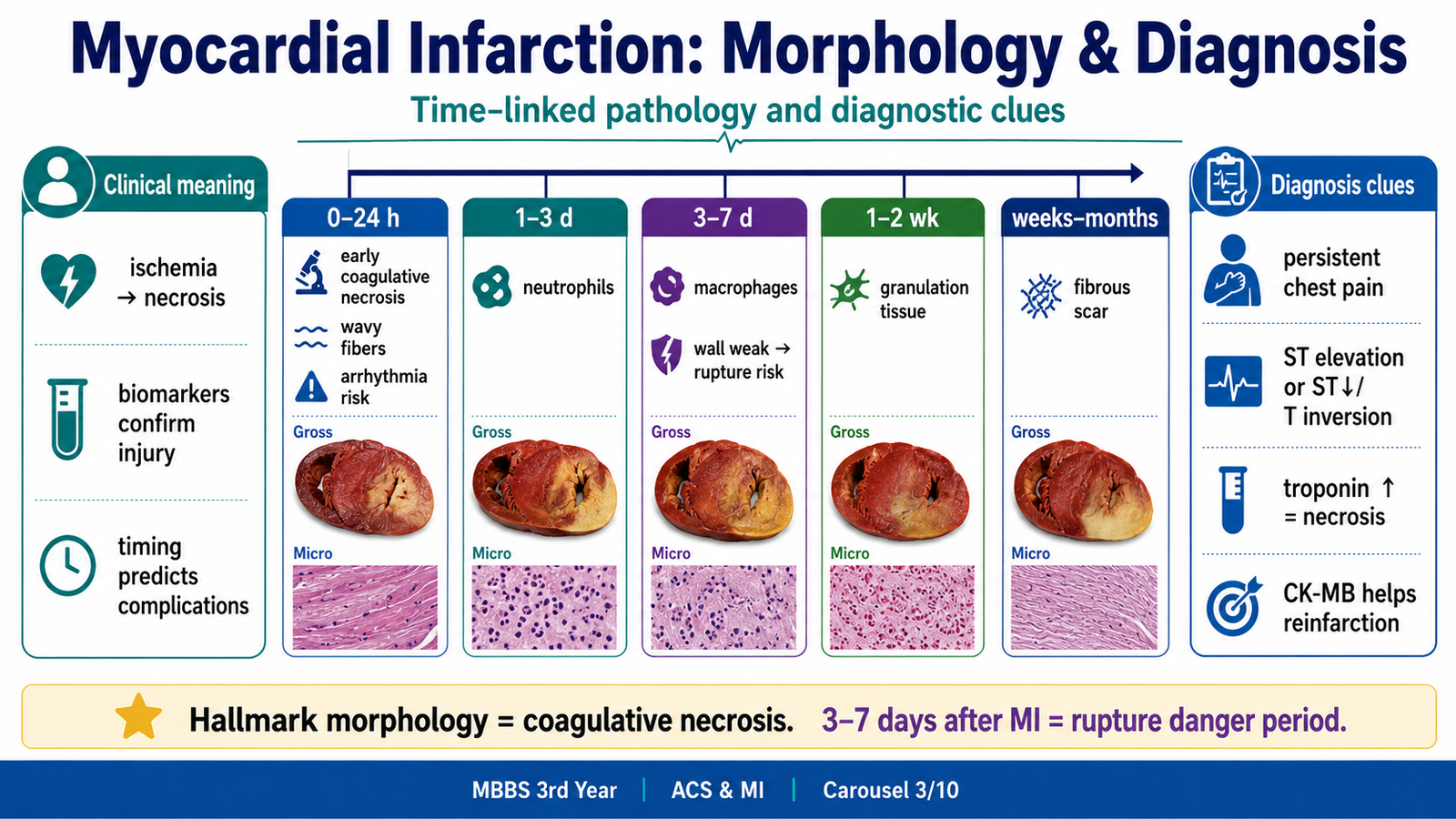
Early MI may show no obvious gross change. Later, the infarcted area becomes pale, then yellow-soft, and eventually fibrotic.
| Time After MI | Gross Change | Microscopic Change | Clinical Importance |
|---|---|---|---|
| 0–4 hours | Usually no gross change | Early reversible/irreversible injury may be difficult to see | ECG/troponin more useful |
| 4–24 hours | Dark mottling may appear | Coagulative necrosis begins, wavy fibers, early neutrophils | Arrhythmias common early |
| 1–3 days | Mottled infarct | Heavy neutrophilic infiltration | Acute inflammation |
| 3–7 days | Yellow soft center | Macrophages remove dead tissue | Wall is weak |
| 1–2 weeks | Red-gray depressed margins | Granulation tissue | Healing phase |
| Weeks to months | Firm white scar | Dense collagen scar | Permanent loss of contractile tissue |
⭐ Hallmark morphology: myocardial infarction shows coagulative necrosis of cardiac muscle.
⭐ Dangerous complication timing: myocardial wall rupture is classically feared around 3–7 days, when macrophages have weakened the infarcted wall.
C. Clinical Features and Diagnosis of Myocardial Infarction
The patient’s symptoms are explained by ischemic myocardial injury and sympathetic activation.
| Clinical Feature | Explanation |
|---|---|
| Severe central chest pressure | Ischemia stimulates cardiac pain fibers |
| Radiation to left arm/jaw | Shared spinal segments for referred pain |
| Sweating and anxiety | Sympathetic discharge |
| Nausea/vomiting | Inferior wall MI may stimulate vagal pathways |
| Dyspnea | LV dysfunction or pulmonary congestion |
| Hypotension | Pump failure, RV infarction, arrhythmia, shock |
| Palpitations/syncope | Ischemia-induced arrhythmia |
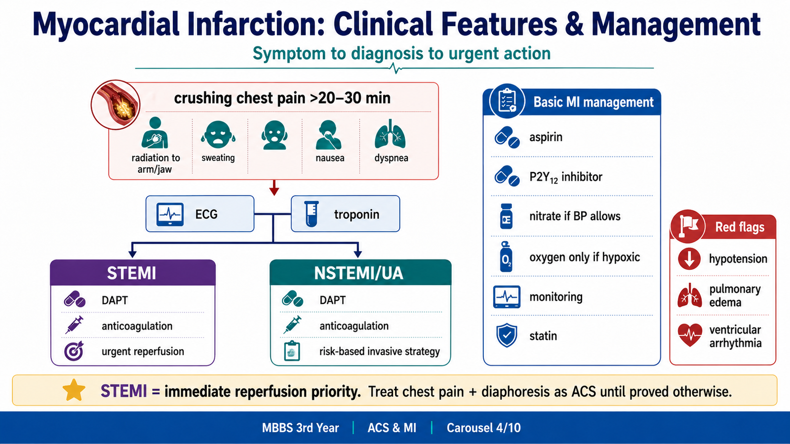
⭐ Most likely diagnosis clue: severe retrosternal crushing chest pain lasting more than 20–30 minutes with sweating should be treated as ACS until proven otherwise.
F. Diagnosis and Interpretation Clues in MI
The diagnosis is built from three pillars:
- Clinical symptoms.
- ECG changes.
- Cardiac biomarkers.
ECG clues
- ST elevation suggests STEMI.
- ST depression or T-wave inversion may suggest NSTEMI/unstable angina.
- New conduction abnormality can be serious.
- Normal ECG does not fully exclude ACS if symptoms are convincing.
Biomarker clues
Cardiac troponins are the most important markers of myocardial necrosis.
- Troponin I and T rise after myocardial injury.
- They remain elevated for several days.
- CK-MB may help in suspected reinfarction because it returns to normal earlier than troponin.
⭐ Diagnostic clue: raised cardiac troponin means myocardial necrosis, not just ischemia.
D. Risk Stratification in Myocardial Infarction
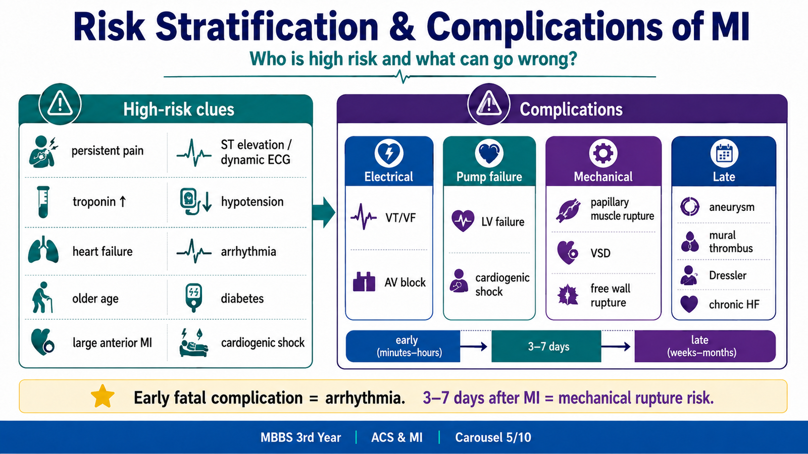
Risk stratification means identifying which MI patient is more likely to deteriorate or die, so urgent care can be prioritized.
Important risk markers include:
- Persistent chest pain.
- ST elevation or dynamic ECG changes.
- Raised troponin.
- Hypotension.
- Tachycardia or bradycardia.
- Heart failure signs.
- Arrhythmias.
- Diabetes, older age, renal disease.
- Previous MI or known coronary artery disease.
- Large anterior wall MI.
- Cardiogenic shock.
Clinically, STEMI is automatically treated as high-risk because it usually indicates acute coronary occlusion needing urgent reperfusion.
NSTEMI patients are risk-stratified using clinical condition, ECG, troponin, and scoring systems such as TIMI or GRACE at basic undergraduate awareness level.
⭐ Emergency point: hypotension, pulmonary edema, altered consciousness, or malignant arrhythmia in MI means high-risk ACS.

E. Management of Acute Coronary Syndrome and Myocardial Infarction
The management goal is simple:
Save myocardium, prevent thrombus extension, restore coronary flow, control pain, prevent complications, and reduce future risk.
Immediate management logic
| Step | Why It Is Done |
|---|---|
| Rapid assessment and ECG | STEMI needs urgent reperfusion |
| Aspirin | Blocks platelet thromboxane A₂ |
| P2Y12 inhibitor | Adds stronger platelet inhibition |
| Anticoagulant | Prevents fibrin-rich thrombus extension |
| Nitrates if appropriate | Relieves ischemic pain by reducing preload and coronary spasm |
| Oxygen only if hypoxic | Avoid unnecessary oxygen in non-hypoxic patients |
| Reperfusion in STEMI | Opens the occluded coronary artery |
| Monitoring | Detect arrhythmias and shock early |
| Statin and risk-factor control | Prevent recurrence |
Reperfusion may be by primary PCI or thrombolytic therapy depending on availability, timing, and contraindications.
⭐ Drug logic: ACS treatment combines antiplatelet therapy for platelet-rich arterial thrombus plus anticoagulation for coagulation cascade/fibrin control.
F. Antiplatelet and Anticoagulant Therapy
Platelets are central in arterial thrombosis. When plaque ruptures, platelets adhere, activate, release mediators, and aggregate through GP IIb/IIIa receptors.
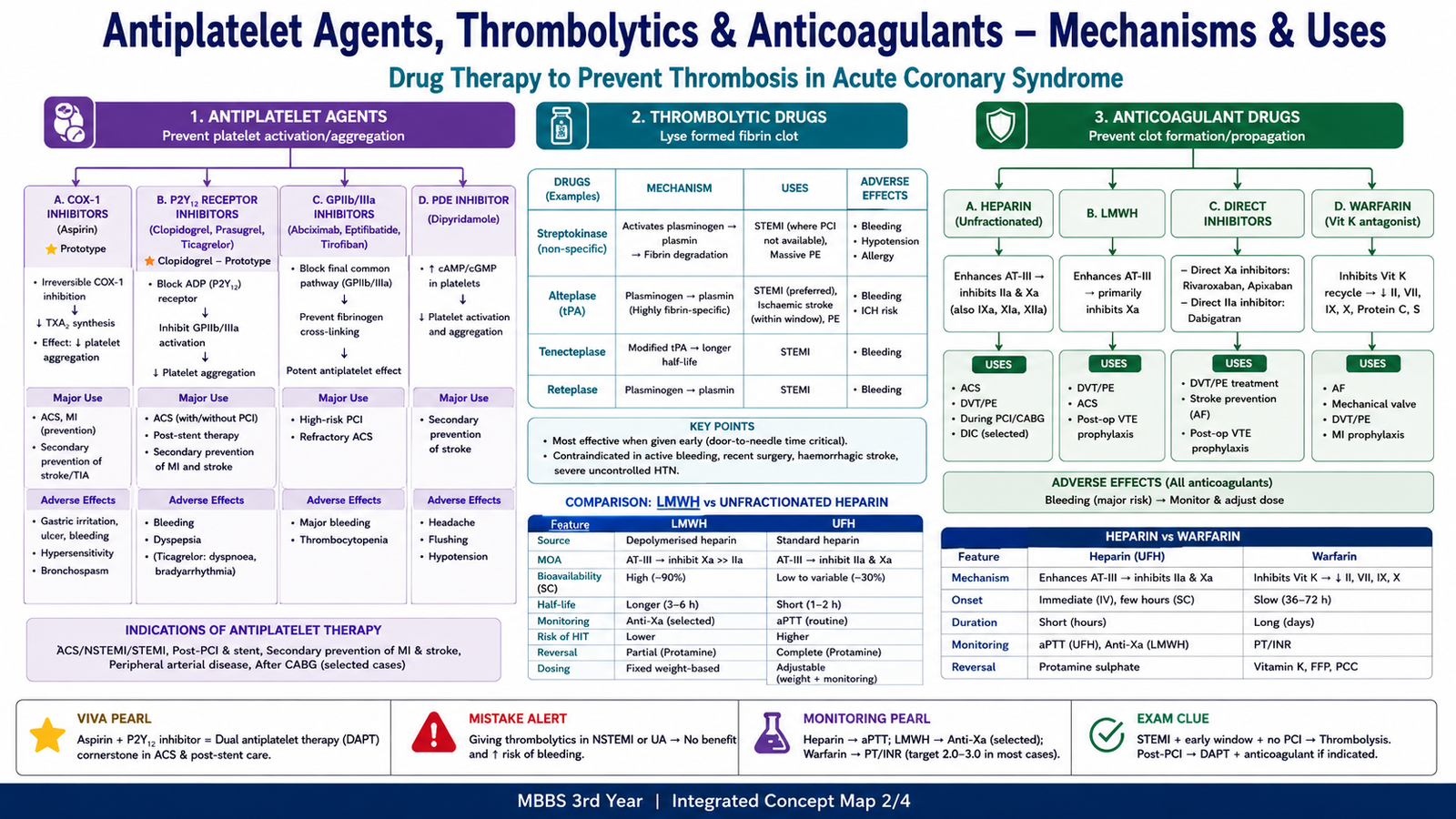
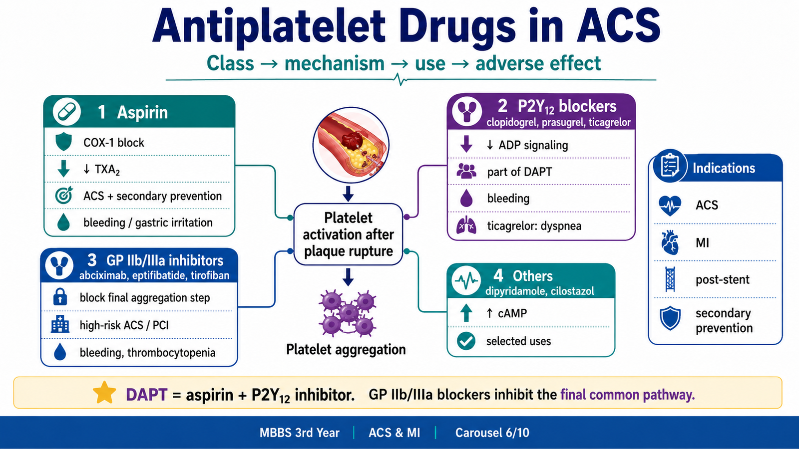
Classification of Antiplatelet Drugs
| Class | Examples | Main Action |
|---|---|---|
| COX inhibitor | Aspirin | Inhibits thromboxane A₂ formation |
| P2Y12 ADP receptor blockers | Clopidogrel, prasugrel, ticagrelor | Prevent ADP-mediated platelet activation |
| GP IIb/IIIa inhibitors | Abciximab, eptifibatide, tirofiban | Block final common pathway of platelet aggregation |
| Phosphodiesterase inhibitors | Dipyridamole, cilostazol | Increase cAMP in platelets, reduce aggregation |
Indications of Antiplatelet Therapy
Antiplatelets are used in:
- Acute coronary syndrome.
- Myocardial infarction.
- After PCI/stent placement.
- Secondary prevention after MI.
- Stroke/TIA prevention where appropriate.
- Peripheral arterial disease.
⭐ Most important clinical use: in ACS, aspirin is given early unless contraindicated.
Aspirin
Class: COX inhibitor.
Prototype: Aspirin.
Mechanism: Irreversibly inhibits COX-1 in platelets → decreases thromboxane A₂ → reduces platelet aggregation.
Use in ACS: Immediate antiplatelet therapy and long-term secondary prevention.
Adverse effects:
- Gastric irritation and peptic ulcer bleeding.
- Hypersensitivity/bronchospasm in sensitive patients.
- Increased bleeding risk.
- Tinnitus at higher doses.
- Reye syndrome risk in children.
Contraindications/cautions:
- Active bleeding.
- Severe aspirin allergy.
- Active peptic ulcer bleeding.
- Bleeding disorders.
⭐ Prototype/high-yield: aspirin irreversibly inhibits platelet COX, so its effect lasts for the platelet lifespan.
P2Y12 Receptor Inhibitors
Examples:
- Clopidogrel.
- Prasugrel.
- Ticagrelor.
Mechanism: Block ADP-mediated activation of platelets by inhibiting P2Y12 receptors → reduce GP IIb/IIIa activation → reduce aggregation.
Uses:
- ACS.
- Dual antiplatelet therapy with aspirin.
- After coronary stenting.
- Secondary prevention in selected patients.
Important differences:
| Drug | Key Point |
|---|---|
| Clopidogrel | Prodrug; variable activation |
| Prasugrel | More potent; higher bleeding risk |
| Ticagrelor | Reversible; not a prodrug |
Adverse effects:
- Bleeding.
- GI upset.
- Rash.
- Ticagrelor may cause dyspnea.
- Rare thrombotic thrombocytopenic purpura with some agents.
Contraindications/cautions:
- Active bleeding.
- Prasugrel is avoided in previous stroke/TIA.
- Caution before surgery due to bleeding risk.
⭐ Common KMU trap: DAPT means aspirin + P2Y12 inhibitor, not aspirin + warfarin.
GP IIb/IIIa Inhibitors
Examples:
- Abciximab.
- Eptifibatide.
- Tirofiban.
Mechanism: Block GP IIb/IIIa receptors on platelets. This prevents fibrinogen from cross-linking platelets, blocking the final common pathway of platelet aggregation.
Uses:
- High-risk ACS in selected cases.
- During PCI in selected patients.
Adverse effects:
- Serious bleeding.
- Thrombocytopenia.
⭐ High-yield mechanism: GP IIb/IIIa inhibitors block the final common pathway of platelet aggregation.
Dipyridamole and Cilostazol
Mechanism: Increase platelet cAMP, reducing platelet activation.
Uses:
- Dipyridamole may be used with aspirin in some stroke prevention settings.
- Cilostazol is used in intermittent claudication.
Adverse effects:
- Headache.
- Flushing.
- Palpitations.
- Hypotension.
These are not first-line emergency ACS drugs but are included in antiplatelet classification.
J. Anticoagulant Therapy in ACS
Anticoagulants mainly interfere with the coagulation cascade and fibrin formation. In ACS, they prevent thrombus propagation after plaque rupture.
Classification of Anticoagulant Drugs
| Class | Examples |
|---|---|
| Parenteral indirect thrombin/Xa inhibitors | Unfractionated heparin, low molecular weight heparin |
| Synthetic factor Xa inhibitor | Fondaparinux |
| Direct factor Xa inhibitors | Rivaroxaban, apixaban, edoxaban |
| Direct thrombin inhibitor | Dabigatran, bivalirudin, argatroban |
| Vitamin K antagonist | Warfarin |
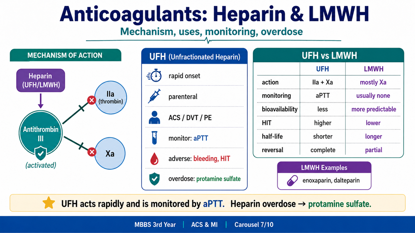
Unfractionated Heparin
Class: Parenteral anticoagulant.
Mechanism: Binds antithrombin III and accelerates its action. This inhibits thrombin factor IIa and factor Xa.
Uses:
- ACS/MI anticoagulation.
- Venous thromboembolism treatment/prevention.
- Pulmonary embolism.
- During procedures where rapid anticoagulation is needed.
- Safe anticoagulant option in pregnancy because it does not cross the placenta.
Monitoring:
UFH is monitored mainly by aPTT.
Adverse effects:
- Bleeding.
- Heparin-induced thrombocytopenia.
- Osteoporosis with prolonged use.
- Hypersensitivity.
- Hyperkalemia rarely.
Treatment of overdose:
- Stop heparin.
- Give protamine sulfate if significant bleeding occurs.
⭐ Antidote: protamine sulfate reverses heparin overdose.
⭐ Serious adverse effect: heparin-induced thrombocytopenia can cause thrombosis despite low platelets.
Low Molecular Weight Heparin vs Unfractionated Heparin
Examples of LMWH:
- Enoxaparin.
- Dalteparin.
| Feature | UFH | LMWH |
|---|---|---|
| Main action | Inhibits IIa and Xa | More anti-Xa than anti-IIa |
| Route | Parenteral | Subcutaneous |
| Monitoring | aPTT needed | Usually no routine monitoring |
| Bioavailability | Less predictable | More predictable |
| Half-life | Shorter | Longer |
| HIT risk | Higher | Lower |
| Reversal by protamine | More complete | Partial |
⭐ Common comparison: LMWH has more predictable action and usually does not need routine aPTT monitoring.
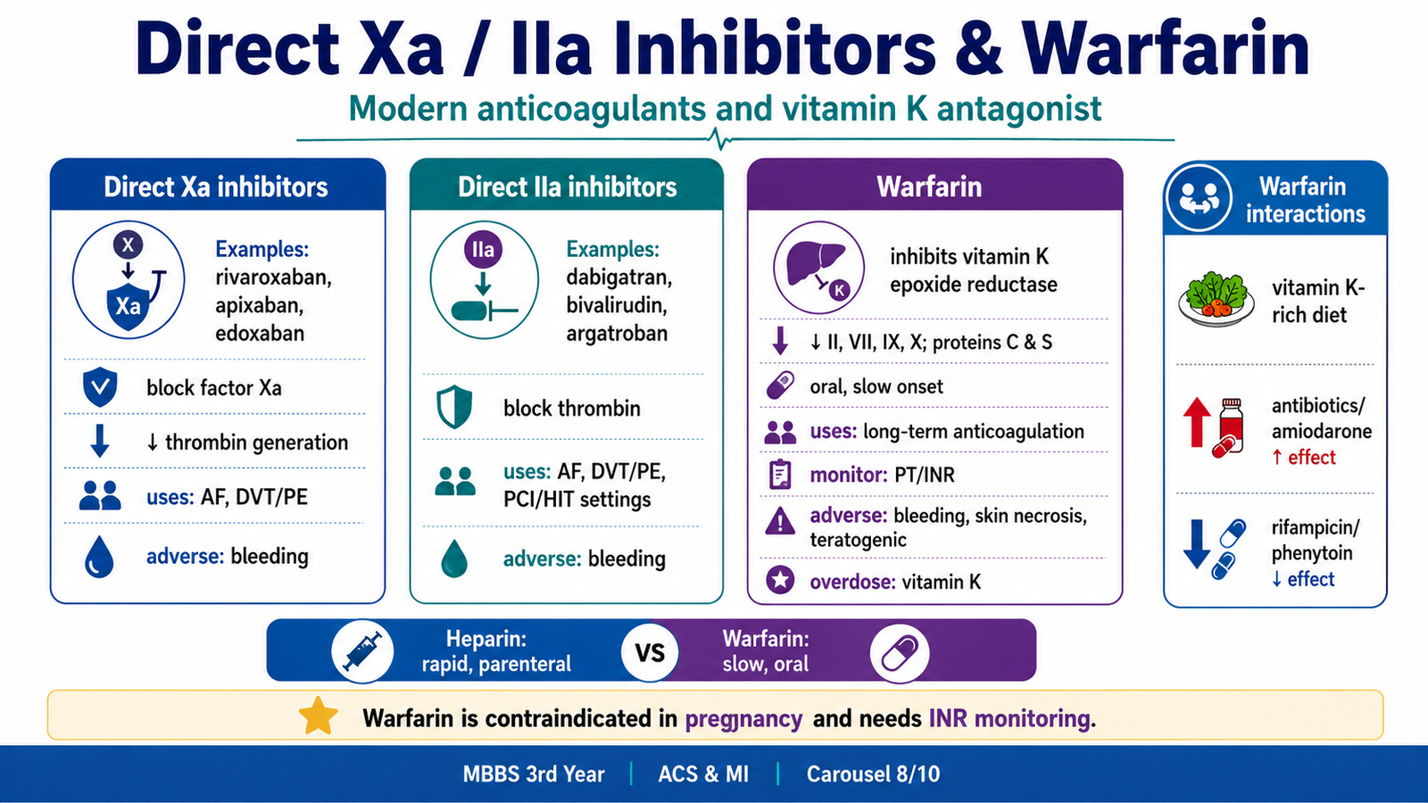
Direct Factor Xa and Direct Thrombin Inhibitors
Direct Factor Xa inhibitors
Examples:
- Rivaroxaban.
- Apixaban.
- Edoxaban.
Mechanism: Directly inhibit factor Xa → reduce thrombin generation → reduce fibrin clot formation.
Uses:
- Atrial fibrillation stroke prevention.
- DVT/PE treatment and prevention.
- Selected cardiovascular indications depending on clinical context.
Adverse effects:
- Bleeding.
- GI bleeding risk.
- Caution in renal impairment.
Direct Thrombin Inhibitors
Examples:
- Dabigatran.
- Bivalirudin.
- Argatroban.
Mechanism: Directly inhibit thrombin factor IIa → prevent conversion of fibrinogen to fibrin.
Uses:
- Atrial fibrillation stroke prevention.
- DVT/PE treatment.
- Bivalirudin may be used during PCI.
- Argatroban is useful in HIT.
Adverse effects:
- Bleeding.
- Dabigatran may cause dyspepsia.
⭐ Mechanism trap: direct Xa inhibitors block Xa; direct thrombin inhibitors block IIa.
Warfarin
Class: Vitamin K antagonist.
Mechanism: Inhibits vitamin K epoxide reductase → decreases activation of factors II, VII, IX, X and proteins C and S.
Uses:
- Long-term anticoagulation.
- Atrial fibrillation.
- Mechanical heart valves.
- DVT/PE treatment and prevention.
- Selected post-MI situations with thrombus risk.
Onset:
Warfarin has slow onset because it affects synthesis of new clotting factors, not already circulating active clotting factors.
Monitoring:
Warfarin is monitored by PT/INR.
Adverse effects:
- Bleeding.
- Teratogenicity.
- Skin necrosis due to early protein C depletion.
- Purple toe syndrome rarely.
- Many drug and food interactions.
Treatment of overdose:
- Stop warfarin.
- Vitamin K for reversal.
- Fresh frozen plasma or prothrombin complex concentrate in serious bleeding depending on urgency and setting.
⭐ Antidote: vitamin K reverses warfarin effect; severe bleeding may need clotting factor replacement.
⭐ Major contraindication: warfarin is contraindicated in pregnancy because it crosses the placenta and is teratogenic.
Heparin vs Warfarin
| Feature | Heparin | Warfarin |
|---|---|---|
| Main mechanism | Activates antithrombin III | Inhibits vitamin K recycling |
| Factors affected | Mainly IIa and Xa | II, VII, IX, X, proteins C and S |
| Onset | Rapid | Slow |
| Route | Parenteral | Oral |
| Monitoring | aPTT for UFH | PT/INR |
| Pregnancy | Safer | Contraindicated |
| Antidote | Protamine sulfate | Vitamin K ± clotting factors |
⭐ Common KMU trap: heparin acts immediately; warfarin has delayed onset and needs INR monitoring.
Monitoring of Anticoagulant Therapy
| Drug | Monitoring |
|---|---|
| UFH | aPTT |
| LMWH | Usually no routine monitoring; anti-Xa in selected cases |
| Warfarin | PT/INR |
| Direct oral anticoagulants | Usually no routine monitoring |
Monitoring matters because under-anticoagulation fails to prevent thrombosis, while over-anticoagulation causes bleeding.
Warfarin Diet and Drug Interactions
Warfarin is affected by vitamin K intake and many drugs.
Diet interaction
Vitamin K-rich foods can reduce warfarin effect if intake suddenly increases. The key advice is not complete avoidance but consistency.
Examples:
- Green leafy vegetables.
- Spinach.
- Kale.
- Broccoli.
Drug interactions
Warfarin effect may increase with:
- Broad-spectrum antibiotics.
- Metronidazole.
- Macrolides.
- Amiodarone.
- Azole antifungals.
- Some NSAIDs due to bleeding risk.
Warfarin effect may decrease with enzyme inducers such as:
- Rifampicin.
- Carbamazepine.
- Phenytoin.
- Barbiturates.
⭐ Prescription point: patients on warfarin need INR monitoring and counseling about consistent vitamin K intake and bleeding warning signs.
G. Thrombolytic Therapy in STEMI
Thrombolytics dissolve fibrin clots by activating plasminogen to plasmin.
Thrombolytic Drugs
Examples:
- Streptokinase.
- Alteplase.
- Reteplase.
- Tenecteplase.
Mechanism of Action
Thrombolytics convert plasminogen to plasmin. Plasmin breaks down fibrin, helping dissolve the thrombus.
Uses
- STEMI when PCI is not available in appropriate time and there are no contraindications.
- Selected massive pulmonary embolism with hemodynamic instability.
- Selected ischemic stroke within strict criteria.
Adverse Effects
- Bleeding.
- Intracranial hemorrhage.
- Hypotension.
- Allergic reaction with streptokinase.
- Reperfusion arrhythmias after successful reperfusion.
Contraindication logic:
Thrombolytics should be avoided when bleeding risk is dangerously high, especially previous hemorrhagic stroke, active bleeding, or recent major surgery/head trauma.
⭐ Dangerous adverse effect: intracranial hemorrhage is the most feared complication of thrombolytic therapy.
⭐ Clinical trap: thrombolytics are used for STEMI when PCI is not available in time, not for uncomplicated unstable angina.

H. Complications of Acute Myocardial Infarction
Complications are understood by linking necrosis, inflammation, electrical instability, and scar formation.
Early complications
| Complication | Mechanism |
|---|---|
| Arrhythmias | Ischemic myocardium becomes electrically unstable |
| Cardiogenic shock | Large LV infarction causes pump failure |
| Acute heart failure | Reduced contractility causes pulmonary congestion |
| Papillary muscle dysfunction | Ischemia affects valve support |
| Pericarditis | Inflammation extends to pericardium |
⭐ Most common early fatal complication: arrhythmia, especially ventricular arrhythmia.
Mechanical complications
| Complication | Usual Logic |
|---|---|
| Papillary muscle rupture | Acute severe mitral regurgitation |
| Ventricular septal rupture | New harsh systolic murmur + shock |
| Free wall rupture | Cardiac tamponade and sudden death |
| Ventricular aneurysm | Late scar bulging, heart failure, thrombus |
Late complications
- Ventricular aneurysm.
- Mural thrombus and embolism.
- Chronic heart failure.
- Dressler syndrome.
- Recurrent MI.
⭐ Dangerous complication: free wall rupture can cause hemopericardium, tamponade, and sudden death.
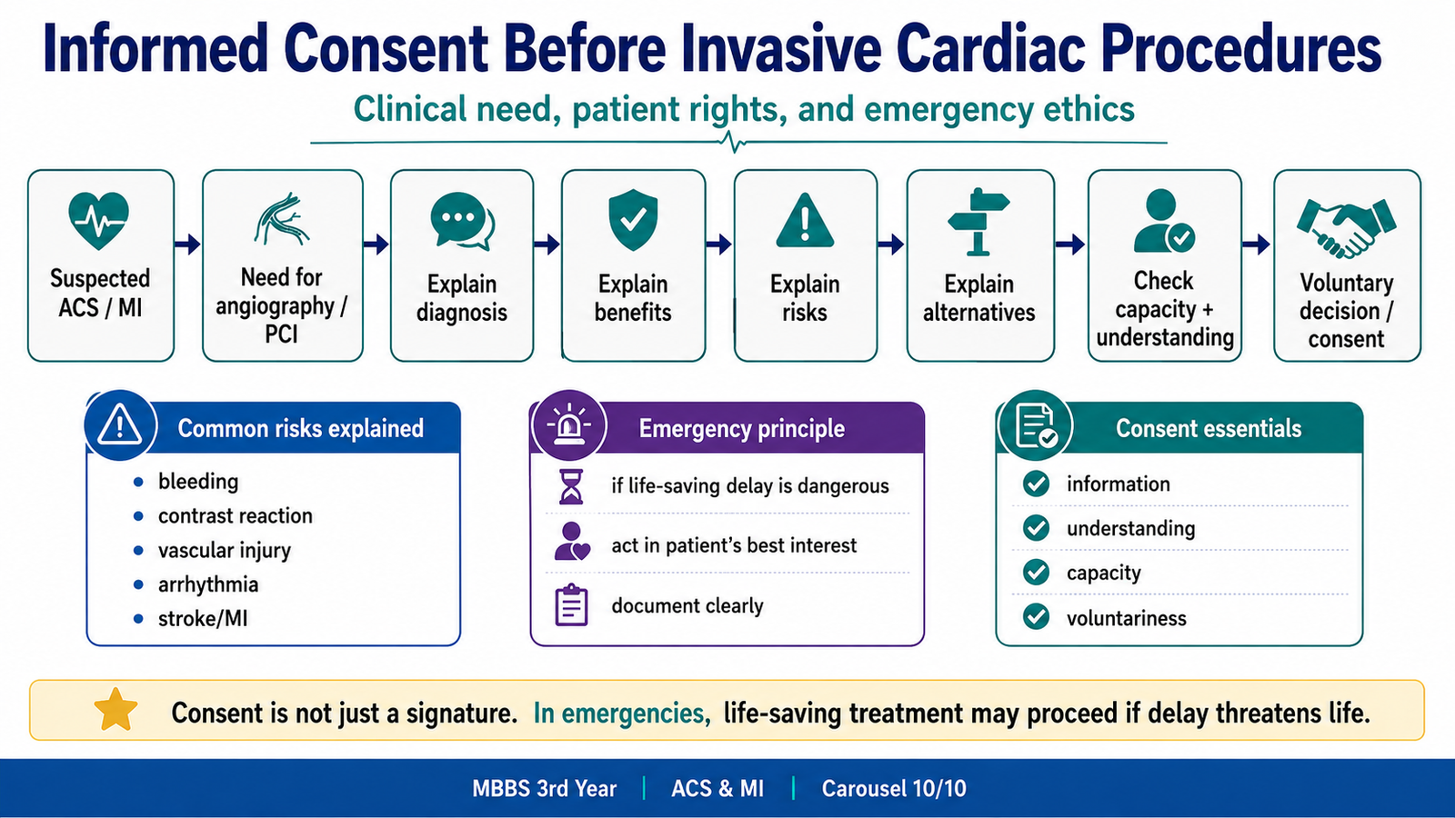
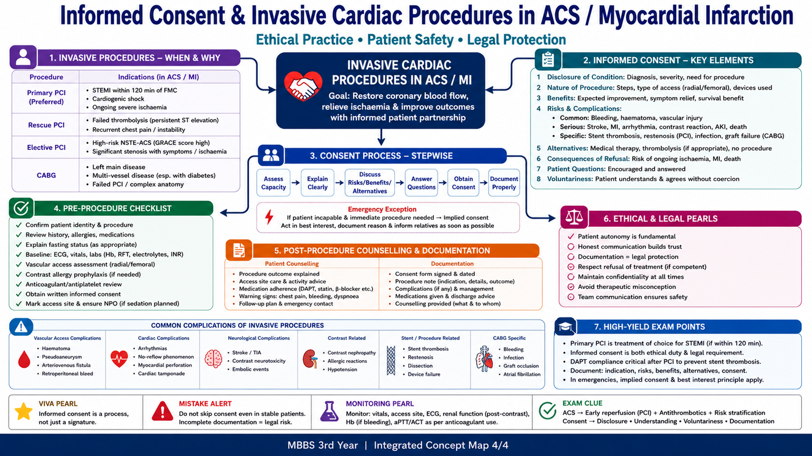
M. Informed Consent Before Invasive Cardiac Procedures
ACS management may require invasive cardiac procedures such as coronary angiography and PCI.
At 3rd-year level, the key ethical and clinical logic is:
- Explain the suspected diagnosis.
- Explain why the procedure is needed.
- Explain benefits, risks, and alternatives.
- Explain possible complications such as bleeding, contrast reaction, vascular injury, arrhythmia, MI, stroke, or need for emergency intervention.
- Confirm patient understanding where possible.
- In emergency life-saving situations, urgent treatment may proceed according to emergency consent principles if the patient lacks capacity and delay threatens life.
⭐ Medicolegal must-know: informed consent is not just a signature; it requires explanation, understanding, voluntariness, and capacity.
Integrated Mechanism Flow
→ platelet adhesion, activation and aggregation
→ coagulation cascade activation and fibrin formation
→ partial or complete coronary occlusion
→ myocardial ischemia and ATP depletion
→ irreversible myocyte injury and coagulative necrosis
→ clinical ACS, complications and need for reperfusion plus antithrombotic therapy
AIM High-Yield Review
- ACS includes unstable angina, NSTEMI and STEMI; troponin is normal in unstable angina but raised in myocardial infarction.
- The usual initiating event is rupture or erosion of an unstable atherosclerotic plaque with superadded thrombosis.
- Prolonged ischemia causes ATP depletion, calcium overload, membrane damage and irreversible myocyte necrosis.
- The subendocardium is affected first because it is least well perfused and has high oxygen demand.
- The characteristic pattern of myocardial necrosis is coagulative necrosis.
- Cardiac troponin is the most important biomarker of myocardial necrosis; CK-MB can help identify reinfarction.
- Hypotension, pulmonary edema, altered consciousness, serious arrhythmias and cardiogenic shock indicate high-risk MI.
- Aspirin irreversibly inhibits COX-1 and reduces thromboxane A₂; P2Y12 blockers prevent ADP-mediated platelet activation.
- GP IIb/IIIa inhibitors block the final common pathway of platelet aggregation.
- Unfractionated heparin acts rapidly through antithrombin and is monitored with aPTT; protamine sulfate treats significant overdose.
- LMWH has more predictable action, less routine monitoring and relatively greater anti-Xa activity than unfractionated heparin.
- Warfarin inhibits vitamin K-dependent clotting-factor synthesis, has a delayed onset, is monitored with PT/INR and has important diet and drug interactions.
- Direct Xa and direct thrombin inhibitors act directly on their target clotting factors and usually do not require routine coagulation monitoring.
- Thrombolytic drugs activate plasminogen to plasmin and dissolve fibrin; major bleeding, especially intracranial hemorrhage, is the most serious adverse effect.
- The myocardium is mechanically weakest around 3–7 days after MI, when macrophages remove dead tissue and rupture may occur.
🎥 Video Learning: Acute Myocardial Infarction and Its Management
Watch this video after completing the learning material to reinforce the clinical recognition and stepwise management of acute myocardial infarction.
AIM Study Focus: Relate coronary thrombosis and myocardial ischemia to the roles of antiplatelet therapy, anticoagulation and urgent reperfusion.