
Study tip: This chapter follows the KMU learning outcomes in a logical sequence. First understand how cardiac electrical activity becomes abnormal, then connect the ECG pattern with clinical management and drug action. Use the final AIM High-Yield Review only after reading the explanations.
KMU Curriculum
AIM Learning Cycle
📖 AIM Learning Material · CVS
Topic 13 — Arrhythmias, Myocarditis, Pericardial Disease and Antiarrhythmic Therapy
A connected introduction to abnormal cardiac rhythms, atrial fibrillation, ventricular arrhythmias, heart blocks, antiarrhythmic drugs, myocarditis, pericarditis and pericardial effusion.
Topic Introduction
Normal cardiac rhythm depends on orderly impulse formation in the sinoatrial node and controlled conduction through the atria, atrioventricular node and ventricles. An arrhythmia develops when impulses arise abnormally, circulate through a re-entry pathway or fail to conduct normally. This chapter first explains how arrhythmias and heart blocks are classified, then applies this framework to atrial fibrillation and ventricular arrhythmias. It also connects antiarrhythmic drug classes with phases of the cardiac action potential. Finally, it explains how inflammation of the myocardium or pericardium can disturb rhythm, contraction or ventricular filling, and how pericardial fluid may progress to cardiac tamponade.
A. Arrhythmias and Heart Blocks: Classification, Mechanisms and Initial Assessment
An arrhythmia is an abnormality in the rate, rhythm, site of origin or conduction of the cardiac impulse. A patient may present with palpitations, dizziness, syncope, chest discomfort, breathlessness, hypotension or sudden cardiac arrest.
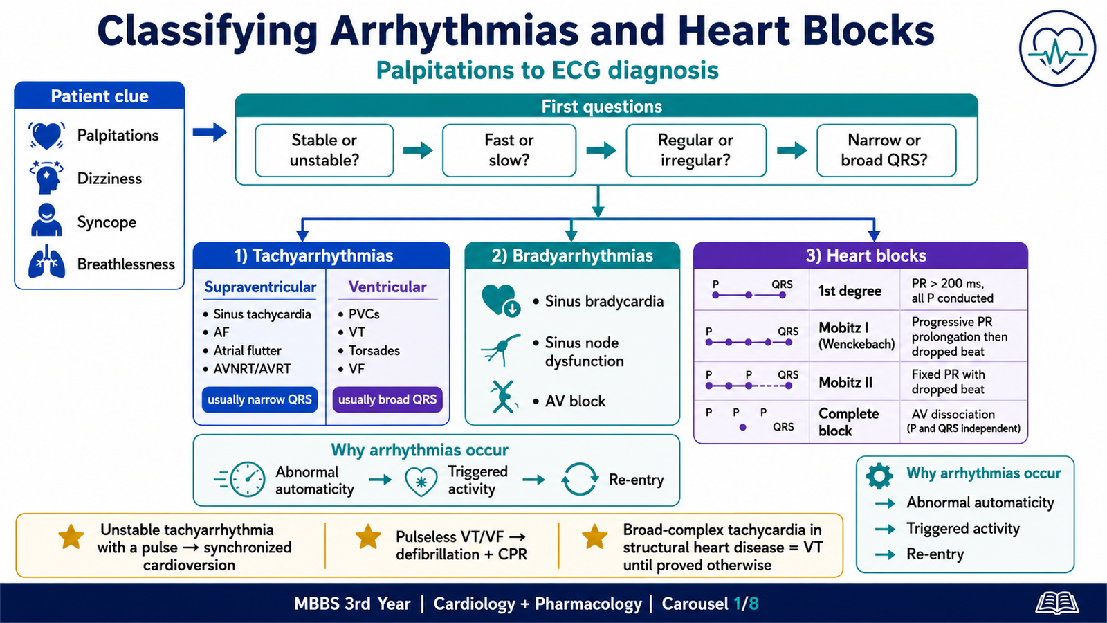
Before naming an arrhythmia, the clinician should answer four basic questions:
- Is the patient hemodynamically stable?
- Is the rhythm fast, slow or apparently normal in rate?
- Is the rhythm regular or irregular?
- Is the QRS complex narrow or broad?
A. Clinical classification of arrhythmias
1. Tachyarrhythmias
A tachyarrhythmia generally has a ventricular rate above 100/min.
They may be classified according to their site of origin.
Supraventricular tachyarrhythmias
These arise above the ventricles, usually in the atria or atrioventricular junction.
Important examples include:
- Sinus tachycardia
- Premature atrial complexes
- Atrial fibrillation
- Atrial flutter
- Atrioventricular nodal re-entry tachycardia
- Atrioventricular re-entry tachycardia
- Focal atrial tachycardia
- Multifocal atrial tachycardia
Most supraventricular rhythms produce a narrow QRS complex, because ventricular activation still passes through the normal His–Purkinje system.
However, a supraventricular rhythm may occasionally appear broad because of:
- Pre-existing bundle branch block
- Rate-related aberrant conduction
- Pre-excitation through an accessory pathway
Ventricular tachyarrhythmias
These originate within ventricular myocardium or the ventricular conduction system.
Important examples include:
- Premature ventricular complexes
- Nonsustained ventricular tachycardia
- Sustained monomorphic ventricular tachycardia
- Polymorphic ventricular tachycardia
- Torsades de pointes
- Ventricular fibrillation
Ventricular tachyarrhythmias usually produce broad QRS complexes, because ventricular activation spreads abnormally through myocardium rather than rapidly through the normal conduction system.
2. Bradyarrhythmias
A bradyarrhythmia generally has a rate below 60/min.
Important causes include:
- Sinus bradycardia
- Sinus node dysfunction
- Sinoatrial exit block
- Atrioventricular block
- Drug-induced conduction slowing
- Inferior myocardial ischemia
- Electrolyte abnormalities
A low heart rate is not automatically pathological. Athletes, sleeping individuals and patients with high vagal tone may have physiological sinus bradycardia.
B. Classification of heart blocks
Heart block refers to delayed or failed conduction of an impulse, most importantly through the atrioventricular conduction system.
First-degree atrioventricular block
Every atrial impulse is conducted to the ventricles, but conduction is delayed.
ECG finding:
- PR interval greater than 200 milliseconds
- Every P wave is followed by a QRS complex
It is usually asymptomatic and may be associated with increased vagal tone, ischemia or drugs such as beta-blockers, verapamil, diltiazem and digoxin.
Second-degree atrioventricular block: Mobitz type I
There is progressive prolongation of the PR interval until one P wave is not followed by a QRS complex.
This is also called the Wenckebach phenomenon.
It usually reflects block within the AV node and may be transient or vagally mediated.
Second-degree atrioventricular block: Mobitz type II
The PR intervals remain constant, but occasional P waves are suddenly not conducted.
It commonly indicates disease below the AV node in the His–Purkinje system.
⭐ Mobitz type II may progress suddenly to complete heart block and usually requires pacing assessment.
2:1 atrioventricular block
Every second atrial impulse is blocked.
Because there is only one conducted PR interval between blocked beats, distinguishing Mobitz I from Mobitz II may be difficult.
Third-degree or complete atrioventricular block
No atrial impulses are conducted to the ventricles.
The atria and ventricles beat independently, producing atrioventricular dissociation.
The ventricular rhythm is maintained by an escape pacemaker:
- Junctional escape rhythms are usually narrow and relatively faster.
- Ventricular escape rhythms are broad and slower.
⭐ Syncope, hypotension or heart failure in complete heart block is an emergency and may require urgent temporary pacing.
C. Mechanisms producing arrhythmias
Most arrhythmias arise through one or more of three mechanisms.
1. Enhanced or abnormal automaticity
Normally, the sinoatrial node has the fastest spontaneous depolarization and controls the heart rate.
Diseased atrial, junctional or ventricular cells may develop increased automaticity because of:
- Sympathetic stimulation
- Hypoxia
- Ischemia
- Electrolyte disturbance
- Digoxin toxicity
- Myocardial inflammation
2. Triggered activity
Triggered activity occurs when abnormal depolarizations arise during or shortly after an action potential.
Early afterdepolarizations
These occur during phases 2 or 3 and are favored by prolonged repolarization and a prolonged QT interval.
They may produce torsades de pointes.
Delayed afterdepolarizations
These occur after repolarization and are often related to intracellular calcium overload.
They may occur with:
- Digoxin toxicity
- Catecholamine excess
- Heart failure
3. Re-entry
Re-entry occurs when an impulse repeatedly circulates through a conduction pathway.
It requires:
- Two pathways with different conduction or refractory properties
- Unidirectional block in one pathway
- Sufficiently slow conduction to allow previously refractory tissue to recover
Re-entry contributes to:
- AV nodal re-entry tachycardia
- Accessory pathway tachycardia
- Atrial flutter
- Many forms of ventricular tachycardia
Initial management principles
The ECG is essential, but the patient’s stability comes first.
⭐ An arrhythmia associated with hypotension, shock, ongoing ischemic chest pain, acute pulmonary edema or altered consciousness should be treated as hemodynamically unstable.
The exact emergency intervention depends on the rhythm:
- Unstable tachyarrhythmia with a pulse generally requires synchronized cardioversion.
- Pulseless ventricular tachycardia or ventricular fibrillation requires immediate defibrillation and cardiopulmonary resuscitation.
- Symptomatic severe bradycardia may require atropine and pacing.
A broad-complex tachycardia should not automatically be labelled supraventricular tachycardia with bundle branch block.
⭐ In an adult with structural heart disease, a regular broad-complex tachycardia should be considered ventricular tachycardia until proved otherwise.
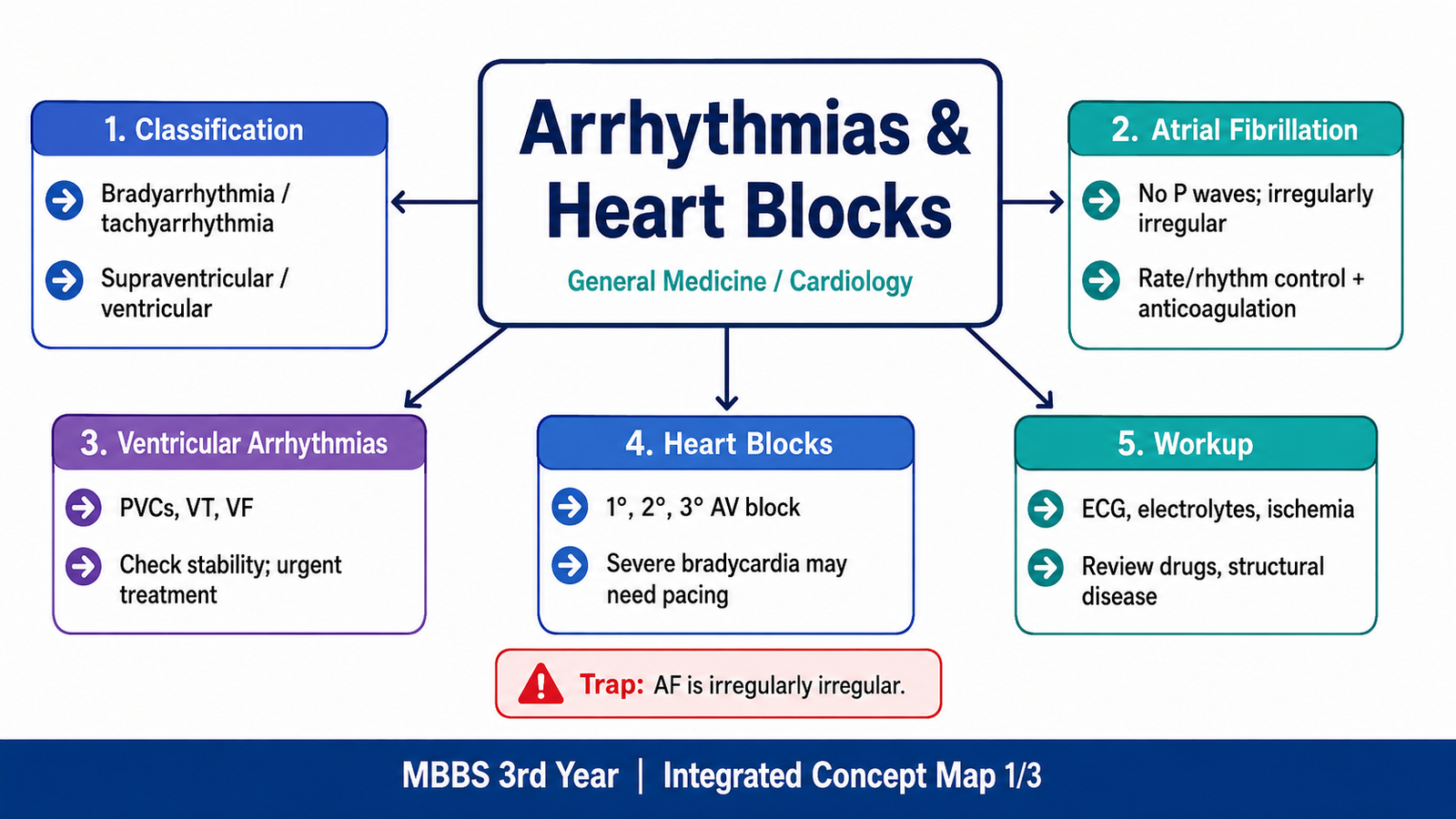
B. Atrial Fibrillation: Causes, ECG Findings, Workup and Management
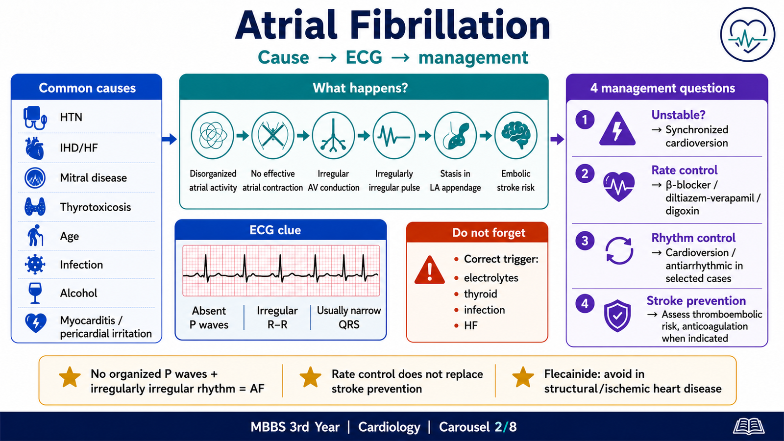
Atrial fibrillation is a supraventricular tachyarrhythmia characterized by uncoordinated atrial electrical activity and ineffective atrial contraction.
Multiple rapidly changing atrial wavelets and focal electrical activity, often originating near the pulmonary veins, prevent organized atrial depolarization.
As a result:
- Effective atrial contraction is lost.
- The AV node receives many irregular impulses.
- Only some impulses pass to the ventricles.
- The ventricular rhythm becomes irregularly irregular.
- Blood may stagnate in the left atrium, especially the left atrial appendage.
- Thrombus formation may lead to systemic embolism and ischemic stroke.
A. Etiology and precipitating factors
Atrial fibrillation is commonly associated with structural or systemic disease.
Important causes include:
- Hypertension
- Ischemic heart disease
- Heart failure
- Mitral valve disease, especially mitral stenosis
- Cardiomyopathy
- Thyrotoxicosis
- Increasing age
- Obesity
- Obstructive sleep apnea
- Diabetes mellitus
- Alcohol excess
- Acute infection or sepsis
- Pulmonary embolism
- Myocarditis
- Pericardial inflammation
- Postoperative states
- Electrolyte disturbances
Acute atrial fibrillation may also be precipitated by stimulants, hypoxia or uncontrolled systemic illness.
B. Clinical presentation
Patients may report:
- Palpitations
- Breathlessness
- Fatigue
- Reduced exercise tolerance
- Chest discomfort
- Dizziness
- Presyncope
Some patients remain asymptomatic and are diagnosed during routine pulse examination or ECG.
The pulse is classically:
- Irregularly irregular
- Variable in volume
- Sometimes associated with a pulse deficit
C. ECG findings
The characteristic ECG findings are:
- Absence of discrete, consistent P waves
- Irregularly irregular R–R intervals
- Fibrillatory baseline activity
- Usually narrow QRS complexes unless aberrant conduction or bundle branch block is present
⭐ Atrial fibrillation means no organized P waves plus an irregularly irregular ventricular rhythm.
D. Basic diagnostic workup
The workup is directed toward confirming the rhythm, identifying its cause and assessing complications.
Important investigations include:
- Twelve-lead ECG
- Full blood count
- Serum electrolytes, especially potassium and magnesium
- Renal and liver function
- Thyroid function tests
- Blood glucose
- Cardiac biomarkers when ischemia or myocarditis is suspected
- Echocardiography to assess chamber size, ventricular function and valvular disease
- Ambulatory ECG monitoring for intermittent symptoms
- Chest imaging when pulmonary disease or heart failure is suspected
E. The four management questions
Atrial fibrillation management becomes easier when separated into four decisions.
1. Is the patient unstable?
Features of instability include:
- Hypotension or shock
- Ongoing myocardial ischemia
- Severe pulmonary edema
- Markedly impaired consciousness
- Severe symptoms directly attributable to the rapid rhythm
⭐ Hemodynamically unstable atrial fibrillation requires urgent synchronized electrical cardioversion.
Anticoagulation considerations remain important, but emergency stabilization should not be delayed when the arrhythmia is causing life-threatening instability.
2. Does the ventricular rate require control?
A rapid ventricular rate shortens diastole, increases myocardial oxygen demand and may worsen heart failure.
Rate-control drugs reduce conduction through the AV node.
Common options include:
- Beta-blockers
- Diltiazem or verapamil
- Digoxin in selected patients
The choice depends on ventricular function, blood pressure, associated disease and urgency.
Beta-blockers
Useful when sympathetic drive is contributing to the rapid rate.
Avoid or use cautiously in severe acute decompensated heart failure, marked bradycardia, advanced AV block and severe bronchospastic disease.
Verapamil or diltiazem
These slow AV nodal conduction.
⭐ Non-dihydropyridine calcium-channel blockers should generally be avoided in significant systolic heart failure because of their negative inotropic effect.
Digoxin
Digoxin increases vagal influence on the AV node and may help control resting heart rate, particularly in selected patients with heart failure.
It is less effective during exercise or high sympathetic activity.
3. Should sinus rhythm be restored?
Rhythm control may be considered when:
- Symptoms persist despite adequate rate control
- Atrial fibrillation is recent in onset
- The arrhythmia is contributing to heart failure
- A reversible precipitant has been corrected
- Maintaining sinus rhythm is considered clinically advantageous
Rhythm control may involve:
- Electrical cardioversion
- Pharmacological cardioversion
- Long-term antiarrhythmic therapy
- Catheter ablation in selected patients
Drugs used for cardioversion or maintenance may include flecainide, propafenone, amiodarone, sotalol or other agents depending on structural heart disease and specialist assessment.
⭐ Flecainide should not be used in significant structural or ischemic heart disease because of proarrhythmic risk.
4. Does the patient require stroke prevention?
Loss of effective atrial contraction allows blood stasis, especially in the left atrial appendage.
Stroke-prevention assessment is therefore separate from the decision to control rate or rhythm.
Risk-based anticoagulation is considered using validated clinical factors such as:
- Previous stroke or transient ischemic attack
- Increasing age
- Hypertension
- Diabetes mellitus
- Heart failure
- Vascular disease
- Sex-related risk context
Bleeding risk should also be assessed and modifiable risk factors corrected. A high bleeding-risk score should prompt caution and risk-factor management rather than automatic denial of anticoagulation. Current guideline-based AF care emphasizes assessment of thromboembolic risk, modification of bleeding risks, management of comorbidities and selection between rate and rhythm-control strategies according to clinical context.
F. Cardioversion and the thromboembolic problem
If atrial fibrillation has been present for a prolonged or uncertain duration, an atrial thrombus may already have formed.
Restoring organized atrial contraction may dislodge that thrombus.
Therefore, elective cardioversion may require:
- Appropriate anticoagulation before and after cardioversion, or
- Imaging-based exclusion of left atrial thrombus in selected pathways
This is different from emergency cardioversion in an unstable patient, where immediate stabilization takes priority.
- A normal ventricular rate does not exclude atrial fibrillation.
- Rate control does not remove the need to assess stroke risk.
- Restoration of sinus rhythm does not automatically remove anticoagulation requirements.
- Digoxin is not the best drug for rapid rate control during marked sympathetic activity.
- AV nodal blockers may be dangerous in atrial fibrillation with certain pre-excitation patterns because conduction through an accessory pathway may accelerate.
C. Ventricular Arrhythmias: Types, Workup and Management
Ventricular arrhythmias range from isolated premature ventricular complexes to ventricular fibrillation and sudden cardiac arrest.
Their importance depends on:
- Duration
- Symptoms
- Ventricular function
- Structural heart disease
- Reversible triggers
- Risk of degeneration into ventricular fibrillation
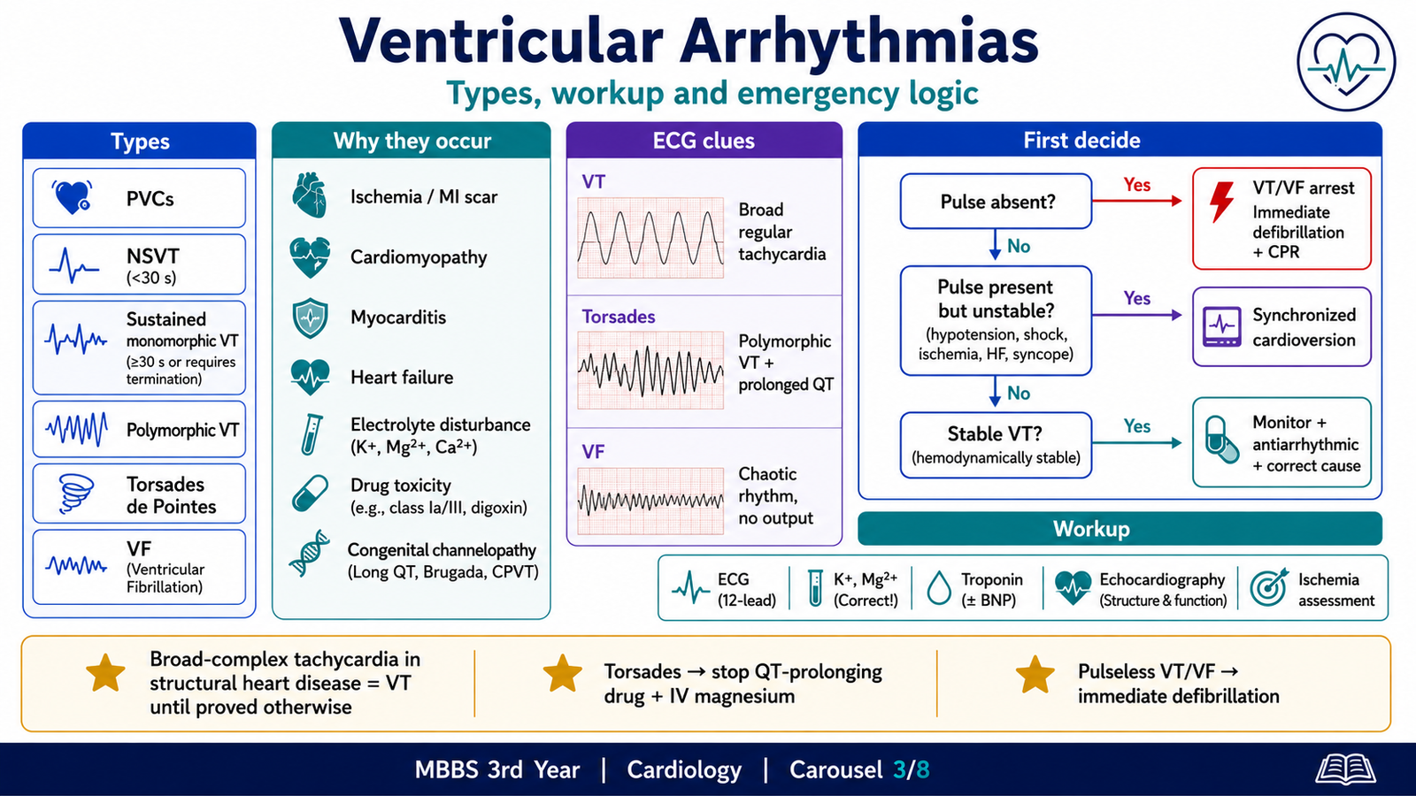
A. Types of ventricular arrhythmias
Premature ventricular complexes
A premature ventricular complex is an early beat arising from the ventricle.
The ECG usually shows:
- Premature broad QRS complex
- No preceding normal P wave
- Discordant T wave
- Often a compensatory pause
Occasional premature ventricular complexes may occur in healthy people, but frequent or complex ectopy may be associated with structural heart disease, stimulants, hypoxia or electrolyte disturbance.
Nonsustained ventricular tachycardia
This is a run of ventricular tachycardia that terminates spontaneously within a short period and does not produce prolonged instability.
Its significance depends heavily on whether structural heart disease or impaired ventricular function is present.
Sustained monomorphic ventricular tachycardia
This is a broad-complex tachycardia with similar QRS morphology from beat to beat.
A common mechanism is re-entry around myocardial scar, particularly after myocardial infarction.
Clinical effects include:
- Palpitations
- Dizziness
- Syncope
- Hypotension
- Chest pain
- Acute heart failure
- Cardiac arrest
Polymorphic ventricular tachycardia
The QRS morphology and axis vary from beat to beat.
It may occur with:
- Acute myocardial ischemia
- Severe structural disease
- Prolonged QT interval
- Electrolyte abnormalities
Torsades de pointes
Torsades de pointes is a form of polymorphic ventricular tachycardia associated with a prolonged QT interval.
The QRS complexes appear to twist around the baseline.
Important causes include:
- Congenital long-QT syndromes
- Class IA antiarrhythmics
- Class III antiarrhythmics
- Certain antimicrobials and psychotropic drugs
- Hypokalemia
- Hypomagnesemia
- Marked bradycardia
⭐ Intravenous magnesium is a key immediate treatment for torsades de pointes, even when the measured serum magnesium is not markedly low.
The offending QT-prolonging drug should be stopped and electrolyte abnormalities corrected.
Ventricular fibrillation
Ventricular fibrillation consists of chaotic ventricular electrical activity with no effective ventricular contraction or cardiac output.
The patient is:
- Unconscious
- Pulseless
- Not breathing normally
⭐ Ventricular fibrillation and pulseless ventricular tachycardia require immediate unsynchronized defibrillation and cardiopulmonary resuscitation.
B. Etiology
Important causes include:
- Acute myocardial ischemia or infarction
- Previous infarction with ventricular scar
- Cardiomyopathies
- Myocarditis
- Heart failure
- Electrolyte abnormalities
- Hypoxia
- Acidosis
- Drug toxicity
- Congenital channelopathies
- Severe valvular disease
- Catecholamine excess
- Mechanical cardiac irritation
- Idiopathic ventricular arrhythmia
C. Workup
The workup should determine whether the arrhythmia is immediately dangerous and whether an underlying reversible or structural cause exists.
Immediate assessment
- Airway, breathing and circulation
- Pulse and blood pressure
- Consciousness
- Signs of shock
- Chest pain
- Pulmonary edema
- Twelve-lead ECG if this does not delay emergency treatment
Laboratory assessment
- Potassium
- Magnesium
- Calcium
- Renal function
- Blood glucose
- Cardiac biomarkers
- Acid–base status when indicated
- Toxicology or drug-level assessment when relevant
Structural and ischemic evaluation
- Echocardiography
- Assessment for acute coronary syndrome
- Ambulatory rhythm monitoring
- Cardiac imaging in selected patients
- Exercise testing in selected stable patients
- Electrophysiological assessment when required
D. Management based on pulse and stability
Pulseless VT or ventricular fibrillation
Management requires:
- Immediate cardiopulmonary resuscitation
- Defibrillation
- Correction of reversible causes
- Adrenaline according to resuscitation protocol
- Amiodarone in refractory shockable rhythms according to protocol
VT with a pulse but hemodynamic instability
Features include:
- Hypotension
- Altered consciousness
- Ischemic chest pain
- Acute heart failure
- Shock
⭐ Unstable ventricular tachycardia with a pulse requires synchronized electrical cardioversion.
Stable sustained monomorphic VT
Management may involve:
- Intravenous antiarrhythmic therapy
- Continuous monitoring
- Correction of electrolyte abnormalities
- Assessment for ischemia and structural disease
- Specialist-directed cardioversion or ablation planning
Amiodarone, procainamide or lidocaine may be considered depending on clinical context and local protocols.
Long-term prevention
Long-term management may include:
- Treatment of ischemia or heart failure
- Removal of proarrhythmic drugs
- Catheter ablation
- Implantable cardioverter-defibrillator
- Disease-specific treatment
An implantable cardioverter-defibrillator does not prevent every arrhythmia from beginning. It detects and terminates dangerous ventricular rhythms and is used in selected patients at high risk of sudden cardiac death. Current ESC guidance emphasizes identification of structural disease, inherited syndromes and reversible causes, together with appropriate use of ablation and implantable defibrillator therapy in selected patients.
A patient with ventricular tachycardia may remain conscious and have a palpable pulse. Ventricular tachycardia does not always mean cardiac arrest.
The immediate distinction is:
- VT with a pulse
- Pulseless VT
D. Antiarrhythmic Drugs and the Cardiac Action Potential
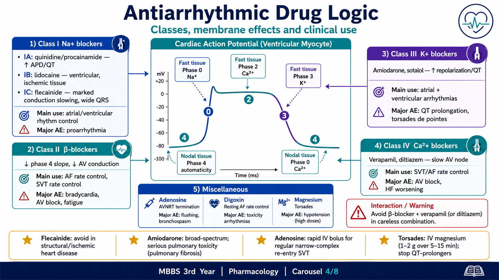
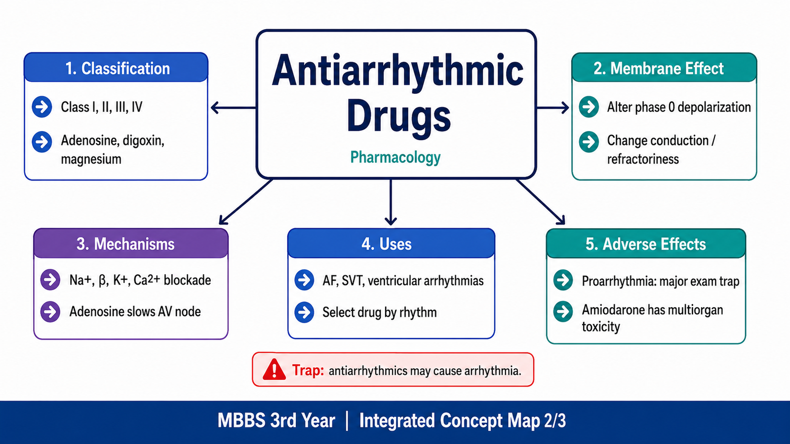
Antiarrhythmic drugs work by altering:
- Ion movement through cardiac cell membranes
- Action-potential duration
- Conduction velocity
- Automaticity
- Refractory period
- AV nodal transmission
However, the same electrical changes that suppress one arrhythmia may create another.
⭐ The major shared danger of antiarrhythmic therapy is proarrhythmia.
A. Two cardiac action potentials must be distinguished
Fast-response action potential
This occurs mainly in:
- Atrial myocytes
- Ventricular myocytes
- His–Purkinje fibres
Its major phases are:
Phase 0: Rapid sodium entry
Phase 1: Brief initial repolarization
Phase 2: Calcium entry balanced by potassium exit
Phase 3: Potassium-mediated repolarization
Phase 4: Stable resting membrane potential
Slow-response action potential
This occurs mainly in:
- Sinoatrial node
- Atrioventricular node
Its main features are:
Phase 4: Spontaneous pacemaker depolarization
Phase 0: Calcium-mediated depolarization
Phase 3: Potassium-mediated repolarization
The AV node depends more on calcium channels than fast sodium channels. Therefore:
- Beta-blockers reduce sympathetic pacemaker and AV nodal activity.
- Verapamil and diltiazem directly suppress calcium-dependent AV nodal conduction.
- Fast sodium-channel blockers have stronger effects on atrial, ventricular and His–Purkinje tissue.
B. Vaughan Williams classification
The traditional classification includes:
- Class I: Sodium-channel blockers
- Class II: Beta-adrenoceptor blockers
- Class III: Potassium-channel blockers
- Class IV: Non-dihydropyridine calcium-channel blockers
- Miscellaneous agents: adenosine, digoxin, magnesium and others
This classification is useful for undergraduate learning, although some drugs have actions belonging to more than one class.
Class I — Sodium-channel blockers
Class I drugs block fast sodium channels in atrial, ventricular and His–Purkinje tissue.
Their overall effect is to reduce the rapid phase 0 upstroke and slow impulse conduction.
They are divided into IA, IB and IC according to sodium-channel blockade and effects on repolarization.
Class IA
Prototypes: Quinidine, procainamide, disopyramide
Membrane-potential effect
- Moderate sodium-channel blockade
- Slower phase 0 depolarization
- Prolonged repolarization because of additional potassium-channel blockade
- Increased action-potential duration
- Increased effective refractory period
- Prolonged QT interval
Mechanism-to-clinical logic
Slowing conduction and prolonging refractoriness can interrupt re-entry circuits. However, prolonging repolarization increases the risk of early afterdepolarizations and torsades de pointes.
Clinical uses
Use is now limited by adverse effects and safer alternatives. Procainamide may be used in selected stable tachyarrhythmias under monitored conditions.
Adverse effects
Class effects include:
- QT prolongation
- Torsades de pointes
- Hypotension
- Conduction block
Drug-specific adverse effects:
Quinidine
- Cinchonism: tinnitus, headache and dizziness
- Gastrointestinal upset
- Thrombocytopenia
- QT prolongation
Procainamide
- Drug-induced lupus-like syndrome
- Agranulocytosis
- Hypotension
- Torsades de pointes
⭐ Procainamide may cause a lupus-like syndrome, particularly with prolonged exposure.
Disopyramide
- Antimuscarinic effects
- Urinary retention
- Dry mouth
- Blurred vision
- Negative inotropic effect
- Worsening heart failure
Contraindications and cautions
- Prolonged QT interval
- History of torsades de pointes
- Severe conduction disease without pacing
- Significant heart failure, especially with disopyramide
- Electrolyte abnormalities that increase torsades risk
Class IB
Prototypes: Lidocaine, mexiletine
Membrane-potential effect
- Weak sodium-channel blockade in normal tissue
- Greater effect in depolarized or ischemic ventricular tissue
- Reduced automaticity
- Shortened action-potential duration in ventricular tissue
- Little effect on atrial tissue
Mechanism-to-clinical logic
Ischemic ventricular cells remain relatively depolarized, allowing lidocaine to bind preferentially to inactivated sodium channels.
Therefore, lidocaine is more useful for ventricular than atrial arrhythmias.
Clinical uses
- Acute ventricular arrhythmias, especially in ischemic settings
- Selected ventricular arrhythmias when other options are unsuitable
Pharmacokinetic point
Lidocaine undergoes extensive hepatic metabolism and is given intravenously for acute arrhythmia treatment.
Reduced hepatic blood flow, liver disease or heart failure may increase toxicity.
Adverse effects
- Drowsiness
- Paresthesia
- Tremor
- Confusion
- Seizures
- Hypotension at excessive doses
⭐ Lidocaine toxicity is mainly neurological, including confusion, tremor and seizures.
Contraindications and cautions
- Severe sinoatrial or AV nodal dysfunction without pacing
- Significant hepatic dysfunction requiring dose adjustment
- Previous serious hypersensitivity to amide local anesthetics
Class IC
Prototypes: Flecainide, propafenone
Membrane-potential effect
- Strong sodium-channel blockade
- Marked reduction in phase 0 upstroke
- Marked slowing of conduction
- Little direct effect on action-potential duration
- Widening of QRS complex
Their sodium-channel blocking effect is often use dependent: conduction slowing becomes more pronounced at faster heart rates.
Clinical uses
- Rhythm control in selected patients with atrial fibrillation
- Certain supraventricular arrhythmias
- Selected patients without major structural heart disease
Adverse effects
- Ventricular proarrhythmia
- Conduction block
- QRS widening
- Worsening heart failure
- Dizziness and visual symptoms
⭐ Flecainide is contraindicated in significant ischemic or structural heart disease because it can increase life-threatening ventricular arrhythmias.
Propafenone also has some beta-blocking activity and may worsen bradycardia or bronchospasm in susceptible patients.
Prescription point
Class IC therapy should be selected carefully, with ECG and structural-heart assessment rather than prescribed merely because the rhythm is supraventricular.
Class II — Beta-adrenoceptor blockers
Prototypes: Metoprolol, propranolol, esmolol
Membrane-potential effect
Beta-blockers mainly affect nodal tissue by reducing sympathetic stimulation.
They:
- Reduce the slope of phase 4 spontaneous depolarization
- Reduce sinoatrial node automaticity
- Slow AV nodal conduction
- Increase AV nodal refractoriness
- Prolong the PR interval
- Reduce catecholamine-induced triggered activity
Mechanism-to-clinical logic
Blocking beta-1 receptors reduces cyclic AMP and calcium-dependent activity.
This slows the sinus rate and makes it harder for rapid atrial impulses to cross the AV node.
Clinical uses
- Ventricular rate control in atrial fibrillation or flutter
- AV nodal re-entry tachycardia
- Catecholamine-related arrhythmias
- Post-myocardial-infarction arrhythmia prevention
- Some ventricular arrhythmias
- Congenital long-QT syndromes
- Symptomatic adrenergic palpitations
⭐ Beta-blockers are important for rate control in atrial fibrillation when there is no major contraindication.
Adverse effects
- Bradycardia
- AV block
- Hypotension
- Fatigue
- Reduced exercise tolerance
- Bronchospasm with non-selective agents
- Masking of hypoglycemia symptoms
- Worsening of acute decompensated heart failure if introduced inappropriately
Contraindications and cautions
- Severe bradycardia
- Advanced AV block without a pacemaker
- Cardiogenic shock
- Severe acute decompensated heart failure
- Severe bronchospastic disease, particularly with non-selective drugs
Drug interactions
Combining a beta-blocker with verapamil or diltiazem can produce marked bradycardia, AV block and myocardial depression.
⭐ Avoid careless combination of beta-blockers with verapamil because severe bradycardia, heart block or cardiac depression may occur.
Toxicity and antidotal logic
Severe beta-blocker overdose may cause bradycardia, hypotension, heart block, hypoglycemia and shock.
⭐ Glucagon is an important antidotal treatment in serious beta-blocker toxicity because it raises cyclic AMP through a receptor-independent pathway.
Supportive care and specialist toxicology management are also required.
Class III — Potassium-channel blockers
Class III drugs prolong repolarization by reducing outward potassium currents.
Prototypes: Amiodarone, sotalol, dofetilide, ibutilide
Membrane-potential effect
- Prolonged phase 3 repolarization
- Increased action-potential duration
- Increased effective refractory period
- Prolonged QT interval
This may interrupt re-entry by keeping tissue refractory for longer.
However, excessive QT prolongation can cause early afterdepolarizations and torsades de pointes.
Amiodarone
Amiodarone has actions belonging to several classes:
- Potassium-channel blockade
- Sodium-channel blockade
- Beta-blocking effect
- Calcium-channel blocking effect
Clinical uses
- Atrial arrhythmias
- Ventricular tachyarrhythmias
- Rhythm control in selected patients with structural heart disease
- Refractory ventricular fibrillation or pulseless VT within resuscitation protocols
⭐ Amiodarone is a broad-spectrum antiarrhythmic useful for both atrial and ventricular arrhythmias.
Pharmacokinetic features
- Very lipid soluble
- Large volume of distribution
- Accumulates in tissues
- Very long and variable half-life
- Effects and toxicity may continue after discontinuation
Adverse effects
- Pulmonary pneumonitis and fibrosis
- Thyroid dysfunction
- Hepatotoxicity
- Corneal deposits
- Optic neuropathy
- Photosensitivity
- Blue-grey skin pigmentation
- Bradycardia
- QT prolongation
- Peripheral neuropathy
- Gastrointestinal disturbance
⭐ Pulmonary toxicity is one of the most serious adverse effects of amiodarone.
Amiodarone contains iodine and may cause either hypothyroidism or hyperthyroidism.
Although it prolongs QT, torsades occurs less frequently with amiodarone than with many other pure potassium-channel blockers.
Drug interactions
Amiodarone can increase the effects or concentrations of drugs such as:
- Warfarin
- Digoxin
- Some statins
- Other bradycardia-producing drugs
- Other QT-prolonging drugs
Prescription and monitoring point
Patients receiving long-term amiodarone require appropriate monitoring, which may include:
- Thyroid function
- Liver function
- Pulmonary assessment
- ECG
- Review of drug interactions
Sotalol
Sotalol combines:
- Non-selective beta-blockade
- Potassium-channel blockade
It slows heart rate and prolongs repolarization.
Clinical uses
- Selected atrial arrhythmias
- Selected ventricular arrhythmias
Adverse effects
- Bradycardia
- Bronchospasm
- QT prolongation
- Torsades de pointes
⭐ Sotalol is both a beta-blocker and a class III antiarrhythmic.
Renal function is important because accumulation may increase QT prolongation and proarrhythmia.
Ibutilide and dofetilide
These agents are used in selected atrial arrhythmias under monitored conditions.
Their major concern is QT prolongation and torsades de pointes.
Class IV — Non-dihydropyridine calcium-channel blockers
Prototypes: Verapamil, diltiazem
These drugs block L-type calcium channels, particularly in nodal tissue.
Membrane-potential effect
- Reduced phase 0 upstroke in the AV node
- Slowed AV nodal conduction
- Increased AV nodal refractory period
- Prolonged PR interval
- Reduced heart rate
- Negative inotropic effect
Clinical uses
- Ventricular rate control in atrial fibrillation or flutter
- Termination or prevention of some AV nodal re-entry tachycardias
- Other selected supraventricular tachyarrhythmias
⭐ Verapamil and diltiazem act mainly on the AV node and are used for supraventricular, not ventricular, rhythm control.
Adverse effects
- Bradycardia
- AV block
- Hypotension
- Worsening heart failure
- Constipation, especially with verapamil
- Peripheral edema
Contraindications and cautions
- Significant systolic heart failure
- Severe hypotension
- Advanced AV block without pacing
- Marked bradycardia
- Certain broad-complex tachycardias
- Atrial fibrillation with an accessory pathway
⭐ Giving verapamil to an undifferentiated broad-complex tachycardia may cause severe hypotension or cardiovascular collapse.
Miscellaneous antiarrhythmic agents
Adenosine
Adenosine activates adenosine receptors in the AV node, increasing potassium efflux and reducing calcium-dependent conduction.
Its effect is:
- Extremely rapid in onset
- Very short in duration
- A brief AV nodal block
Clinical use
⭐ Adenosine is the drug of choice for acute termination of many regular narrow-complex AV node-dependent re-entry tachycardias in stable patients.
It may also temporarily reveal underlying atrial activity in some diagnostic situations.
Adverse effects
- Flushing
- Chest pressure
- Breathlessness
- Sense of impending doom
- Transient bradycardia
- Bronchospasm
Because of its very short half-life, most effects resolve rapidly.
Contraindications and cautions
- Severe asthma or active bronchospasm
- Advanced AV block without pacing
- Sick sinus syndrome without pacing
- Irregular or polymorphic broad-complex tachycardia
Drug interactions
- Dipyridamole enhances adenosine action.
- Methylxanthines such as caffeine and theophylline oppose its effect.
- Carbamazepine may increase conduction-blocking effects.
Prescription point
⭐ Adenosine must be administered as a rapid intravenous bolus followed immediately by a saline flush because its plasma half-life is only a few seconds.
Digoxin
Digoxin indirectly slows AV nodal conduction by increasing vagal tone.
It is useful for ventricular rate control in selected patients, particularly at rest.
Adverse effects and toxicity
- Nausea and vomiting
- Visual disturbances
- Confusion
- Bradyarrhythmias
- AV block
- Atrial tachycardia with block
- Ventricular arrhythmias
- Hyperkalemia in severe acute toxicity
Factors increasing toxicity include:
- Renal impairment
- Hypokalemia
- Drug interactions
- Advanced age
- Excessive dosing
⭐ Digoxin can treat an arrhythmia but can also cause almost any type of arrhythmia in toxicity.
⭐ Digoxin-specific antibody fragments are used in severe, life-threatening digoxin toxicity.
Magnesium
Magnesium stabilizes cardiac electrical activity and helps suppress early afterdepolarizations.
⭐ Intravenous magnesium is especially important in torsades de pointes and some arrhythmias associated with hypomagnesemia.
Final pharmacological principle
Antiarrhythmic drugs should not be selected simply according to whether the heart rate is fast.
Selection depends on:
- Site of origin
- Mechanism
- QRS width
- QT interval
- Ventricular function
- Presence of ischemic or structural heart disease
- Renal and hepatic function
- Interaction with other drugs
- Risk of proarrhythmia
E. Myocarditis: Causes, Clinical Effects and Management
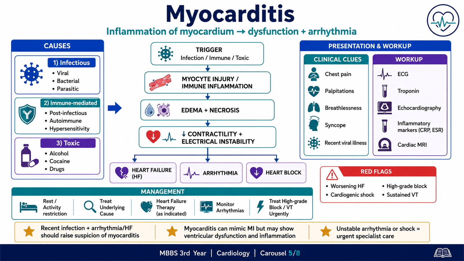
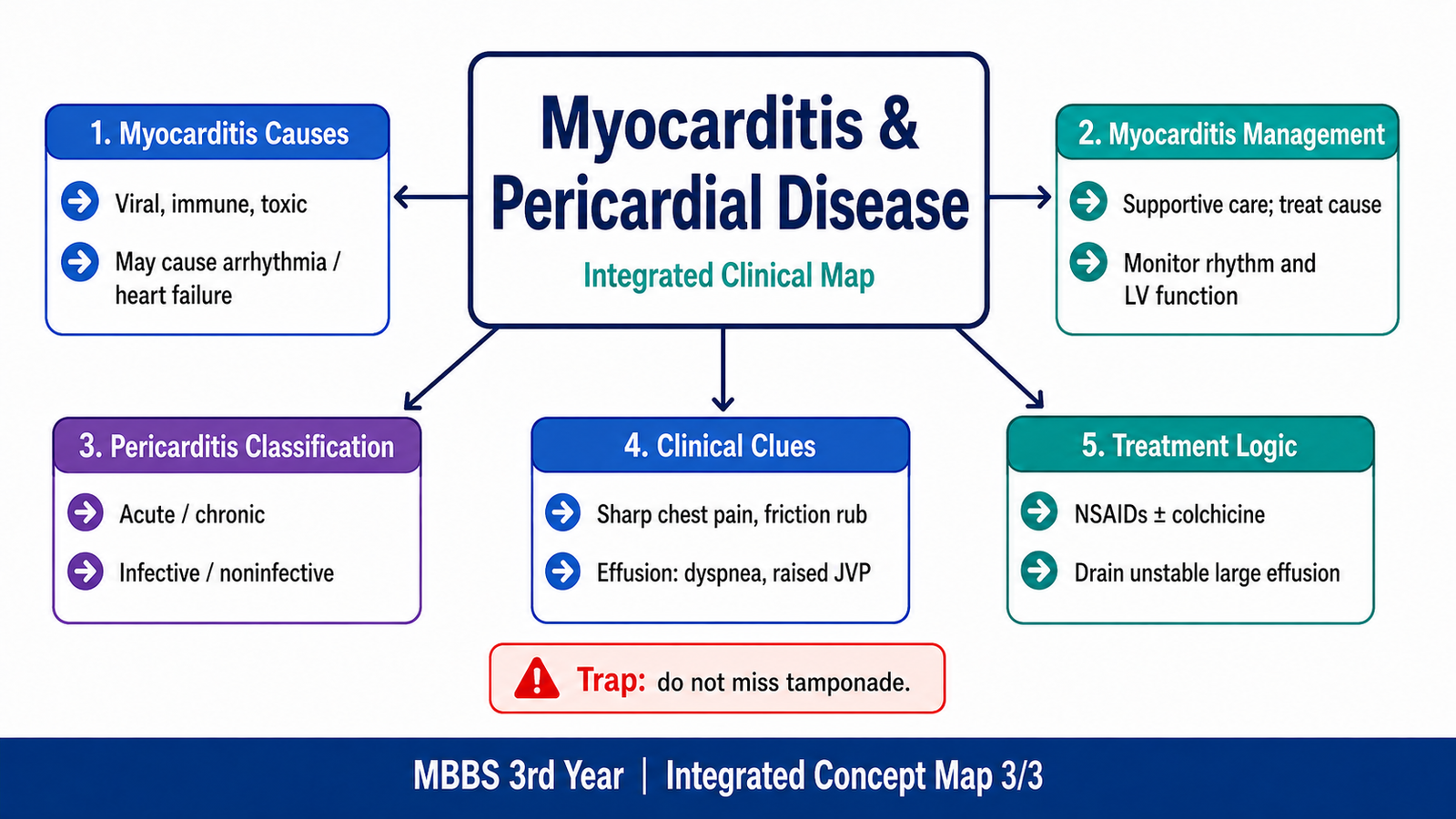
Myocarditis is an inflammatory disease of the myocardium. It can injure cardiac myocytes, impair contractility and create electrically unstable areas that produce arrhythmias.
The presentation varies from mild chest discomfort to severe heart failure, ventricular arrhythmia or cardiogenic shock.
A. Causes of myocarditis
Infectious causes
Viruses are common causes and may include enteroviruses, adenoviruses, influenza-associated viruses and other cardiotropic or systemic viral infections.
Other infectious causes include:
- Bacterial infections
- Spirochetal infections
- Rickettsial disease
- Fungal infections
- Protozoal infections
- Parasitic infections
The likely organism varies according to geography, exposure and immune status.
Immune-mediated causes
- Post-infectious immune injury
- Autoimmune disease
- Hypersensitivity myocarditis
- Sarcoidosis
- Giant-cell myocarditis
- Immune reactions associated with certain drugs or therapies
Toxic causes
- Alcohol
- Cocaine
- Some chemotherapeutic drugs
- Other cardiotoxic agents
B. Pathophysiological sequence
A simplified sequence is:
Trigger or infection
→ myocardial injury or immune activation
→ inflammation and myocyte damage
→ reduced contractility and myocardial edema
→ ventricular dysfunction
→ electrical instability
→ arrhythmia, heart block or heart failure
The inflammatory process may resolve completely, persist or lead to dilated ventricular dysfunction.
C. Clinical findings
Patients may present with:
- Recent flu-like or febrile illness
- Fatigue
- Chest pain
- Palpitations
- Breathlessness
- Reduced exercise tolerance
- Syncope
- Heart failure
- Ventricular arrhythmia
- Conduction block
Chest pain may resemble myocardial infarction or may occur with associated pericardial involvement.
⭐ Myocarditis should be considered when a previously healthy patient develops chest pain, heart failure, arrhythmia or conduction block after a recent infection.
D. Basic diagnostic approach
Investigations may show:
- ECG abnormalities
- Elevated cardiac troponin
- Raised inflammatory markers
- Ventricular dysfunction on echocardiography
- Regional or global wall-motion abnormalities
- Cardiac magnetic resonance evidence of myocardial inflammation
- Arrhythmias on monitoring
Coronary disease may need to be excluded when the presentation resembles acute coronary syndrome.
Endomyocardial biopsy is not required in every patient. It is reserved for selected situations in which identifying a particular inflammatory pattern would alter management.
The modern diagnostic approach increasingly combines clinical presentation, biomarkers, ECG, echocardiography and cardiac magnetic resonance, with biopsy used selectively.
E. Management logic
Management depends on severity and cause.
General measures
- Restrict strenuous physical activity during active disease
- Treat heart failure when present
- Monitor for arrhythmias
- Correct electrolytes
- Stop cardiotoxic or potentially causative drugs
- Treat a specific infection or systemic disease when appropriate
Heart failure management
Patients with ventricular dysfunction may require standard heart-failure therapy, adjusted for blood pressure, renal function and clinical stability.
Arrhythmia management
Arrhythmias are treated according to:
- Type of rhythm
- Hemodynamic stability
- Ventricular function
- Reversible factors
Marked conduction block may require temporary pacing.
Life-threatening ventricular arrhythmias require emergency resuscitation and specialist management.
Immunosuppressive treatment
Routine immunosuppression is not automatically given to every presumed viral myocarditis case.
It is considered in selected immune-mediated or biopsy-defined forms under specialist care.
⭐ Unexplained ventricular arrhythmia, high-grade heart block, rapidly worsening heart failure or cardiogenic shock in suspected myocarditis is a red flag requiring urgent specialist care.
Myocarditis is not diagnosed simply because the patient had a recent viral illness and chest pain. The diagnosis requires evidence suggesting myocardial injury or dysfunction, such as troponin elevation, ECG change, impaired ventricular function or supportive imaging.
F. Pericarditis: Classification, Clinical Findings and Treatment
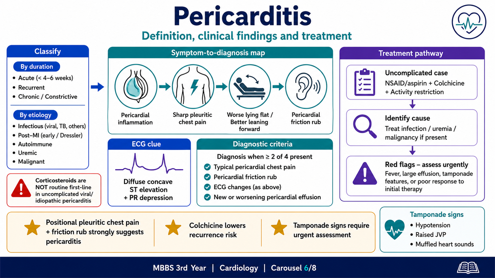
Pericarditis is inflammation of the pericardium, the fibroserous sac surrounding the heart.
It may occur alone or with myocardial involvement.
A. Classification of pericarditis
Pericarditis may be classified by clinical course:
- Acute pericarditis
- Incessant pericarditis
- Recurrent pericarditis
- Chronic pericarditis
- Constrictive pericarditis
It may also be classified by etiology:
- Idiopathic or presumed viral
- Infectious
- Post-myocardial infarction
- Post-cardiac injury
- Autoimmune or inflammatory
- Uremic
- Malignant
- Radiation-induced
- Drug-related
- Traumatic
- Metabolic
B. Mechanism of symptoms
Inflamed pericardial layers rub against one another, producing pain and sometimes a friction rub.
The pain is usually:
- Sharp
- Pleuritic
- Worse during inspiration or coughing
- Worse when lying supine
- Improved by sitting up and leaning forward
- Sometimes referred to the shoulder or trapezius ridge
The positional nature of the pain helps differentiate it from typical ischemic chest pressure.
C. Clinical findings
Pericardial chest pain
This is the most common symptom.
Pericardial friction rub
A pericardial rub is a superficial, scratchy or grating sound.
It is best heard:
- Near the left lower sternal border
- With the diaphragm of the stethoscope
- While the patient leans forward
- Sometimes during held expiration
It may be transient, so its absence does not exclude pericarditis.
⭐ A pericardial friction rub is highly suggestive of acute pericarditis.
ECG changes
Classical acute pericarditis may produce:
- Widespread concave ST-segment elevation
- PR-segment depression
- Reciprocal PR elevation and ST depression in lead aVR
- Later normalization and T-wave inversion
Unlike acute myocardial infarction, the changes are usually diffuse rather than confined to one coronary territory.
Diagnostic criteria
Acute pericarditis is generally diagnosed when at least two of the following are present:
- Typical pericardial chest pain
- Pericardial friction rub
- New widespread ST elevation or PR depression
- New or worsening pericardial effusion
Supportive findings include raised inflammatory markers and evidence of pericardial inflammation on imaging.
D. Basic investigation
- ECG
- Echocardiography
- Full blood count
- Inflammatory markers
- Renal function
- Cardiac troponin
- Chest radiograph where indicated
- Etiology-specific testing according to clinical context
Troponin may rise when there is associated myocardial involvement.
E. Treatment
First-line anti-inflammatory treatment
For uncomplicated idiopathic or presumed viral acute pericarditis, treatment usually includes:
- A nonsteroidal anti-inflammatory drug or aspirin
- Colchicine
- Gastroprotection where appropriate
- Restriction of strenuous activity
⭐ Colchicine reduces persistence and recurrence of acute pericarditis when appropriately added to anti-inflammatory treatment.
The selected NSAID should be given at an effective anti-inflammatory regimen and tapered according to clinical response and inflammatory activity.
Corticosteroids
Corticosteroids are not routine first-line therapy for uncomplicated presumed viral pericarditis.
They may be considered when:
- NSAIDs and colchicine are contraindicated or ineffective
- A specific autoimmune indication exists
- Another specialist-defined indication is present
⭐ Early unnecessary corticosteroid use may increase the risk of recurrent pericarditis.
Treat the cause
- Bacterial pericarditis requires urgent antimicrobial and drainage-based management.
- Tuberculous pericardial disease requires appropriate antituberculous therapy.
- Uremic pericarditis requires management of renal failure, including dialysis optimization.
- Malignant pericardial disease requires oncological and drainage assessment.
F. Red flags requiring hospital assessment
Features suggesting higher risk include:
- Fever
- Subacute onset
- Large effusion
- Cardiac tamponade
- Failure to improve with initial anti-inflammatory therapy
- Immunosuppression
- Trauma
- Anticoagulant treatment
- Associated myocardial involvement
⭐ Pericarditis with hypotension, raised jugular venous pressure or evidence of chamber compression must be assessed urgently for cardiac tamponade.
ST elevation in pericarditis is usually widespread and concave, whereas ST elevation in acute myocardial infarction follows a coronary territory and is commonly associated with reciprocal ischemic changes.
G. Pericardial Effusion and Cardiac Tamponade
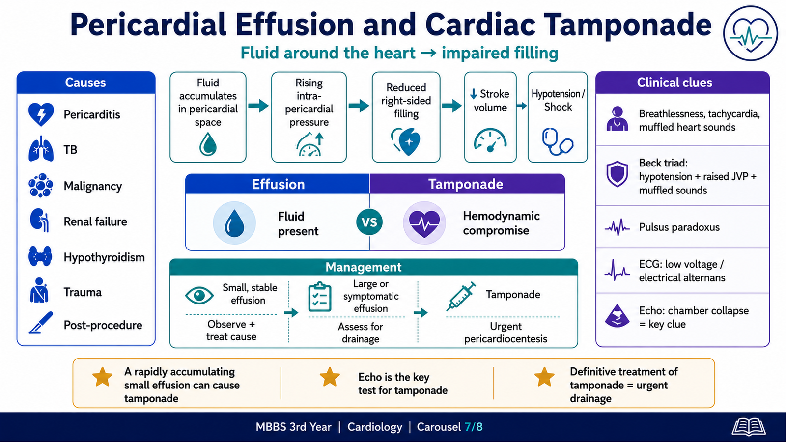
Pericardial effusion means accumulation of fluid within the pericardial cavity.
The clinical effect depends not only on the amount of fluid but also on:
- Rate of accumulation
- Compliance of the pericardium
- Intracardiac pressures
- Underlying cardiac disease
A slowly developing large effusion may produce fewer acute symptoms than a rapidly accumulating small effusion.
A. Etiology
Important causes include:
- Acute pericarditis
- Tuberculosis
- Malignancy
- Renal failure
- Hypothyroidism
- Autoimmune disease
- Trauma
- Cardiac procedures
- Post-myocardial infarction complications
- Aortic dissection
- Bacterial infection
- Radiation
- Heart failure
- Drug-related disease
B. Clinical findings
Small effusions may be asymptomatic.
Larger effusions may cause:
- Breathlessness
- Chest pressure
- Cough
- Fatigue
- Tachycardia
- Distant heart sounds
- Enlarged cardiac silhouette when chronic and large
C. Progression to cardiac tamponade
Cardiac tamponade occurs when intrapericardial pressure becomes high enough to impair cardiac filling.
The sequence is:
Fluid accumulates
→ intrapericardial pressure rises
→ right atrial and right ventricular filling becomes restricted
→ stroke volume falls
→ sympathetic tachycardia develops
→ hypotension and shock may follow
Because right-sided chambers have lower pressure, they are commonly compressed first.
D. Clinical clues to tamponade
Classical findings include:
- Hypotension
- Raised jugular venous pressure
- Muffled heart sounds
This combination is known as Beck triad, but all three features are not always present.
Other clues include:
- Tachycardia
- Breathlessness
- Narrow pulse pressure
- Pulsus paradoxus
- Poor peripheral perfusion
- Electrical alternans in a large effusion
- Chamber collapse on echocardiography
Pulsus paradoxus is an exaggerated inspiratory fall in systolic blood pressure.
⭐ The absence of the complete Beck triad does not exclude cardiac tamponade.
E. Diagnostic approach
Echocardiography is central because it can show:
- Size and location of effusion
- Right atrial or right ventricular collapse
- Respiratory variation in filling
- Inferior vena cava plethora
- Evidence of hemodynamic compromise
ECG may show:
- Low QRS voltage
- Sinus tachycardia
- Electrical alternans in a large swinging heart
Chest radiography may show a globular enlarged cardiac silhouette in a large, slowly developing effusion, but it is not a reliable test for acute tamponade.
F. Management
Stable small effusion
Management may include:
- Treatment of the underlying cause
- Clinical monitoring
- Repeat echocardiography according to risk
- Anti-inflammatory therapy when associated with inflammatory pericarditis
Large or symptomatic effusion
The cause must be investigated, and drainage may be required when there is:
- Hemodynamic compromise
- Suspected bacterial disease
- Suspected malignancy requiring diagnosis
- Persistent significant symptoms
- Large or progressive effusion
Cardiac tamponade
⭐ Cardiac tamponade is a medical emergency requiring urgent pericardial drainage, usually by image-guided pericardiocentesis or surgical drainage according to the cause and situation.
While urgent drainage is arranged:
- Oxygen and monitoring are provided.
- Hypotension may require cautious intravenous fluid support.
- Drugs that markedly reduce preload should be avoided.
- Positive-pressure ventilation may worsen venous return and requires careful specialist management.
Diuretics may reduce venous return and worsen hemodynamics in tamponade. The definitive treatment is relief of pericardial pressure, not routine diuresis.
H. Integrated Clinical Pathway: Electrical and Inflammatory Cardiac Disease
The opening patient has three connected findings:
- An irregularly irregular rhythm consistent with atrial fibrillation
- Positional pleuritic chest pain and a pericardial rub consistent with pericarditis
- A small pericardial effusion
A recent viral illness may trigger inflammation involving:
- Pericardium alone: pericarditis
- Myocardium alone: myocarditis
- Both myocardium and pericardium: overlapping inflammatory myocardial and pericardial disease
Inflammation may produce arrhythmias through:
- Atrial irritation
- Myocyte injury
- Altered conduction
- Increased sympathetic activity
- Ventricular dysfunction
- Electrolyte or metabolic disturbance during systemic illness
Integrated clinical approach
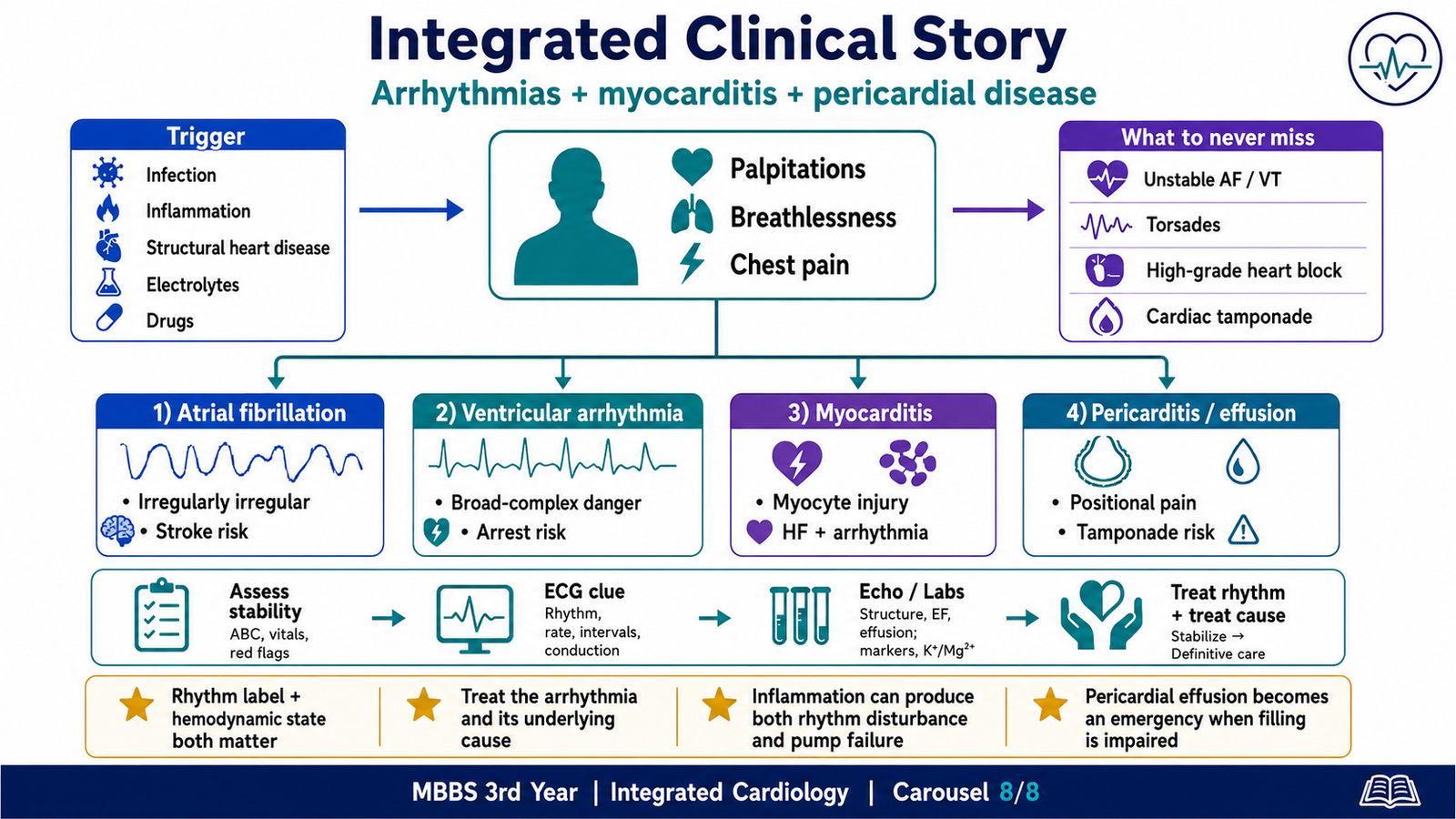
Step 1: Assess immediate danger
Look for:
- Hypotension
- Shock
- Syncope
- Severe pulmonary edema
- Ongoing ischemic chest pain
- Sustained ventricular arrhythmia
- High-grade AV block
- Tamponade
Step 2: Identify the rhythm
Use:
- Rate
- Regularity
- P waves
- PR interval
- QRS width
- QT interval
- Relationship between atrial and ventricular activity
Step 3: Search for the underlying cause
Consider:
- Ischemia
- Structural heart disease
- Myocarditis
- Pericarditis
- Thyroid disease
- Infection
- Electrolyte disturbance
- Drug toxicity
- Hypoxia
- Pulmonary embolism
Step 4: Treat both the rhythm and its substrate
Suppressing an arrhythmia without treating the cause may fail.
Examples:
- Correct hypokalemia before repeatedly escalating antiarrhythmic therapy.
- Treat myocarditis-associated heart failure and inflammation-related complications.
- Relieve tamponade rather than treating compensatory tachycardia alone.
- Assess stroke risk in atrial fibrillation even after rate control.
- Stop a QT-prolonging drug in torsades rather than merely adding another rhythm drug.
Final integrated exam principle
A rhythm is not just an ECG label. It is the electrical expression of an underlying physiological or pathological problem.
AIM High-Yield Review
- Classify an arrhythmia by rate, regularity, QRS width, site of origin and the patient’s hemodynamic stability.
- Most supraventricular rhythms are narrow-complex because ventricular activation uses the normal His–Purkinje system.
- Mobitz I shows progressive PR prolongation before a dropped beat; Mobitz II has constant PR intervals with sudden dropped QRS complexes and may progress to complete block.
- ⭐ A regular broad-complex tachycardia in an adult with structural heart disease should be treated as ventricular tachycardia until proved otherwise.
- Atrial fibrillation shows absent organized P waves and irregularly irregular R–R intervals; loss of atrial contraction promotes left atrial thrombus and embolic stroke.
- Management of atrial fibrillation is organized around stability, ventricular rate control, rhythm control when appropriate and prevention of thromboembolism.
- Pulseless ventricular tachycardia and ventricular fibrillation require immediate defibrillation and cardiopulmonary resuscitation.
- Class I drugs block sodium channels, class II drugs are beta-blockers, class III drugs mainly prolong repolarization through potassium-channel blockade and class IV drugs block L-type calcium channels.
- ⭐ Many antiarrhythmic drugs are proarrhythmic; excessive slowing of conduction or prolongation of repolarization can create a new dangerous rhythm.
- Myocarditis can impair contractility and create an electrically unstable myocardium, producing heart failure, conduction disease or ventricular arrhythmias.
- Acute pericarditis commonly causes sharp pleuritic pain that worsens on lying flat and improves on sitting forward; a pericardial friction rub is an important clue.
- Pericardial effusion becomes dangerous when rising intrapericardial pressure restricts diastolic filling and causes cardiac tamponade.
- In tamponade, urgent recognition is essential because hypotension and impaired filling may rapidly progress to obstructive shock.
🎥 AIM Video Resource
Antiarrhythmic Drugs and Cardiac Electrical Activity
This lecture explains cardiac action potentials, the Vaughan Williams classification, mechanisms of antiarrhythmic drugs, important clinical uses and major adverse effects.
AIM viewing focus: Relate each drug class to the cardiac action-potential phase or nodal tissue that it affects, and connect this action with its therapeutic use and risk of proarrhythmia.