
Study tip: This chapter follows the KMU learning outcomes in a logical sequence. First understand how a vessel wall becomes weak, split or inflamed, and then use the AIM High-Yield Review for rapid revision.
📖 AIM Learning Material
Aneurysms, Aortic Dissection and Vasculitis
CVS Module — Vascular wall weakness, dissection and inflammatory vessel injury
Topic Introduction
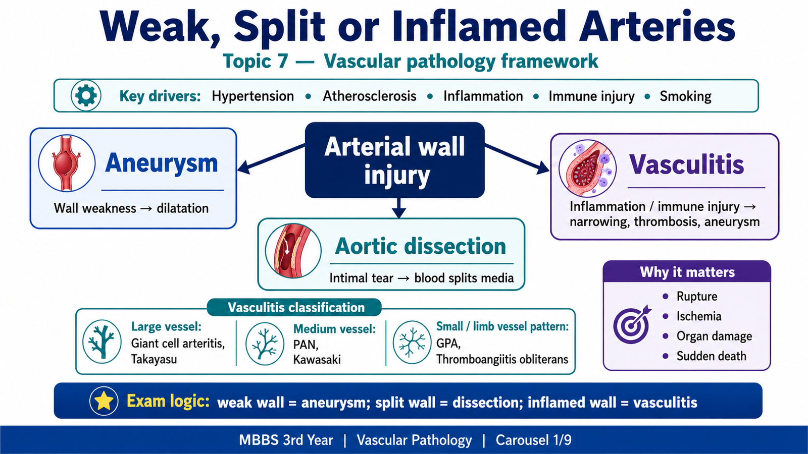
Arteries must remain strong enough to tolerate blood pressure while keeping their lumen open. This topic explains three major ways in which that normal structure can fail. In an aneurysm, a weakened vessel wall dilates. In an aortic dissection, blood enters the aortic wall and separates its layers. In vasculitis, inflammation damages vessels and may cause narrowing, thrombosis, ischemia, aneurysm or rupture. You will learn the causes, pathogenesis, morphology and manifestations of aneurysms and dissection, followed by the classification, immune mechanisms and characteristic features of the major vasculitides required by KMU.
A. The Common Vascular Wall Problem
A normal artery depends on intact smooth muscle cells, elastic tissue, collagen and extracellular matrix. These components allow the wall to resist pressure and preserve blood flow. Disease develops when the wall becomes weak, when blood enters and splits the wall, or when inflammation damages the wall and lumen.
Core pathological sequence:
Cause or risk factor → vessel-wall injury → structural change → disturbed flow, ischemia or rupture → clinical manifestation
- Aneurysm: abnormal localized dilatation caused by loss of wall strength.
- Aortic dissection: blood enters through an intimal tear and separates the layers of the media.
- Vasculitis: inflammation of a vessel wall that may narrow, thrombose, weaken or rupture the vessel.
Vessel size is useful for organizing vasculitis because different diseases preferentially affect different vascular territories and therefore produce different clinical patterns.
| Vessel group | Important examples in this chapter |
|---|---|
| Large vessels | Giant cell arteritis, Takayasu arteritis |
| Medium vessels | Polyarteritis nodosa, Kawasaki disease |
| Small vessels | Granulomatosis with polyangiitis |
| Small and medium limb vessels | Thromboangiitis obliterans |
B. Vascular and Aortic Aneurysms
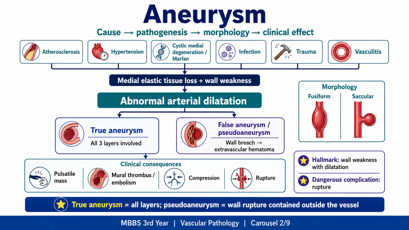
An aneurysm is a localized abnormal dilatation of a blood vessel or cardiac chamber. Vascular aneurysms form when the arterial wall loses enough structural support that intraluminal pressure gradually stretches it. Damage to smooth muscle, elastic tissue, collagen or extracellular matrix therefore lies at the center of aneurysm formation.
Etiology and pathogenesis
Different diseases weaken the wall by different routes. Atherosclerosis causes inflammation and matrix degradation, especially in the abdominal aorta. Hypertension increases mechanical stress and contributes to medial injury. Cystic medial degeneration weakens elastic tissue and smooth muscle, particularly in aging and Marfan syndrome. Infection, trauma and vasculitis may directly destroy the wall. Syphilitic aortitis damages the vasa vasorum, reducing the blood supply to the aortic media.
- Atherosclerosis: important in abdominal aortic aneurysm.
- Hypertension: adds wall stress and promotes medial injury.
- Cystic medial degeneration: associated with aging and Marfan syndrome.
- Infection: may produce a mycotic aneurysm.
- Trauma: may produce a false aneurysm.
- Vasculitis: weakens the wall through inflammatory destruction.
- Syphilitic aortitis: classically affects the thoracic aorta through vasa-vasorum injury.
Types
A true aneurysm includes all three layers of the vessel wall: intima, media and adventitia. A false aneurysm, or pseudoaneurysm, is a wall defect that allows blood to form an extravascular hematoma communicating with the lumen. By shape, a fusiform aneurysm dilates the full circumference of a long segment, whereas a saccular aneurysm forms a localized outpouching on one side.
Morphology and manifestations
Grossly, an aneurysm appears as a dilated vessel segment with a thinned wall. Atherosclerotic plaques, calcification and a mural thrombus may be present. Microscopically, there is loss or fragmentation of elastic tissue, medial thinning and loss of smooth muscle cells. In infectious or vasculitic aneurysms, inflammation is also present.
Aneurysms may remain silent until they enlarge. Their effects occur because the enlarged sac compresses nearby structures, promotes stagnant flow and thrombosis, or finally ruptures.
- Pressure on adjacent structures may produce pain or organ-specific symptoms.
- Mural thrombosis may develop because flow becomes abnormal and sluggish.
- Fragments of thrombus may embolize and cause distal ischemia.
- Progressive wall thinning may end in rupture and massive hemorrhage.
- An abdominal aortic aneurysm may present as a pulsatile abdominal mass.
Abdominal and thoracic aortic aneurysms
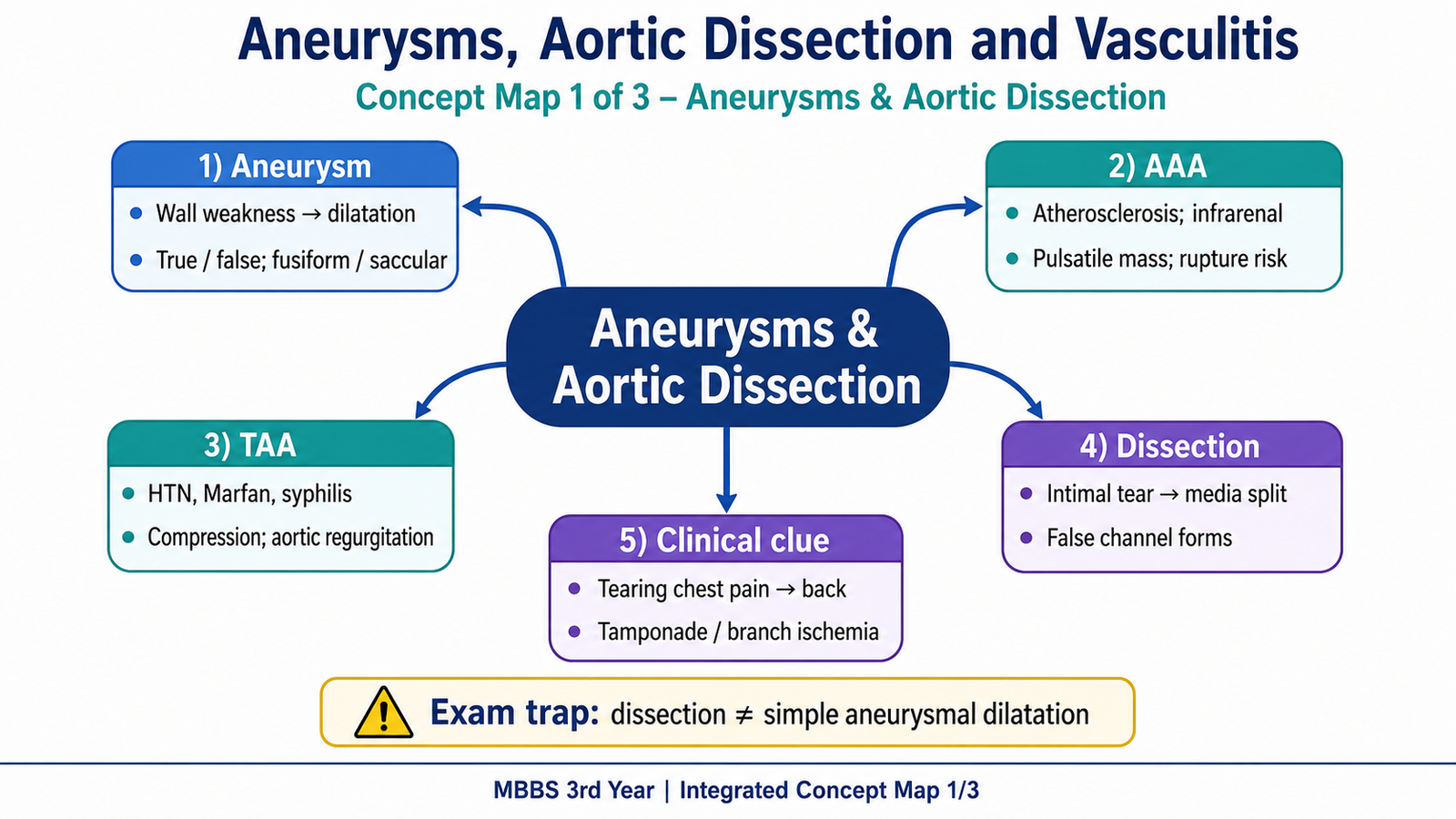
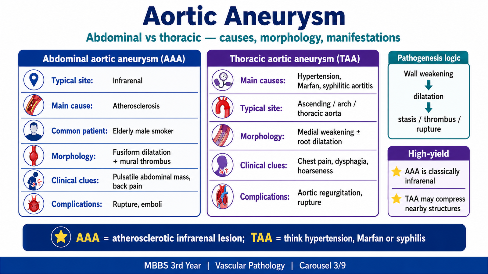
Abdominal aortic aneurysm (AAA) most often develops below the renal arteries and is strongly associated with atherosclerosis. Plaque-related inflammation and matrix degradation weaken the media, while smoking, age, male sex and hypertension increase risk. Grossly, the lesion is often fusiform, with severe atherosclerosis, wall thinning, calcification and mural thrombus. It may cause a pulsatile abdominal mass, abdominal or back pain, distal embolization or rupture with severe hypotension.
Thoracic aortic aneurysm is more closely associated with hypertension, cystic medial degeneration, Marfan syndrome and syphilitic aortitis. Enlargement may compress the esophagus and cause dysphagia, compress the recurrent laryngeal nerve and cause hoarseness, or compress airways and cause breathing difficulty. Dilatation of the aortic root may also produce aortic regurgitation.
| Feature | Abdominal aortic aneurysm | Thoracic aortic aneurysm |
|---|---|---|
| Typical association | Atherosclerosis | Hypertension, medial degeneration, Marfan syndrome, syphilitic aortitis |
| Typical site | Usually infrarenal | Thoracic aorta or aortic root |
| Main manifestations | Pulsatile mass, abdominal/back pain, embolization, rupture | Chest symptoms, dysphagia, hoarseness, aortic regurgitation |
C. Aortic Dissection
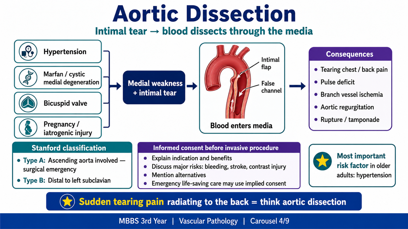
Aortic dissection is different from an aneurysm. In dissection, blood enters the aortic wall through an intimal tear and travels within the media, creating a false channel. The process can extend proximally toward the heart, distally along the aorta or into major branch arteries.
Causes and pathogenesis
Hypertension is the most important association in older adults because long-term pressure damages and weakens the media. In younger patients, connective-tissue disorders such as Marfan syndrome may produce medial degeneration. Other associations include bicuspid aortic valve, pregnancy, trauma and iatrogenic injury.
Mechanism: Hypertension or connective-tissue defect → medial degeneration → intimal tear → blood enters the media → false channel extends → rupture or branch-vessel obstruction
Morphology
Grossly, an intimal tear is usually present in the ascending aorta. Blood separates the medial layers and may create a double-barrel appearance with true and false lumina. Microscopically, the media may show fragmentation and loss of elastic tissue, reduced smooth muscle cells and pools of mucoid extracellular matrix. Severe atherosclerosis is not required for dissection.
Clinical features and complications
The typical presentation is sudden, severe tearing or ripping chest pain that may radiate to the back. Unequal pulses or blood pressures occur when the dissection obstructs a branch vessel. The danger comes from extension into vital structures.
- Rupture into the pericardial sac may cause cardiac tamponade.
- Extension to the aortic root may produce acute aortic regurgitation.
- Coronary artery involvement may cause myocardial ischemia.
- Carotid involvement may cause neurologic deficits.
- Renal, mesenteric or limb-vessel obstruction may cause organ or limb ischemia.
- Rupture into the pleural or peritoneal cavity may cause fatal hemorrhage.
Classification logic: Stanford type A involves the ascending aorta and is the more dangerous proximal form. Stanford type B begins distal to the left subclavian artery and does not involve the ascending aorta.
D. Vasculitis: Classification and Immune-Mediated Injury
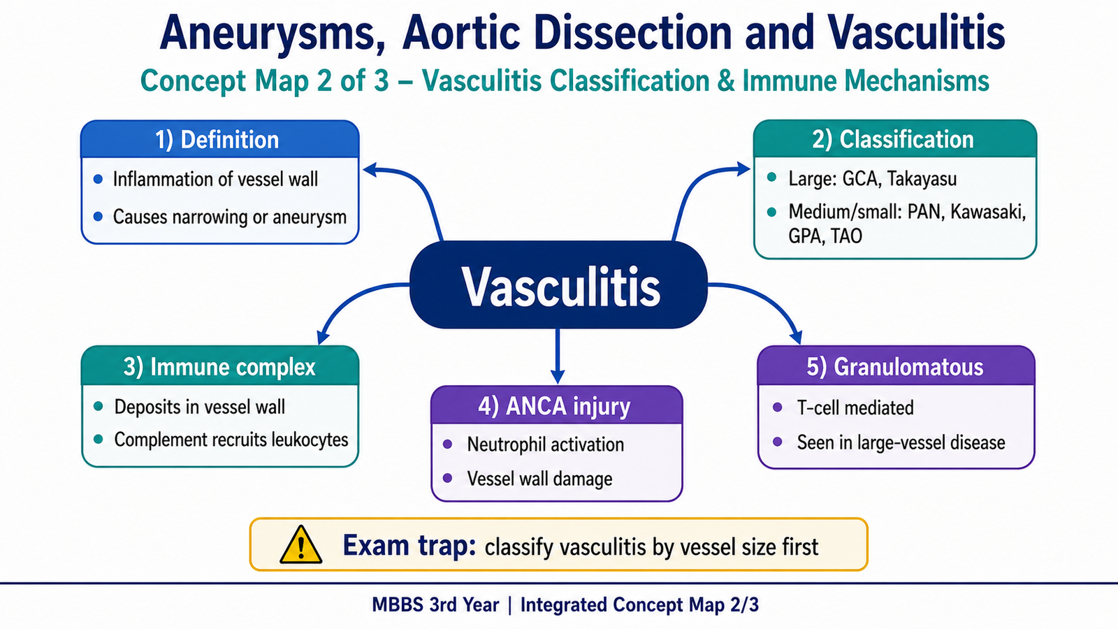
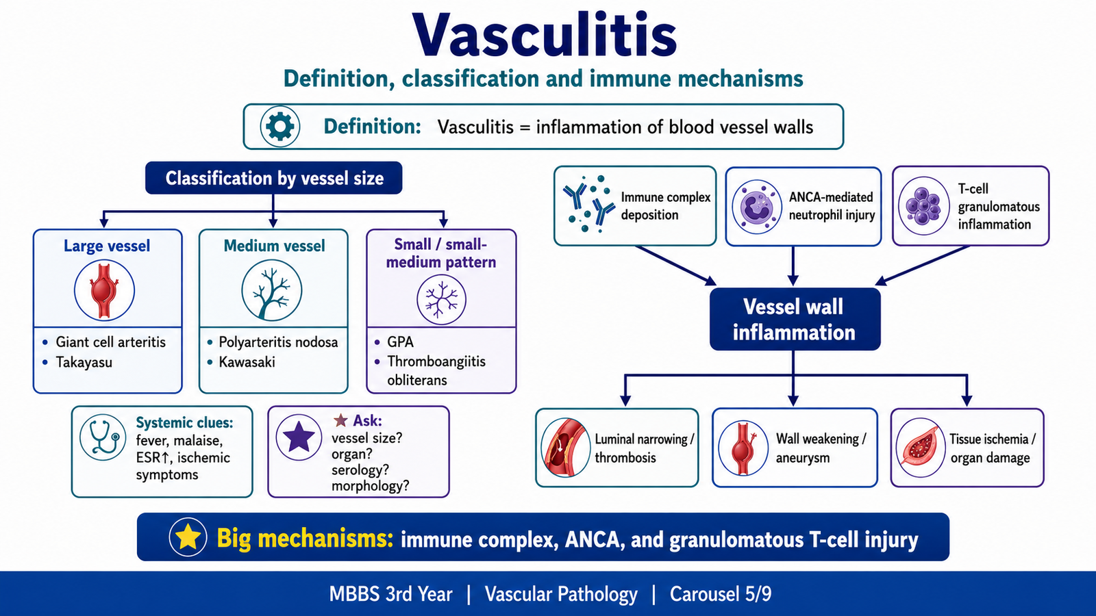
Vasculitis means inflammation of blood-vessel walls. The inflammation may damage the endothelium and media, narrow or occlude the lumen, promote thrombosis, or weaken the wall enough to form an aneurysm or rupture. The affected organ therefore depends on which vessels are involved.
Classification
Vasculitides are mainly classified according to the size of the vessels most commonly affected. Large-vessel diseases mainly involve the aorta and its major branches. Medium-vessel diseases affect major visceral arteries and their branches. Small-vessel diseases involve arterioles, capillaries and venules, although overlap can occur.
Immunological mechanisms of non-infectious vasculitis
Most non-infectious vasculitides result from an abnormal immune response. The main mechanisms are immune-complex deposition, antineutrophil cytoplasmic antibody–related injury and T-cell–mediated granulomatous inflammation.
- Immune-complex deposition: antigen–antibody complexes settle in vessel walls, activate complement and recruit neutrophils. Neutrophil enzymes then damage the wall.
- ANCA-mediated injury: ANCAs activate primed neutrophils, which attach to endothelium and release destructive enzymes and reactive oxygen species.
- T-cell–mediated injury: activated T cells and macrophages produce granulomatous inflammation, especially in giant cell arteritis and Takayasu arteritis.
Common consequences include luminal narrowing, thrombosis and ischemia; hemorrhage when the wall is destroyed; and aneurysm when inflammation weakens the wall.
E. Large-Vessel Vasculitis: Giant Cell and Takayasu Arteritis
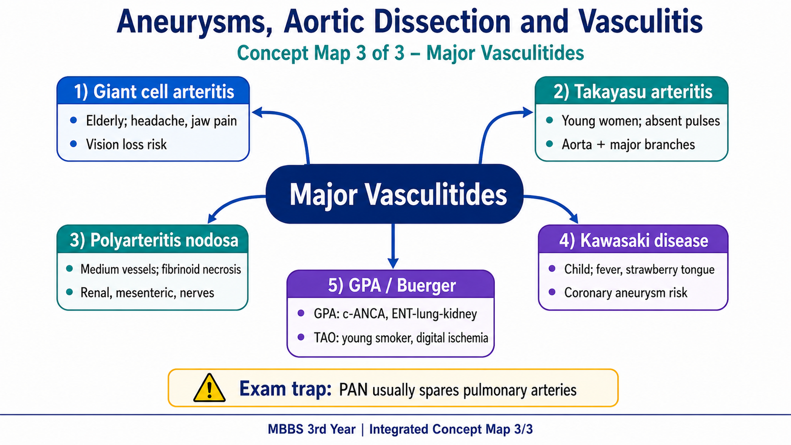
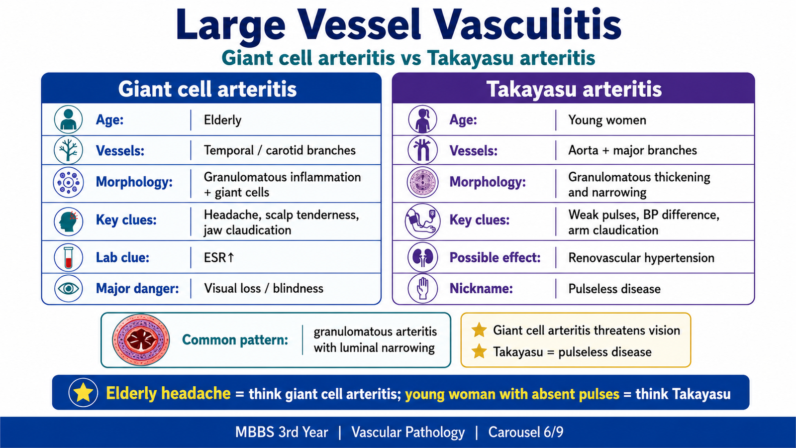
Giant cell arteritis and Takayasu arteritis are granulomatous large-vessel vasculitides. In both conditions, T-cell and macrophage–mediated inflammation damages elastic tissue, thickens the intima and narrows the lumen. Their major distinction is the age group and the vascular territory that produces the dominant symptoms.
Giant cell arteritis
Giant cell arteritis usually affects older adults and commonly involves branches of the carotid artery, especially the temporal artery. Granulomatous inflammation fragments the internal elastic lamina and causes marked intimal thickening. The narrowed lumen reduces blood flow to tissues supplied by the affected artery.
- Morphology: segmental granulomatous inflammation, multinucleated giant cells, disruption of the internal elastic lamina and intimal thickening.
- Clinical features: new headache, tender or thickened temporal artery, scalp tenderness, jaw claudication and constitutional symptoms.
- Important complication: ophthalmic artery involvement may cause sudden irreversible visual loss.
Takayasu arteritis
Takayasu arteritis mainly affects younger women and involves the aortic arch and its major branches. Granulomatous inflammation and later fibrosis thicken the wall and narrow the lumen. Reduced flow to upper limbs explains weak or absent pulses and unequal blood pressures.
- Morphology: granulomatous inflammation, elastic-tissue destruction, intimal thickening, adventitial fibrosis and stenosis of major branches.
- Clinical features: weak or absent upper-limb pulses, unequal arm blood pressures, limb claudication, visual or neurologic symptoms and vascular bruits.
- Classic name: “pulseless disease” because major arterial narrowing reduces detectable pulses.
| Feature | Giant cell arteritis | Takayasu arteritis |
|---|---|---|
| Typical patient | Older adult | Young woman |
| Main vessels | Temporal and other carotid branches | Aortic arch and major branches |
| Key clue | Headache, jaw claudication, visual symptoms | Absent pulses, unequal arm pressures |
F. Medium-Vessel Vasculitis: Polyarteritis Nodosa and Kawasaki Disease
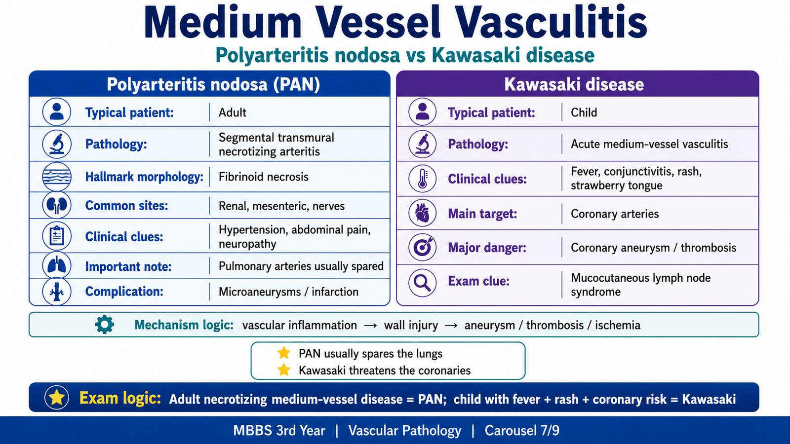
Polyarteritis nodosa and Kawasaki disease both affect medium-sized arteries, but their patients and dominant complications are different. Polyarteritis nodosa causes systemic necrotizing arteritis and organ ischemia, whereas Kawasaki disease is a childhood vasculitis with a special tendency to involve coronary arteries.
Polyarteritis nodosa
Polyarteritis nodosa (PAN) is a segmental transmural necrotizing inflammation of medium and small muscular arteries. Immune-mediated wall injury causes fibrinoid necrosis. Because lesions occur at different stages in different segments, affected vessels may show active inflammation beside healing fibrosis. Weak points may dilate into microaneurysms, while thrombosis and narrowing reduce organ blood flow.
- Morphology: segmental transmural necrotizing inflammation with fibrinoid necrosis, thrombosis and microaneurysm formation.
- Clinical features: fever, malaise, weight loss, hypertension from renal-artery involvement, abdominal pain from mesenteric ischemia, skin lesions, muscle pain and peripheral neuropathy.
- Important distinction: classic PAN usually spares pulmonary circulation and does not cause glomerulonephritis.
Kawasaki disease
Kawasaki disease is an acute febrile vasculitis of children. Immune activation produces inflammation of medium-sized arteries, especially coronary arteries. Destruction of the arterial wall may form coronary aneurysms, which can thrombose and lead to myocardial ischemia.
- Morphology: transmural inflammation of medium arteries with possible coronary aneurysm and thrombosis.
- Clinical features: prolonged fever, conjunctival redness, oral and tongue changes, rash, cervical lymph-node enlargement and changes in hands and feet.
- Major complication: coronary artery aneurysm with thrombosis or rupture.
| Feature | Polyarteritis nodosa | Kawasaki disease |
|---|---|---|
| Typical patient | Adult | Child |
| Main pattern | Systemic organ ischemia and microaneurysms | Mucocutaneous illness with coronary arteritis |
| Major danger | Renal, bowel or neurologic ischemia | Coronary aneurysm and thrombosis |
G. Granulomatosis with Polyangiitis and Thromboangiitis Obliterans
Granulomatosis with polyangiitis
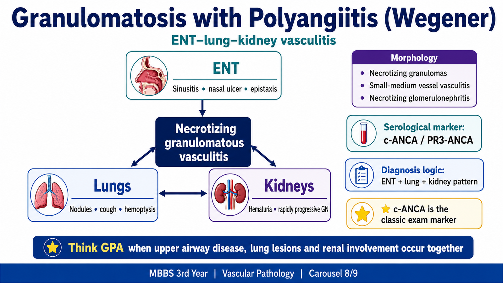
Granulomatosis with polyangiitis (GPA), formerly called Wegener granulomatosis, is a necrotizing granulomatous vasculitis of small to medium vessels. It classically links disease in the upper respiratory tract, lungs and kidneys. ANCA-activated neutrophils injure endothelium, while granulomatous inflammation produces destructive respiratory lesions.
- Serological marker: c-ANCA, usually directed against proteinase-3 (PR3-ANCA).
- Morphology: necrotizing granulomas in the respiratory tract, necrotizing vasculitis and necrotizing crescentic glomerulonephritis.
- Upper airway features: chronic sinusitis, nasal ulceration, epistaxis and otitis media.
- Lung features: cough, hemoptysis, nodular or cavitary lesions.
- Renal features: hematuria, proteinuria and rapidly progressive glomerulonephritis.
The diagnosis is suggested most strongly by the combined pattern of ENT disease, lung lesions, renal involvement and c-ANCA rather than by ANCA alone.
Thromboangiitis obliterans
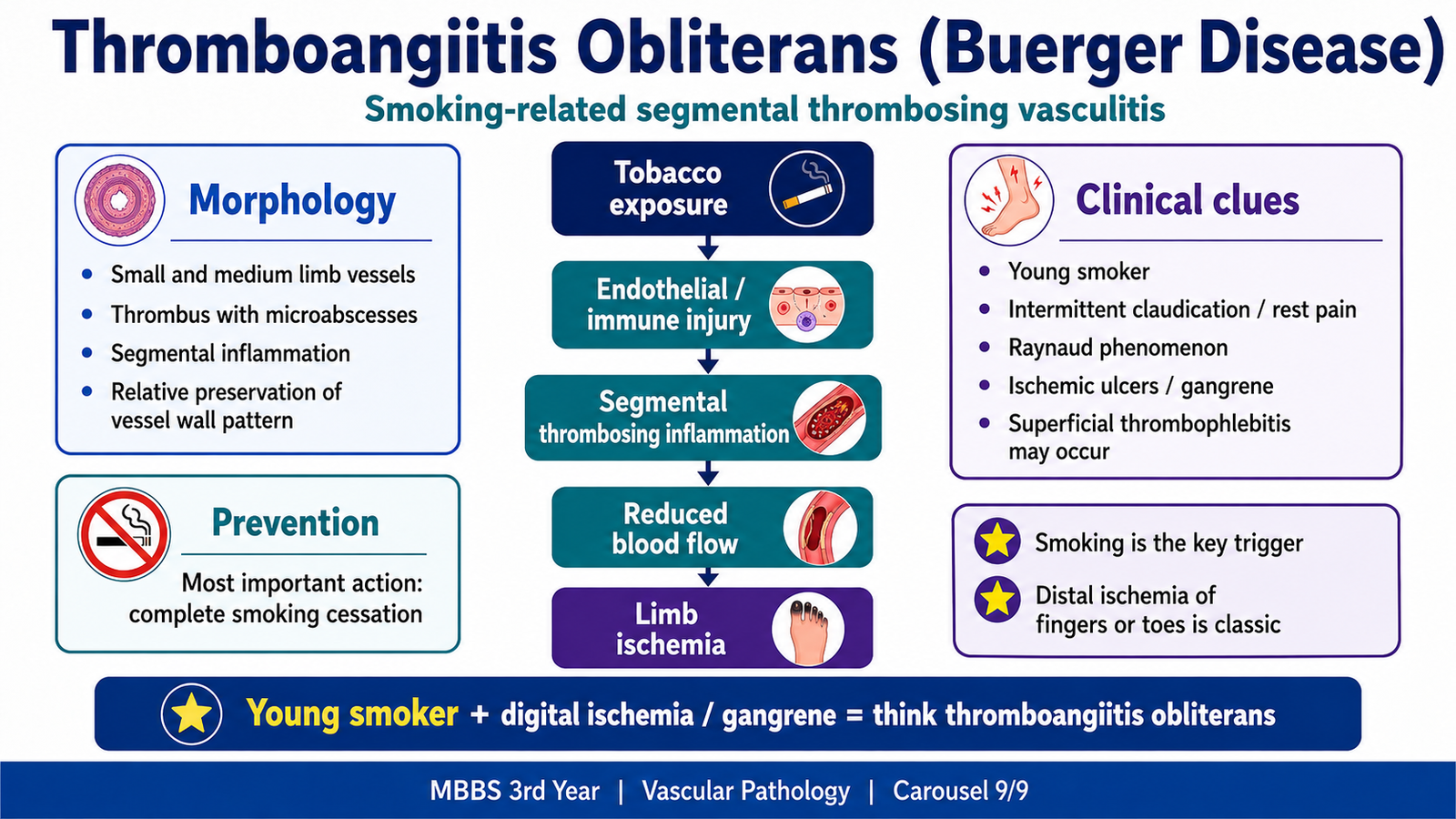
Thromboangiitis obliterans, or Buerger disease, is a segmental inflammatory and thrombosing disease of small and medium vessels of the limbs. It is strongly linked to tobacco exposure. Endothelial injury and immune inflammation promote thrombosis, which reduces distal blood flow and causes ischemic pain, ulcers and gangrene.
- Typical patient: a young smoker with distal limb ischemia.
- Morphology: segmental thrombosing inflammation, microabscesses within the thrombus and relative preservation of the vessel-wall architecture.
- Common vessels: tibial and radial arteries.
- Clinical features: intermittent claudication, Raynaud phenomenon, ischemic ulcers, gangrene of fingers or toes and superficial nodular phlebitis.
- Essential preventive point: complete smoking cessation.
| Feature | Thromboangiitis obliterans | Atherosclerotic peripheral disease |
|---|---|---|
| Age | Younger | Usually older |
| Dominant association | Smoking | Age, diabetes, hyperlipidemia, hypertension |
| Morphology | Segmental thrombosing vasculitis | Atheromatous plaques |
Integrated Mechanism Flow
Wall weakness or immune injury → loss of elastic and muscular support or inflammatory wall damage → dilatation, dissection, luminal narrowing or thrombosis → disturbed flow and tissue ischemia or hemorrhage → clinical clues such as pulsatile mass, tearing pain, absent pulses, visual symptoms, organ ischemia or gangrene → complications including rupture, embolism, infarction and organ failure.
AIM High-Yield Review
- Aneurysm is dilatation caused by wall weakness; dissection is blood splitting the media after an intimal tear.
- A true aneurysm includes all wall layers; a false aneurysm is a communicating extravascular hematoma.
- Abdominal aortic aneurysm is classically infrarenal and strongly associated with atherosclerosis.
- Thoracic aneurysm is associated with hypertension, medial degeneration, Marfan syndrome and syphilitic aortitis.
- ⭐ Hypertension is the major risk factor for aortic dissection in older adults.
- Sudden tearing chest pain radiating to the back with unequal pulses strongly suggests aortic dissection.
- Vasculitis causes ischemia by narrowing or thrombosis, and hemorrhage or aneurysm by wall destruction.
- Giant cell arteritis: older patient, temporal headache, jaw claudication and danger of visual loss.
- Takayasu arteritis: young woman, aortic-arch disease, unequal arm pressures and absent pulses.
- PAN shows segmental transmural necrotizing arteritis, fibrinoid necrosis and microaneurysms; lungs are usually spared.
- Kawasaki disease occurs in children and may cause coronary artery aneurysms.
- GPA combines ENT disease, lung lesions, renal disease and c-ANCA/PR3-ANCA.
- Thromboangiitis obliterans occurs in young smokers and shows segmental thrombosing inflammation with microabscesses.
- Complete smoking cessation is essential in thromboangiitis obliterans.
🎥 AIM Video Learning — Aneurysms, Aortic Dissection and Vasculitis
Watch this pathology lecture after reading the learning material to reinforce the classification, pathogenesis, morphology and clinical features of aneurysms, aortic dissection and major vasculitides.
AIM viewing focus: Differentiate aneurysm from dissection, understand vessel-size classification of vasculitis, and identify the hallmark pathological and clinical features of the major vasculitides.