
Study tip: This chapter follows the KMU learning outcomes and explains shock, sepsis, hemorrhage and DIC in a logical sequence. First understand how reduced tissue perfusion causes cellular injury, then use the AIM High-Yield Review for revision.
KMU Curriculum
AIM Learning Cycle
📖 AIM Learning Material
Shock, Sepsis, Hemorrhage and Disseminated Intravascular Coagulation
CVS Module — Classification, pathophysiology, stages, laboratory findings and major complications
Topic Introduction
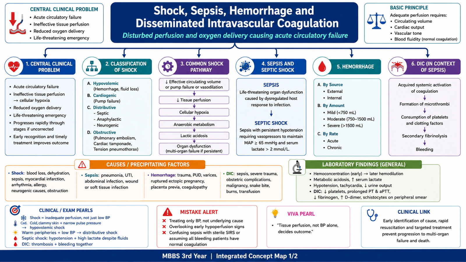
Shock is a state of acute circulatory failure in which tissues do not receive enough blood and oxygen to meet their needs. The central problem is therefore not only low blood pressure, but inadequate tissue perfusion followed by cellular hypoxia and organ dysfunction. Shock may result from loss of circulating volume, failure of the heart, severe infection, loss of vascular tone or mechanical obstruction to blood flow. This chapter explains the main types and stages of shock, how sepsis produces septic shock, how acute blood loss produces hemorrhagic shock, and how severe sepsis may activate coagulation throughout the body to cause disseminated intravascular coagulation, or DIC.
A. Shock: Definition, Central Mechanism and Classification
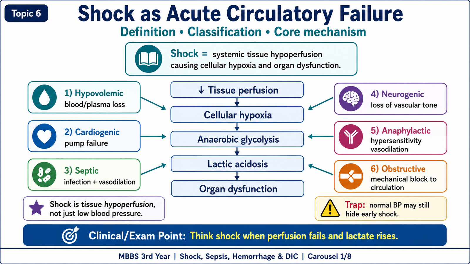
Shock means systemic hypoperfusion severe enough to cause cellular hypoxia and organ dysfunction. The circulation normally supplies oxygen and nutrients to tissues and removes metabolic waste. When effective perfusion falls, cells cannot maintain normal aerobic metabolism. They shift toward anaerobic glycolysis, produce excess lactic acid and generate less ATP.
Reduced ATP impairs energy-dependent cell functions, including the sodium–potassium pump. Sodium and water enter cells, causing cellular swelling. If perfusion is restored early, this injury may remain reversible. Persistent hypoxia causes mitochondrial dysfunction, membrane injury, necrosis and failure of vital organs.
Central mechanism:
Reduced effective perfusion → reduced oxygen delivery → cellular hypoxia → anaerobic glycolysis → lactic acidosis → cellular injury → organ dysfunction.
Classification of shock
Shock is classified according to the main disturbance that reduces tissue perfusion.
- Hypovolemic shock: circulating volume is reduced by blood or fluid loss. Important causes include hemorrhage, severe dehydration, burns, vomiting and diarrhea.
- Cardiogenic shock: the heart cannot pump effectively. Causes include myocardial infarction, severe arrhythmias and myocarditis.
- Septic shock: severe infection produces systemic inflammation, vasodilation, endothelial injury, capillary leakage, myocardial depression and microvascular thrombosis.
- Neurogenic shock: loss of sympathetic vascular tone causes widespread vasodilation, for example after spinal cord injury or anesthesia-related autonomic failure.
- Anaphylactic shock: systemic immediate hypersensitivity produces vasodilation, increased vascular permeability and possible airway compromise.
- Obstructive shock: a mechanical obstruction reduces cardiac filling or forward blood flow. Examples include pulmonary embolism, cardiac tamponade and tension pneumothorax.
The most important pathology types for this topic are hypovolemic or hemorrhagic shock, cardiogenic shock and septic shock. A patient may have significant tissue hypoperfusion before a major fall in blood pressure becomes obvious.
B. Stages of Shock and Organ Injury
Shock usually develops through three stages: compensated, progressive and irreversible. These stages form a continuous process rather than completely separate events. The outcome depends on the severity and duration of hypoperfusion and whether the underlying cause is corrected.
Compensated or non-progressive stage
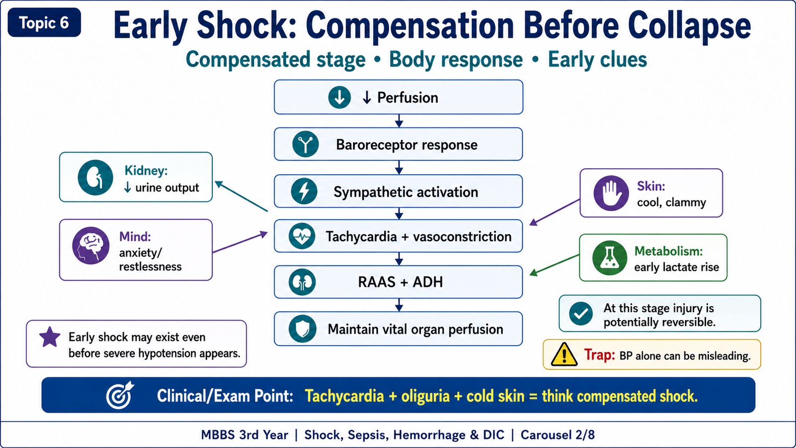
When arterial pressure and tissue perfusion begin to fall, baroreceptors activate the sympathetic nervous system. Catecholamine release increases the heart rate and produces peripheral vasoconstriction. This helps preserve blood flow to the heart and brain while reducing flow to the skin, kidneys and other less immediately vital tissues.
Reduced renal blood flow activates the renin–angiotensin–aldosterone system, while antidiuretic hormone promotes water retention. These responses attempt to restore circulating volume and blood pressure.
- Tachycardia develops to support cardiac output.
- Peripheral vasoconstriction produces cool, pale and clammy skin in most forms of shock.
- Renal vasoconstriction and fluid-conserving hormones reduce urine output.
- Anxiety and restlessness may appear as cerebral perfusion begins to fall.
- Early cellular injury includes reduced ATP, sodium–potassium pump failure, cellular swelling and lactic acid accumulation.
In early septic shock, the skin may initially be warm because widespread vasodilation is prominent. It may become cold and clammy later as circulatory failure progresses.
Progressive stage
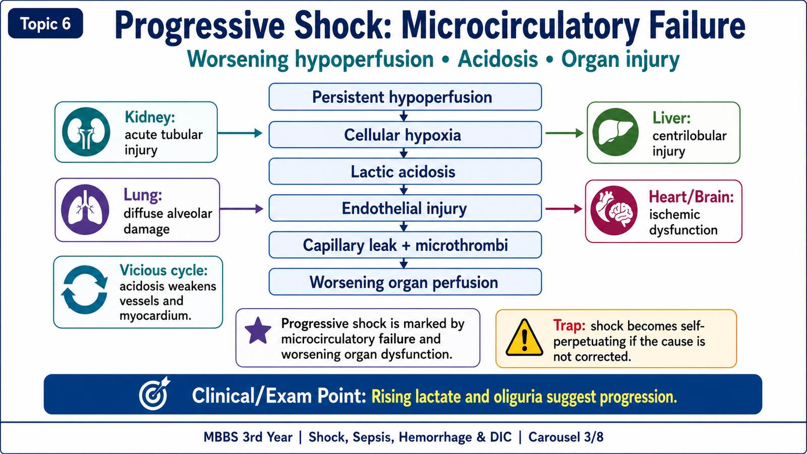
If the cause is not corrected, persistent hypoperfusion increases cellular hypoxia and lactic acidosis. Acidosis reduces vascular responsiveness and weakens myocardial contractility. Blood pressure falls further and tissue perfusion becomes even worse, creating a self-amplifying cycle.
Vicious cycle:
Low perfusion → cellular hypoxia → lactic acidosis → vasodilation and myocardial depression → worse hypotension → further hypoperfusion.
The microcirculation also becomes abnormal. Endothelial injury, capillary leakage, leukocyte adhesion, platelet activation and microthrombi reduce effective oxygen delivery. Fluid movement into tissues produces edema and further reduces the effective circulating volume.
Organ changes in prolonged shock
- Kidneys: ischemia causes acute tubular injury, especially in vulnerable tubular segments. Oliguria and rising creatinine follow.
- Lungs: endothelial and epithelial injury may cause diffuse alveolar damage, leading to hypoxemia and acute respiratory distress.
- Liver: centrilobular, or zone 3, hepatocytes are most vulnerable to hypoxia. Their injury may raise liver enzymes.
- Heart: reduced coronary perfusion and hypoxemia may cause subendocardial ischemia and worsening myocardial function.
- Brain: cerebral hypoperfusion produces confusion, drowsiness, ischemic encephalopathy and, in severe cases, coma.
Irreversible stage
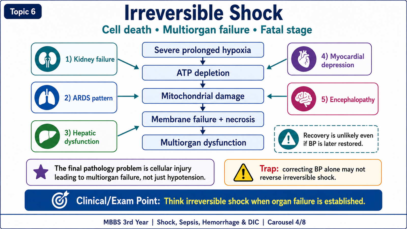
In irreversible shock, cellular and organ injury has become so severe that restoring blood pressure may no longer restore normal tissue function. Marked ATP depletion causes mitochondrial failure, severe membrane damage, lysosomal enzyme leakage and necrosis. Inflammatory mediators and microvascular thrombosis continue to injure tissues.
The major outcome is multiorgan dysfunction syndrome. The kidneys, lungs, liver, heart, brain and coagulation system may fail together. DIC may develop, particularly in severe sepsis.
C. Sepsis and Septic Shock
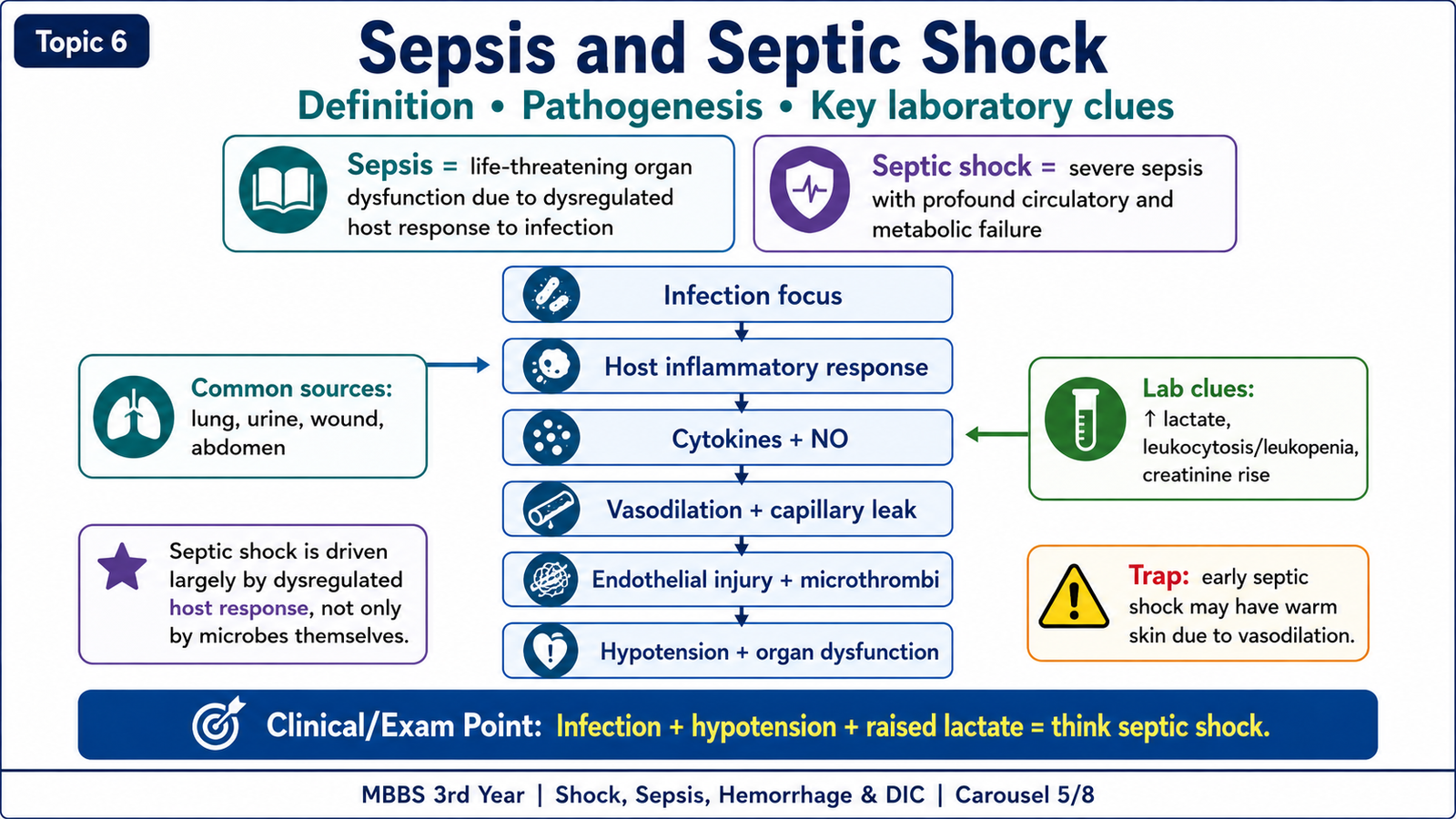
Sepsis is life-threatening organ dysfunction caused by a dysregulated host response to infection. The term dysregulated means that the body’s response is excessive, poorly controlled and damaging to its own tissues. Sepsis is therefore more than the presence of organisms in blood. Severe infection can cause sepsis even when a blood culture is negative.
Septic shock is a severe form of sepsis in which profound circulatory and metabolic abnormalities produce persistent tissue hypoperfusion and a high risk of death.
Causes
Sepsis can begin from many sites of infection. The important principle is that a local infection triggers a systemic host response.
- Pneumonia
- Urinary tract infection
- Infected wounds
- Intra-abdominal infection
- Meningitis
- Infected intravenous lines
- Postoperative or postpartum infection
Pathogenesis
Microbial products activate immune cells and endothelial cells. Inflammatory cytokines and other mediators are released. Nitric oxide and related mediators cause widespread vasodilation, which lowers systemic vascular resistance and blood pressure. At the same time, endothelial activation increases vascular permeability, allowing plasma to leak into tissues. This reduces the effective circulating volume even when the total body fluid has not been lost.
Endothelial injury also changes the vascular surface from an anticoagulant state to a procoagulant state. Tissue factor expression, platelet activation and thrombin generation produce microvascular fibrin thrombi. These thrombi obstruct small vessels and worsen tissue hypoxia. Inflammatory mediators may also depress myocardial contractility, while metabolic abnormalities reduce the ability of cells to use oxygen efficiently.
Septic shock mechanism:
Infection → dysregulated inflammation → vasodilation and capillary leakage → endothelial injury and microthrombi → reduced tissue perfusion → cellular hypoxia, lactic acidosis and organ dysfunction.
Laboratory findings
Laboratory abnormalities reflect infection, tissue hypoperfusion and developing organ injury. No single test explains the complete process, so findings are interpreted together.
- Leukocytosis or leukopenia due to an altered systemic inflammatory response
- Raised lactate due to tissue hypoperfusion and increased anaerobic metabolism
- Metabolic acidosis caused mainly by lactic acid accumulation
- Raised inflammatory markers such as CRP or procalcitonin where used
- Rising creatinine due to renal hypoperfusion and acute tubular injury
- Deranged liver enzymes due to hepatic hypoxic injury
- Thrombocytopenia when platelets are consumed or DIC develops
- Prolonged PT and aPTT, raised D-dimer and reduced fibrinogen when complicated by DIC
D. Hemorrhage and Hemorrhagic Shock
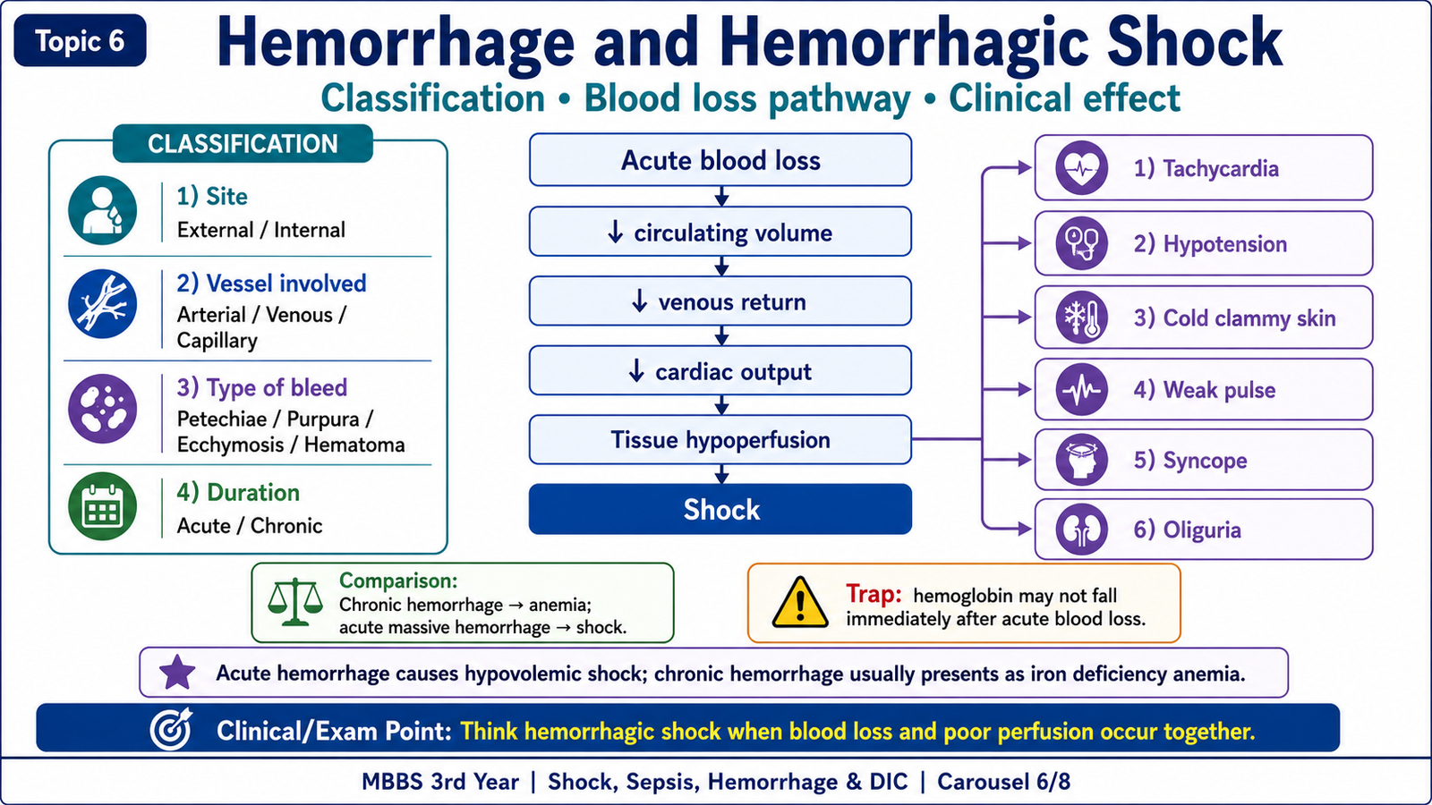
Hemorrhage means escape of blood from the vascular system. Its effects depend on the amount, rate and site of blood loss. Rapid and severe blood loss reduces circulating volume and may cause hypovolemic shock. Slow, chronic blood loss is more likely to cause anemia than immediate circulatory collapse.
Classification of hemorrhage
Hemorrhage can be classified in several useful ways.
According to site:
- External hemorrhage: blood leaves the body through a wound or natural opening.
- Internal hemorrhage: blood collects within tissues or organs.
- Cavity hemorrhage: blood accumulates in a body cavity, as in hemothorax, hemopericardium, hemoperitoneum or hemarthrosis.
According to the vessel involved:
- Arterial hemorrhage
- Venous hemorrhage
- Capillary hemorrhage
According to size and pattern:
- Petechiae: tiny pinpoint hemorrhages, often associated with platelet or small-vessel abnormalities.
- Purpura: slightly larger areas of hemorrhage.
- Ecchymoses: larger subcutaneous bruises.
- Hematoma: a localized collection of blood within tissue.
According to timing:
- Acute hemorrhage
- Chronic hemorrhage
Pathophysiology of hemorrhagic shock
Acute blood loss reduces the volume returning to the heart. Reduced venous return lowers preload, which lowers stroke volume and cardiac output. As cardiac output falls, tissue perfusion and oxygen delivery decrease. Cells then develop hypoxia and lactic acidosis.
Hemorrhagic shock mechanism:
Acute blood loss → reduced circulating volume → reduced venous return and preload → reduced cardiac output → tissue hypoperfusion → cellular hypoxia and lactic acidosis.
The sympathetic response produces tachycardia and peripheral vasoconstriction. Vasoconstriction explains the pale, cold and clammy skin. Reduced cerebral perfusion causes dizziness, syncope or confusion. Renal vasoconstriction reduces urine output. If blood loss is rapid and severe, compensation fails and progressive shock develops.
- Tachycardia
- Weak pulse
- Hypotension
- Cold, pale and clammy skin
- Dizziness or syncope
- Oliguria
- Confusion in severe cases
Chronic hemorrhage usually causes gradual loss of iron and may lead to iron deficiency anemia. In contrast, acute massive hemorrhage mainly threatens the patient through rapid loss of circulating volume and shock.
E. Disseminated Intravascular Coagulation in Sepsis
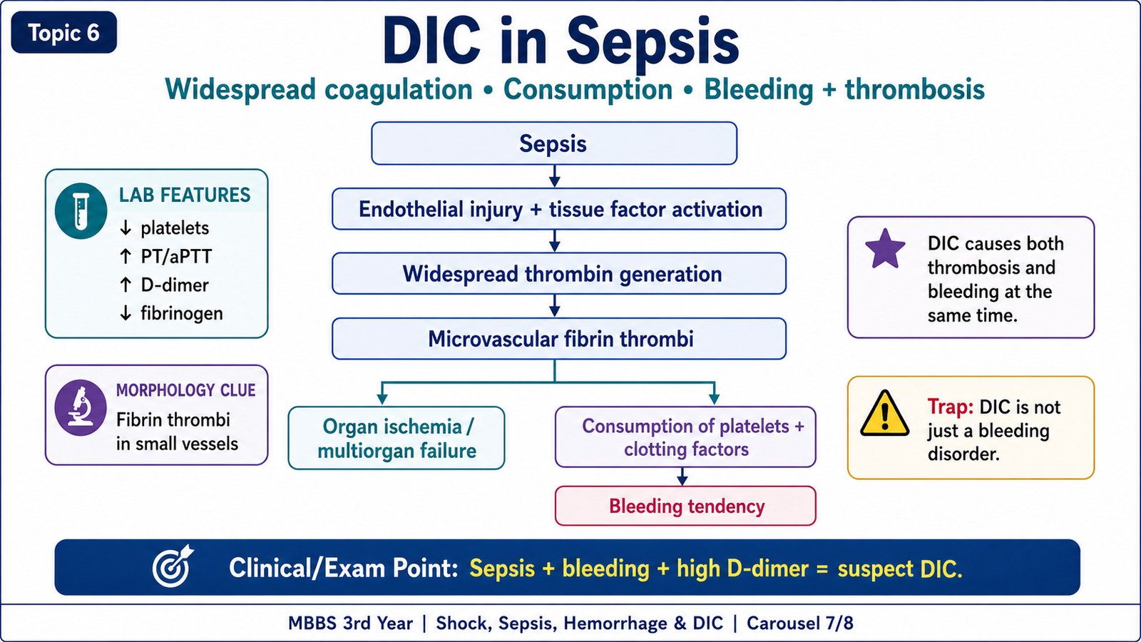
Disseminated intravascular coagulation is an acquired disorder in which coagulation is activated throughout the microcirculation. It is not a primary disease by itself. It develops as a complication of another serious condition, particularly sepsis in this topic.
DIC produces two apparently opposite problems at the same time. Fibrin thrombi form within small vessels and reduce blood flow, causing tissue ischemia and organ injury. At the same time, platelets and clotting factors are consumed faster than they can be replaced. Secondary fibrinolysis also breaks down fibrin. The patient therefore develops a bleeding tendency while microvascular thrombosis is occurring.
Important causes
- Sepsis
- Major trauma or severe tissue injury
- Obstetric complications
- Malignancy
- Severe burns
- Incompatible blood transfusion
Pathogenesis in sepsis
Inflammatory cytokines and endothelial injury increase tissue factor expression and activate the coagulation cascade. Thrombin generation increases, and fibrin is deposited in small vessels. Natural anticoagulant mechanisms become impaired, while fibrinolytic activity later contributes to the breakdown of fibrin.
DIC mechanism:
Sepsis and endothelial injury → tissue factor activation → increased thrombin and fibrin formation → microvascular thrombi → platelet and clotting-factor consumption → secondary fibrinolysis → organ ischemia plus bleeding.
Laboratory findings
The laboratory pattern reflects consumption of coagulation components and increased breakdown of fibrin.
- Reduced platelet count: platelets are consumed during widespread coagulation.
- Prolonged PT and aPTT: clotting factors are consumed.
- Reduced fibrinogen: fibrinogen is converted into fibrin throughout the circulation.
- Raised D-dimer and fibrin degradation products: cross-linked fibrin is being broken down.
- Schistocytes: fragmented red cells may appear because erythrocytes are mechanically damaged while passing through fibrin-rich small vessels.
Morphology and consequences
Fibrin-rich microthrombi may be found in capillaries and small vessels of the kidneys, brain, heart, lungs and other organs. These thrombi cause ischemic injury and contribute to organ failure. Consumption of platelets and clotting factors causes bleeding from puncture sites, wounds, mucosal surfaces and internal organs.
The key pathological feature is therefore the simultaneous presence of microvascular thrombosis and bleeding.
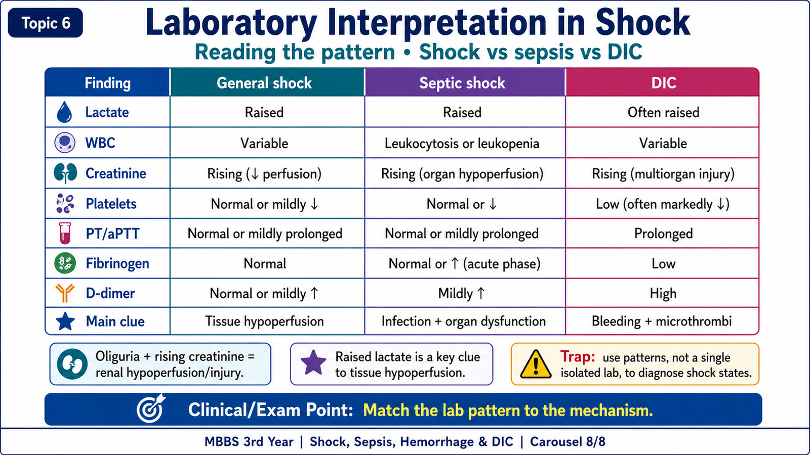
F. Laboratory Interpretation in Shock
Laboratory findings help show the cause of shock, the degree of tissue hypoperfusion and the organs that are becoming injured. They should always be interpreted as part of the complete pathological process rather than as isolated values.
| Finding | Why it occurs | Main significance |
|---|---|---|
| Raised lactate | Reduced tissue perfusion increases anaerobic metabolism. | Supports significant tissue hypoperfusion. |
| Metabolic acidosis | Lactic acid accumulates and renal function may deteriorate. | Suggests progressive metabolic failure. |
| Rising creatinine | Renal hypoperfusion and acute tubular injury reduce filtration. | Indicates kidney injury. |
| Raised liver enzymes | Hepatocytes, especially in zone 3, undergo hypoxic injury. | Indicates hepatic involvement. |
| Leukocytosis or leukopenia | The systemic inflammatory response changes leukocyte production, release and consumption. | Supports severe infection when interpreted clinically. |
| Low platelets, prolonged PT/aPTT, high D-dimer and low fibrinogen | Systemic coagulation consumes platelets, fibrinogen and clotting factors while fibrin is degraded. | Classic pattern of DIC. |
Important interpretation in hemorrhage
In early acute hemorrhage, hemoglobin may not fall immediately because whole blood is lost: red cells and plasma leave the circulation together. The measured hemoglobin becomes more clearly reduced after fluid shifts or fluid replacement dilute the remaining blood. Therefore, an initially near-normal hemoglobin does not exclude major recent blood loss.
Chronic hemorrhage is different. Repeated blood loss gradually removes iron and commonly produces iron deficiency anemia.
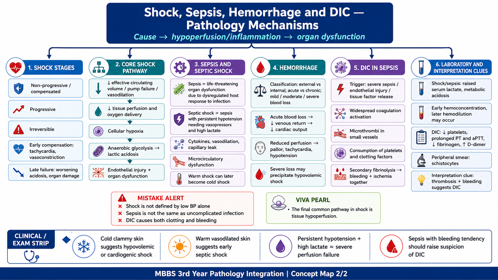
Integrated Mechanism Flow
1. Blood loss, cardiac failure, severe infection, vascular-tone loss or obstruction reduces effective circulation.
2. Tissue perfusion and oxygen delivery fall.
3. Cells shift toward anaerobic glycolysis and produce lactic acid.
4. ATP depletion causes cellular swelling, dysfunction and eventually necrosis.
5. Endothelial injury, capillary leakage and microvascular thrombosis worsen perfusion.
6. Kidney, lung, liver, heart and brain injury develops.
7. Persistent severe shock may progress to DIC, multiorgan dysfunction and irreversible injury.
Important Comparison
| Feature | Hemorrhagic shock | Septic shock | DIC |
|---|---|---|---|
| Main trigger | Acute blood loss | Severe infection with dysregulated host response | Systemic activation of coagulation, often due to sepsis |
| Main circulatory problem | Reduced circulating volume and preload | Vasodilation, capillary leakage, myocardial depression and microthrombi | Microvascular fibrin thrombi with consumption of hemostatic components |
| Typical clue | Acute bleeding, weak pulse, cold clammy skin | Infection, hypotension, raised lactate and organ dysfunction | Bleeding and organ ischemia in the same patient |
| Characteristic laboratory pattern | Hemoglobin may initially remain near normal after acute loss | Raised lactate, acidosis and evidence of organ injury | Low platelets, prolonged PT/aPTT, high D-dimer and low fibrinogen |
⭐ AIM High-Yield Review
- Shock is systemic tissue hypoperfusion causing cellular hypoxia, lactic acidosis and organ dysfunction; it is not simply a low blood-pressure reading.
- The major types are hypovolemic, cardiogenic, septic, neurogenic, anaphylactic and obstructive shock.
- Shock progresses through compensated, progressive and irreversible stages.
- Early compensation includes sympathetic activation, tachycardia, peripheral vasoconstriction, RAAS activation, ADH release and reduced urine output.
- Progressive shock creates a vicious cycle of hypoperfusion, lactic acidosis, myocardial depression and worsening hypotension.
- Prolonged shock commonly injures renal tubules, lungs, centrilobular hepatocytes, subendocardial myocardium and the brain.
- Sepsis is life-threatening organ dysfunction caused by a dysregulated host response to infection.
- Septic shock involves vasodilation, capillary leakage, endothelial injury, microvascular thrombosis, myocardial depression and metabolic dysfunction.
- Raised lactate indicates significant tissue hypoperfusion and increased anaerobic metabolism.
- Acute hemorrhage causes hypovolemic shock by reducing venous return, preload and cardiac output; chronic hemorrhage more commonly causes iron deficiency anemia.
- Early acute hemorrhage may not immediately produce a major fall in measured hemoglobin because red cells and plasma are lost together.
- DIC causes both microvascular thrombosis and bleeding because coagulation is activated while platelets and clotting factors are consumed.
- The classic DIC laboratory pattern is thrombocytopenia, prolonged PT and aPTT, raised D-dimer and reduced fibrinogen.
- Bleeding plus organ ischemia in a septic patient strongly suggests DIC.
Concept Not Clear? Ask AIM AI
Use the AIM Learning Assistant to clarify difficult concepts from this topic before proceeding to assessment.
You may ask AIM AI to:
- Explain a difficult concept in simpler words.
- Describe a mechanism step by step.
- Compare two confusing diseases, drugs or findings.
- Provide a clinically relevant example.
- Highlight a high-yield KMU examination point.
- Explain a common MCQ trap related to the topic.
Best use: Ask one focused question at a time. AIM AI supports your understanding but does not replace the complete learning material.