
This chapter follows the KMU learning outcomes and builds the topic in a logical sequence. First understand how normal esophageal movement becomes disturbed, then connect the mechanism with symptoms, investigations and management. Finish with the AIM High-Yield Review for rapid revision.
Esophageal Motility Disorders, Esophagitis, Achalasia and GERD
Understand how disturbed esophageal movement and mucosal injury produce dysphagia, heartburn, regurgitation and related complications, and learn the major investigations and management principles used in clinical practice.
Topic Introduction
The esophagus transports swallowed food from the pharynx to the stomach by coordinated muscular contractions called peristalsis. Symptoms develop when this movement is disturbed, when the lower esophageal sphincter does not function normally, or when the mucosal lining becomes inflamed. Esophageal motility disorders mainly produce dysphagia and regurgitation, while esophagitis and gastroesophageal reflux disease commonly produce heartburn and retrosternal discomfort. Achalasia is an important primary motility disorder in which the lower esophageal sphincter fails to relax normally. This chapter connects these functional abnormalities with their pathology, clinical presentation, investigations, complications and broad management principles.
A. Esophageal Motility and Major Motility Disorders
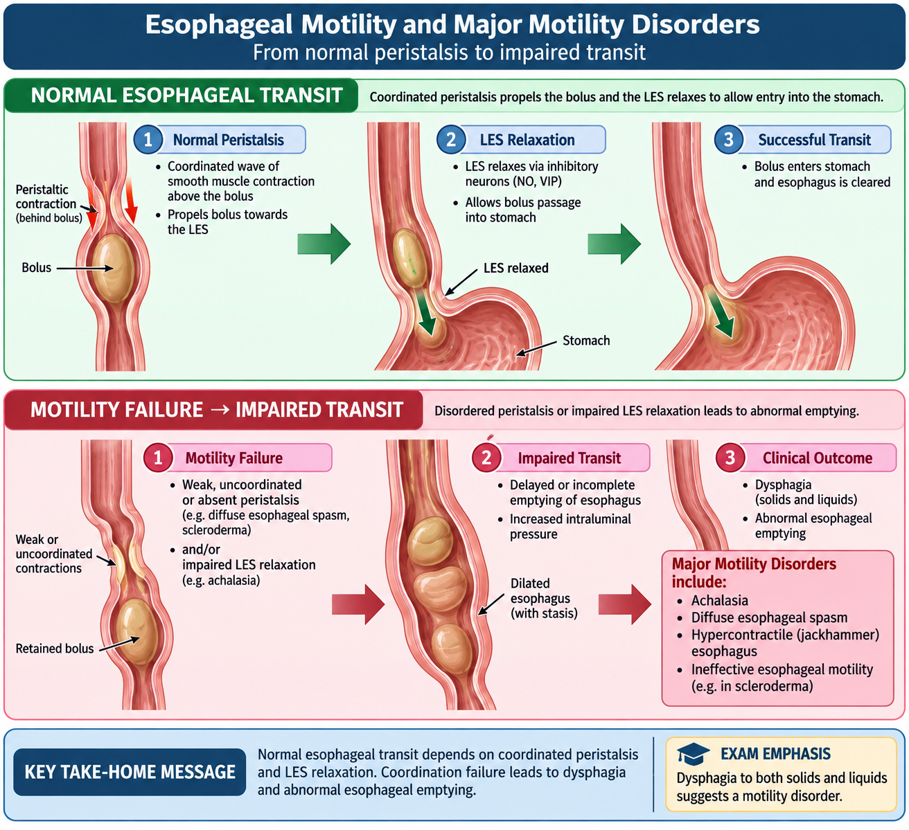
The esophagus is not simply a passive tube. A swallow initiates a coordinated wave of contraction that moves the food bolus downward. At the same time, the lower esophageal sphincter (LES) relaxes so that the bolus can enter the stomach. A motility disorder occurs when contraction of the esophageal body, relaxation of the LES, or coordination between these events becomes abnormal.
Relevant physiology
Normal swallowing depends on coordinated neural control and intact smooth muscle in the distal esophagus. Excitatory neural activity promotes contraction, while inhibitory neural activity permits relaxation. Disturbance of this balance can produce excessive contraction, ineffective contraction or failure of sphincter relaxation.
Swallowing → coordinated peristaltic wave → LES relaxation → passage of food into stomach
Causes and important types
Motility abnormalities may be primary, arising from dysfunction of esophageal neuromuscular control, or secondary to another disease that affects nerves or smooth muscle.
- Achalasia: impaired LES relaxation together with loss of normal esophageal peristalsis.
- Spastic motility disorders: poorly coordinated or excessively forceful contractions can cause intermittent dysphagia and chest pain.
- Hypomotility: weak or ineffective contractions reduce clearance of swallowed material and refluxed gastric contents.
- Secondary motility disturbance: may occur when systemic disease damages esophageal smooth muscle or its neural control.
Clinical features
The dominant symptom is usually dysphagia. Because the problem is functional rather than a fixed physical narrowing, both solids and liquids may be affected, although the exact pattern depends on the disorder. Regurgitation may occur when retained food or fluid remains in the esophagus. Some patients develop retrosternal chest pain because abnormal contractions stretch or strongly contract the esophageal wall.
Investigations
Investigation aims to determine whether the problem is due to abnormal movement or a structural obstruction.
- Upper gastrointestinal endoscopy: assesses the mucosa and helps exclude a mechanical obstruction.
- Contrast swallow: demonstrates esophageal emptying, dilatation, narrowing and abnormalities of bolus transit.
- Esophageal manometry: directly assesses contraction and sphincter function and is the key functional investigation for motility disorders.
Management principles
Management depends on the underlying functional abnormality. Treatment may aim to reduce abnormal sphincter resistance, improve passage of food, control associated reflux, or treat the underlying systemic disease. Persistent or progressive dysphagia requires proper evaluation before being labelled as a functional motility problem.
B. Esophagitis: Classification, Etiology and Pathology
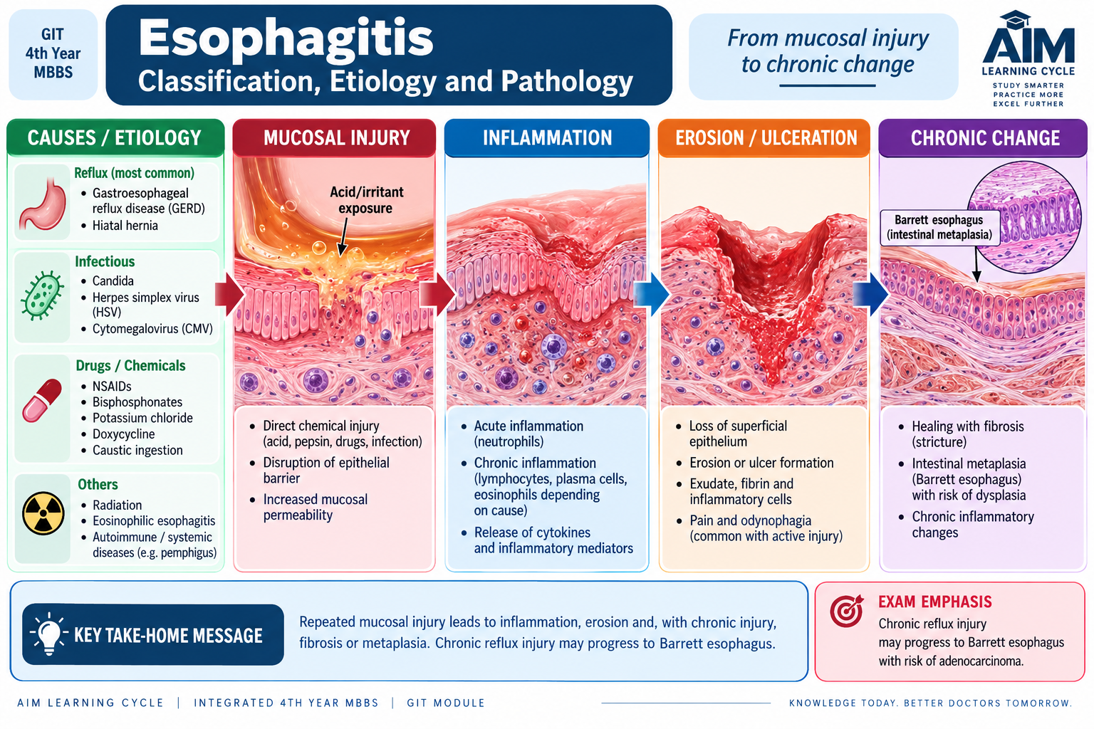
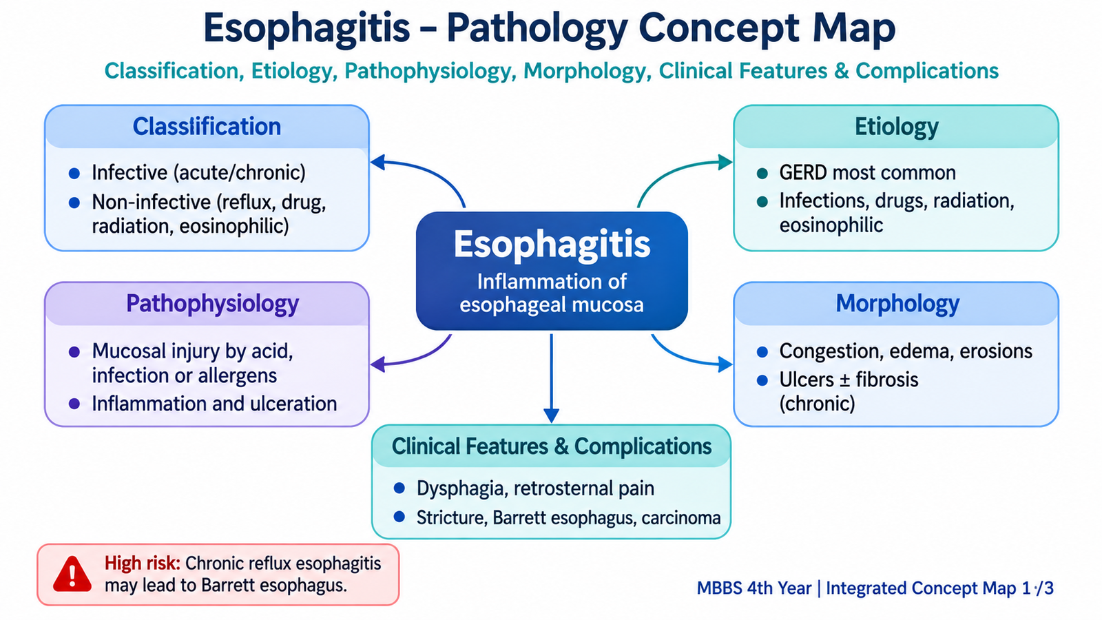
Esophagitis means inflammation and injury of the esophageal mucosa. The mucosa is normally protected from prolonged exposure to damaging substances. Injury develops when these protective mechanisms are overwhelmed by refluxed gastric contents, infection, irritating drugs or other forms of mucosal damage.
Classification and etiology
Esophagitis can be organized according to the cause of mucosal injury.
- Reflux esophagitis: caused by repeated exposure to refluxed gastric contents and is closely related to GERD.
- Infectious esophagitis: occurs particularly when host defenses are impaired.
- Drug- or chemical-associated injury: develops when an irritating substance remains in contact with the mucosa.
- Eosinophilic esophagitis: characterized by eosinophil-rich inflammation and associated with an immune-mediated pattern of mucosal injury.
Pathophysiology
Regardless of cause, persistent mucosal injury disrupts epithelial integrity and initiates inflammation. In reflux disease, repeated contact with gastric refluxate damages the distal esophageal mucosa. The inflammatory response then produces epithelial changes and may lead to ulceration or subsequent healing with fibrosis.
Damaging exposure → epithelial injury → inflammatory response → mucosal erosion or ulceration → healing, fibrosis or metaplastic change in persistent disease
Morphology
Morphological changes vary with the cause and severity. In reflux esophagitis, the distal esophagus is most affected because it lies immediately above the stomach and receives the greatest exposure to refluxed contents.
Gross morphology
- Mucosal redness and congestion may be present.
- More severe injury can produce erosions and ulceration.
- Chronic healing after deep injury may contribute to narrowing by fibrosis.
Microscopic morphology of reflux esophagitis
- Basal-zone hyperplasia: increased regenerative activity thickens the basal epithelial layer.
- Elongation of lamina propria papillae: reflects mucosal injury and repair.
- Inflammatory cells: inflammatory infiltration becomes more prominent with active mucosal damage.
Complications
Persistent inflammation can produce deeper mucosal damage. Ulceration may cause pain or bleeding. Healing by fibrosis can produce an esophageal stricture and therefore progressive dysphagia. Chronic reflux-related injury may also produce Barrett esophagus, in which the normal distal squamous lining is replaced by metaplastic columnar epithelium. This is clinically important because it is associated with an increased risk of esophageal adenocarcinoma.
C. Esophagitis: Clinical Features, Diagnosis and Management
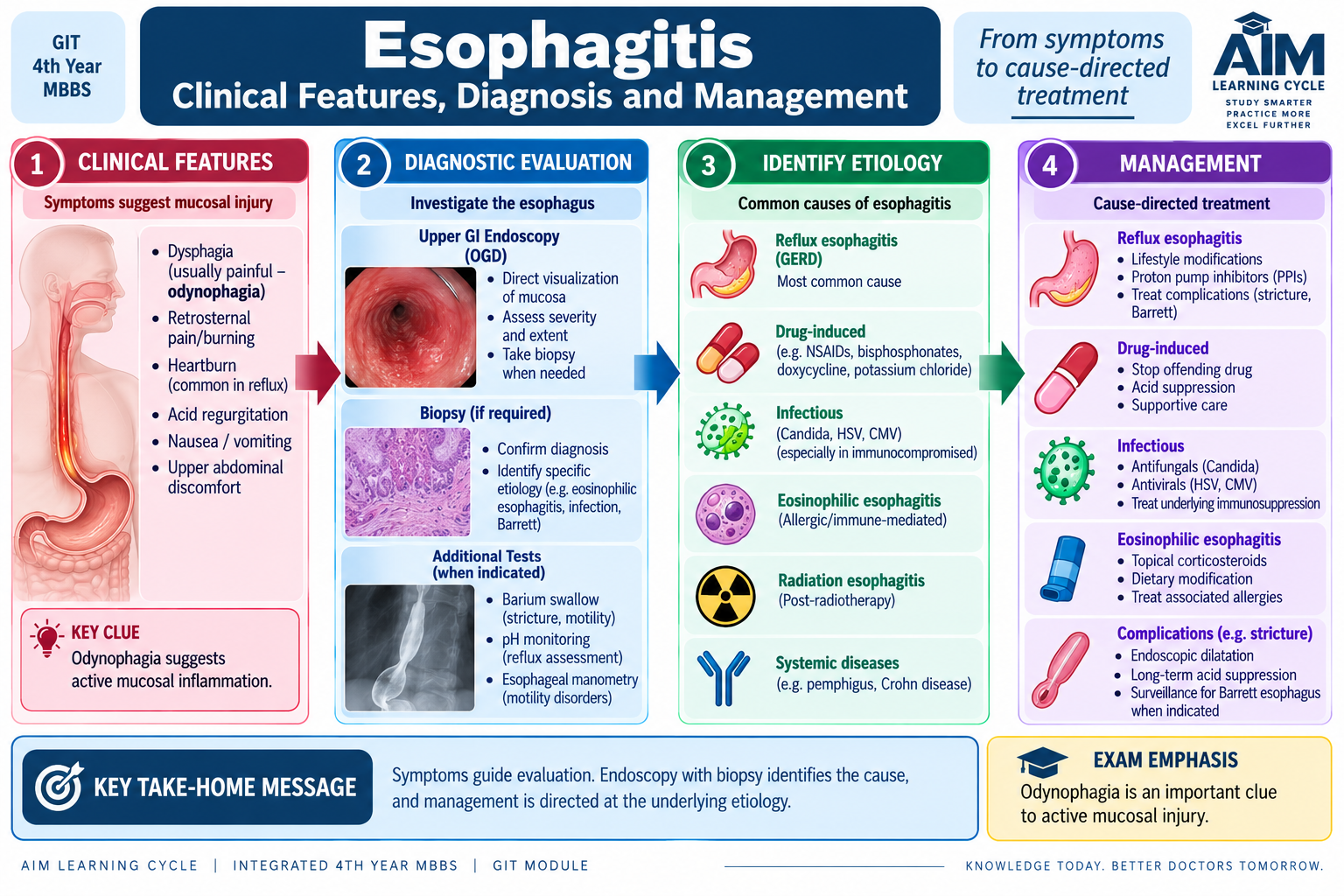
The clinical presentation of esophagitis reflects irritation or disruption of the esophageal mucosa. Symptoms therefore often appear during swallowing or after reflux exposure. The diagnostic approach combines the symptom pattern with appropriate testing to identify the cause and assess mucosal injury.
Clinical features
- Heartburn: a burning retrosternal sensation, particularly when esophagitis is reflux related.
- Odynophagia: painful swallowing occurs when inflamed or ulcerated mucosa is stretched by the food bolus.
- Dysphagia: may result from inflammation, edema, impaired motility or later fibrotic narrowing.
- Retrosternal discomfort: reflects irritation of the esophageal wall.
- Bleeding: may occur when erosions or ulcers damage mucosal vessels.
Diagnostic testing
The investigation is selected according to the suspected cause and severity.
- Upper GI endoscopy: directly assesses mucosal inflammation, erosions, ulceration, narrowing and other structural abnormalities.
- Biopsy: is useful when the cause cannot be determined from appearance alone or when a specific inflammatory pattern or metaplastic change is suspected.
- Reflux testing: may be used when reflux is suspected but the diagnosis remains uncertain.
- Contrast examination: may demonstrate narrowing or altered passage when structural complications are suspected.
Management
Management is directed at the underlying cause while reducing continuing mucosal injury. Reflux-associated inflammation is treated by controlling reflux and reducing gastric acid exposure. Infectious esophagitis requires treatment directed at the responsible infection. When medication-associated injury is suspected, the offending agent or manner of administration should be addressed where appropriate. Significant stricturing, persistent dysphagia, bleeding or diagnostic uncertainty requires further specialist assessment.
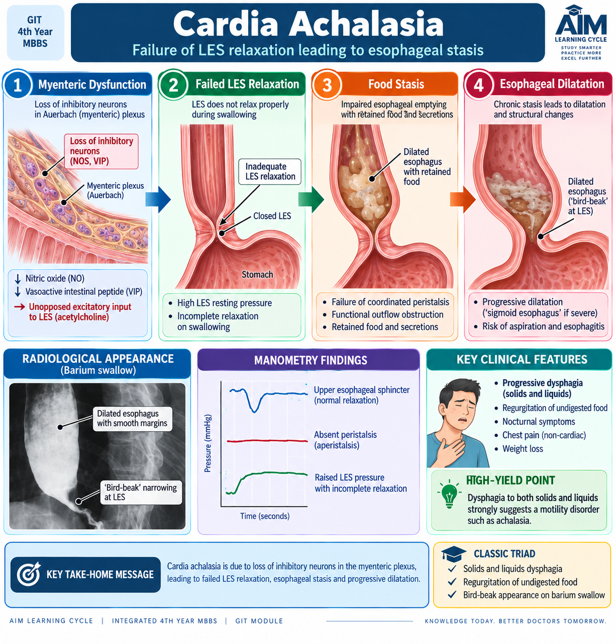
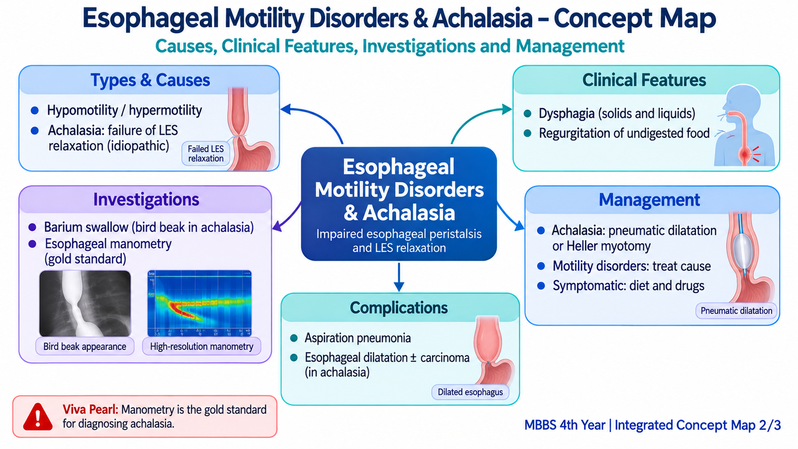
D. Cardia Achalasia
Achalasia is a primary esophageal motility disorder characterized by failure of the lower esophageal sphincter to relax adequately during swallowing and by loss of normal peristalsis in the esophageal body. Food therefore reaches the distal esophagus but cannot pass normally into the stomach. With time, retained material causes progressive dilatation of the esophagus.
Etiology and pathophysiology
The key abnormality is loss or dysfunction of inhibitory neurons in the myenteric plexus. These neurons normally help the LES relax during swallowing. Their loss leaves sphincter tone relatively unopposed and also disrupts coordinated peristalsis.
Loss of inhibitory myenteric function → impaired LES relaxation + absent effective peristalsis → obstruction to esophageal emptying → food retention → progressive esophageal dilatation
Most cases are primary. A similar clinical picture can occur when another disease damages the esophageal neural plexus or mechanically obstructs the gastroesophageal junction; this is commonly termed secondary or pseudoachalasia and must be considered during assessment.
Clinical features
- Dysphagia to both solids and liquids: both are affected because the fundamental problem is failure of coordinated transit rather than a simple fixed narrowing.
- Regurgitation of retained food: material accumulates above the poorly relaxing sphincter.
- Retrosternal discomfort: may result from distension or abnormal contraction.
- Weight loss: may develop when eating becomes progressively difficult.
- Respiratory symptoms: retained material may be regurgitated and aspirated, especially when lying down.
Investigations
Diagnosis combines structural assessment with direct measurement of esophageal function.
- Contrast swallow: may show a dilated esophagus with smooth narrowing at the gastroesophageal junction, classically described as a bird-beak appearance.
- Esophageal manometry: demonstrates impaired LES relaxation together with abnormal or absent normal peristalsis and provides the most direct functional confirmation.
- Upper GI endoscopy: helps exclude a structural lesion causing distal esophageal obstruction or pseudoachalasia.
Management
The aim is to reduce resistance at the LES so that gravity and remaining esophageal contractions can assist passage of food. Treatment does not restore the lost myenteric neurons.
- Endoscopic botulinum toxin: reduces excitatory cholinergic influence and can lower sphincter tone, although the effect may not be permanent.
- Pneumatic dilatation: mechanically reduces sphincter resistance.
- Myotomy: divides LES muscle fibers to improve emptying.
- Selected patients may require specialist endoscopic or surgical intervention according to clinical circumstances.
Complications
Long-standing food stasis can produce marked esophageal dilatation, inflammation and aspiration of retained contents. Chronic achalasia is also associated with an increased risk of esophageal carcinoma, making persistent or changing symptoms clinically important.
E. Gastroesophageal Reflux Disease (GERD)
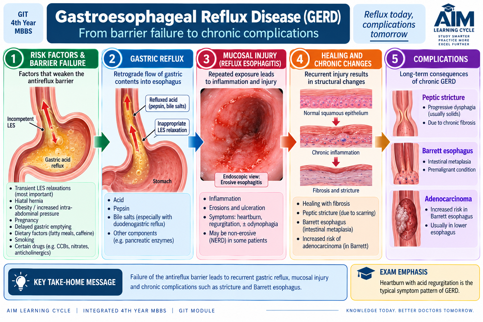
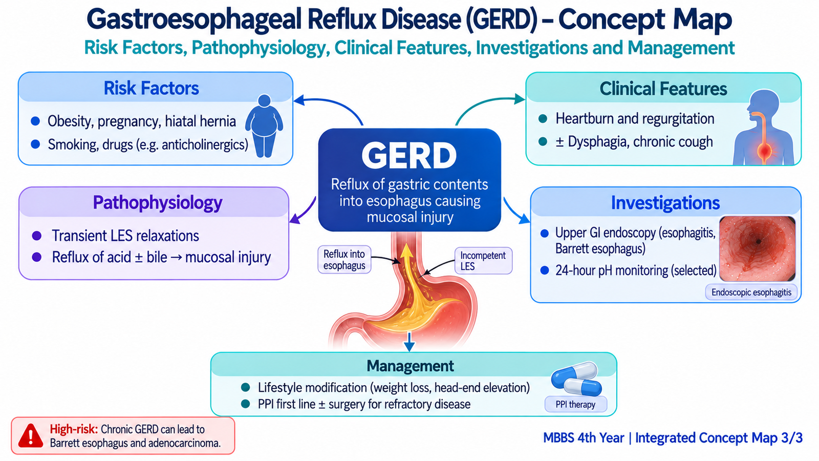
Gastroesophageal reflux disease (GERD) is a condition in which reflux of gastric contents into the esophagus produces troublesome symptoms, mucosal injury or both. Small episodes of reflux can occur physiologically, but disease develops when reflux becomes sufficiently frequent or prolonged to overwhelm the normal clearance and protective mechanisms of the esophagus.
Etiology and risk factors
The most important functional problem is failure of the normal antireflux barrier at the gastroesophageal junction. Reduced LES effectiveness allows gastric contents to move upward into the esophagus.
- Reduced or inappropriate LES competence.
- Hiatal hernia, which can reduce the effectiveness of the gastroesophageal antireflux mechanism.
- Increased intra-abdominal pressure, which promotes upward movement of gastric contents.
- Delayed gastric emptying, which increases the volume available for reflux.
- Reduced esophageal clearance, allowing refluxed material to remain in contact with the mucosa for longer.
Pathophysiology
Normal defense against reflux depends on an effective LES, rapid esophageal clearance and mucosal resistance. When these mechanisms fail, acidic gastric contents repeatedly contact the squamous mucosa. The longer the contact continues, the greater the risk of epithelial injury and reflux esophagitis.
Antireflux barrier failure → gastric reflux → prolonged mucosal exposure → symptoms ± reflux esophagitis → ulceration, stricture or Barrett change in chronic disease
Clinical features
- Heartburn: burning retrosternal discomfort caused by reflux-related mucosal irritation.
- Acid regurgitation: refluxed gastric contents may reach the throat or mouth.
- Retrosternal discomfort: may accompany reflux episodes.
- Dysphagia: requires attention because it may indicate substantial inflammation, a peptic stricture or another structural lesion.
Investigations
Many patients can be recognized from a typical symptom pattern, while investigations become important when symptoms are atypical, persistent, associated with complications, or diagnostically uncertain.
- Upper GI endoscopy: identifies reflux esophagitis, ulceration, strictures and Barrett-related mucosal change and helps exclude other structural disease.
- Ambulatory reflux monitoring: demonstrates abnormal reflux exposure when objective confirmation is required.
- Esophageal manometry: is not primarily a test for mucosal GERD but can assess esophageal motor function when a motility disorder is suspected or before selected interventions.
Complications
- Reflux esophagitis: repeated chemical injury produces inflammation.
- Ulceration and bleeding: deeper mucosal injury can disrupt vessels.
- Peptic stricture: repeated inflammation and healing can produce fibrosis and narrowing.
- Barrett esophagus: chronic injury promotes metaplastic replacement of distal squamous epithelium by columnar-type epithelium.
- Adenocarcinoma risk: Barrett esophagus is a recognized precursor state for esophageal adenocarcinoma.
Management
Treatment aims to reduce reflux exposure, allow inflamed mucosa to heal and prevent complications. Measures that reduce reflux-provoking circumstances may help appropriate patients. Acid suppression, particularly with proton-pump inhibitors, reduces the acidity of refluxed gastric contents and therefore decreases continued chemical injury to the esophageal mucosa. Patients with persistent symptoms, complications or selected mechanical problems may require specialist evaluation and antireflux intervention.
F. Clinical Differentiation and Diagnostic Approach
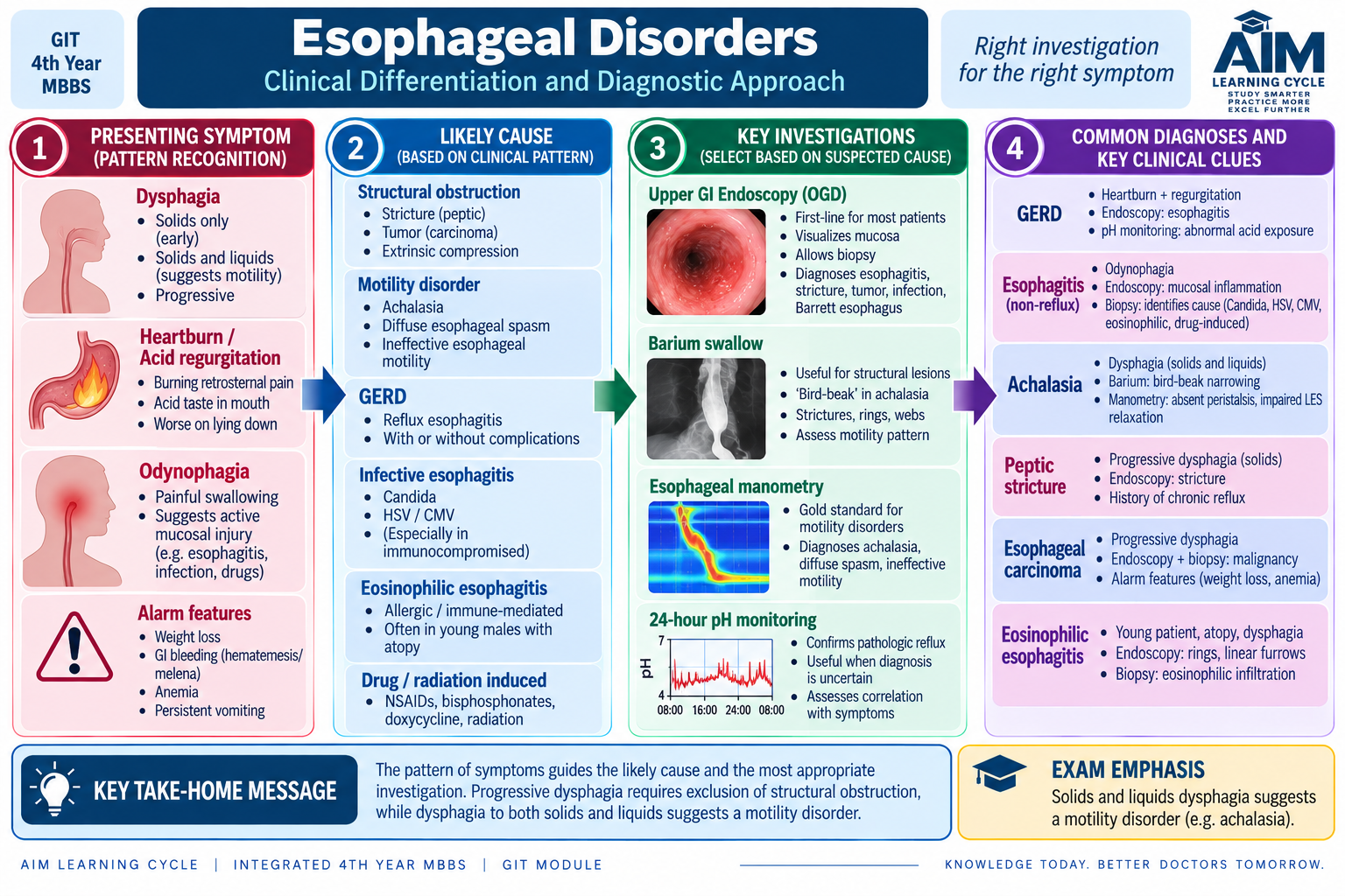
Dysphagia, heartburn, chest discomfort and regurgitation overlap among esophageal disorders, so the pattern of symptoms helps decide which investigation is most useful. A motility disorder should be suspected when swallowing difficulty involves both solids and liquids from an early stage. A reflux-related disorder is more likely when heartburn and acid regurgitation dominate. Painful swallowing suggests active mucosal inflammation or ulceration.
Symptom-to-investigation reasoning
- Solids and liquids both difficult: consider impaired motility; manometry becomes particularly important.
- Heartburn and acid regurgitation: consider GERD; endoscopy or objective reflux testing may be required depending on the clinical situation.
- Odynophagia: consider active mucosal disease; endoscopic assessment is useful when significant disease is suspected.
- Progressive dysphagia: structural obstruction must be excluded rather than assuming a benign motility disorder.
- Regurgitation of undigested retained food: strongly supports esophageal stasis, as occurs in achalasia.
| Feature | Achalasia | GERD | Esophagitis |
|---|---|---|---|
| Central problem | Failed LES relaxation and absent effective peristalsis | Pathological reflux of gastric contents | Inflammation and injury of esophageal mucosa |
| Typical symptom emphasis | Dysphagia to solids and liquids, regurgitation | Heartburn and acid regurgitation | Odynophagia, heartburn or dysphagia |
| Most characteristic investigation | Manometry | Reflux assessment when objective confirmation is needed | Endoscopy ± biopsy |
| Important consequence | Stasis, dilatation and aspiration | Reflux esophagitis and chronic complications | Ulceration, bleeding or stricture |
Integrated Mechanism Flow
Neuromuscular dysfunction or antireflux failure
Abnormal transit or gastric reflux
Food stasis or mucosal exposure
Dysphagia, regurgitation, heartburn or odynophagia
Functional testing or mucosal assessment
Cause-directed management and complication prevention
⭐ AIM High-Yield Review
Esophageal Motility Disorders, Achalasia, GERD & Esophagitis
Watch this video after reading the learning material to reinforce esophageal motility disorders, achalasia, gastroesophageal reflux disease and reflux esophagitis.