
14. Gallstone Disease, Cholecystitis and Gallbladder Carcinoma
Understand how gallstones form, how obstruction produces biliary pain and gallbladder inflammation, how acute and chronic cholecystitis differ, and how long-standing gallbladder disease relates to carcinoma.
Topic Introduction
Gallstone disease is one of the commonest disorders of the gallbladder. Stones develop when components of bile precipitate and form solid material. Many stones remain silent, but obstruction of the cystic duct can produce biliary pain or inflammation of the gallbladder. Acute cholecystitis usually develops after sudden cystic-duct obstruction, whereas chronic cholecystitis results from repeated or persistent injury and fibrosis. Long-standing gallstone-associated inflammation is also an important background risk for gallbladder carcinoma. In this chapter, you will connect stone formation with morphology, clinical presentation, investigations, complications and surgical management, and then study the pathological basis, staging and clinical consequences of gallbladder carcinoma.
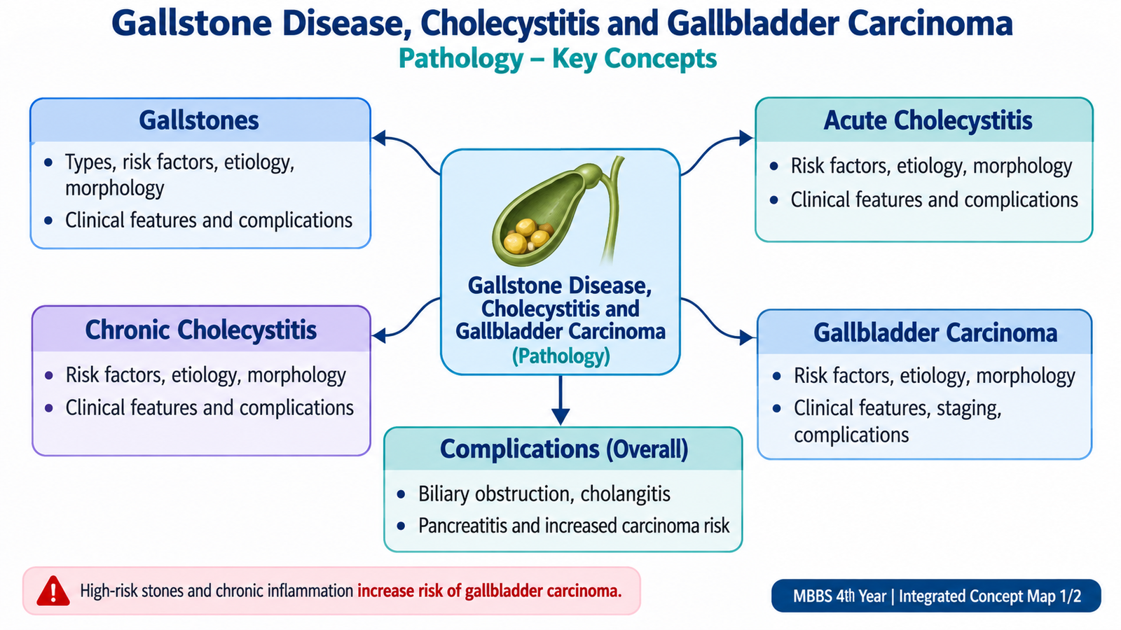
A. Gallstones: Types, Causes and Formation
Gallstones, or cholelithiasis, are solid concretions that form from components of bile. Most develop within the gallbladder, although stones may also occur within the bile ducts. Their formation reflects an imbalance between the substances normally kept dissolved in bile and the factors that permit those substances to precipitate, grow and remain within the biliary system.
Types of gallstones
The major distinction is between cholesterol stones and pigment stones. Pigment stones are further divided into black and brown stones because their formation occurs in different settings.
- Cholesterol stones: contain predominantly cholesterol together with variable amounts of calcium salts, bile pigments and proteins. They are usually pale yellow to yellow-green.
- Black pigment stones: contain calcium salts of unconjugated bilirubin and are typically small, hard and brittle. They are particularly associated with increased bilirubin load, such as chronic hemolysis, and with cirrhosis.
- Brown pigment stones: are soft, greasy stones that tend to form within bile ducts where there is biliary stasis and infection.
How cholesterol stones form
Cholesterol is poorly soluble in water. Bile normally keeps cholesterol dissolved through bile salts and phospholipids. Stones become more likely when bile contains too much cholesterol for these solubilizing substances to handle.
Gallbladder hypomotility contributes because bile remains in the gallbladder for longer, giving crystals time to enlarge and aggregate. Mucin secreted by the gallbladder mucosa can provide a matrix on which crystals accumulate.
Important risk factors
Risk factors are understandable when related to the mechanism of stone formation. Factors that increase cholesterol secretion into bile, reduce the bile-salt pool, or reduce gallbladder emptying favour cholesterol stones.
- Increasing age.
- Female sex and estrogenic influences.
- Obesity and metabolic factors that increase biliary cholesterol secretion.
- Rapid weight loss, which mobilizes cholesterol and may impair gallbladder emptying.
- Pregnancy, because hormonal changes increase cholesterol saturation and reduce gallbladder motility.
- Ileal disease or ileal resection, which reduces bile-salt reabsorption and decreases the bile-salt pool.
- Chronic hemolysis, especially for black pigment stones, because increased bilirubin turnover increases the amount of bilirubin entering bile.
- Biliary stasis and infection, especially for brown pigment stones.
Morphology
Gallstones may be solitary or multiple. Multiple stones may develop faceted surfaces because they press against one another within the gallbladder. Cholesterol stones are generally pale, while pigment stones are dark brown or black. Their clinical importance depends less on their appearance than on whether they migrate and obstruct part of the biliary tract.

B. Gallstone Disease: Presentation, Investigations, Treatment and Complications
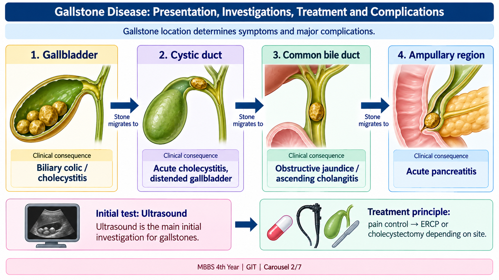
The clinical behaviour of gallstones depends mainly on their location and whether they obstruct bile flow. A stone lying freely inside the gallbladder may cause no symptoms. Symptoms usually begin when a stone obstructs the cystic duct or migrates into the common bile duct.
Clinical features
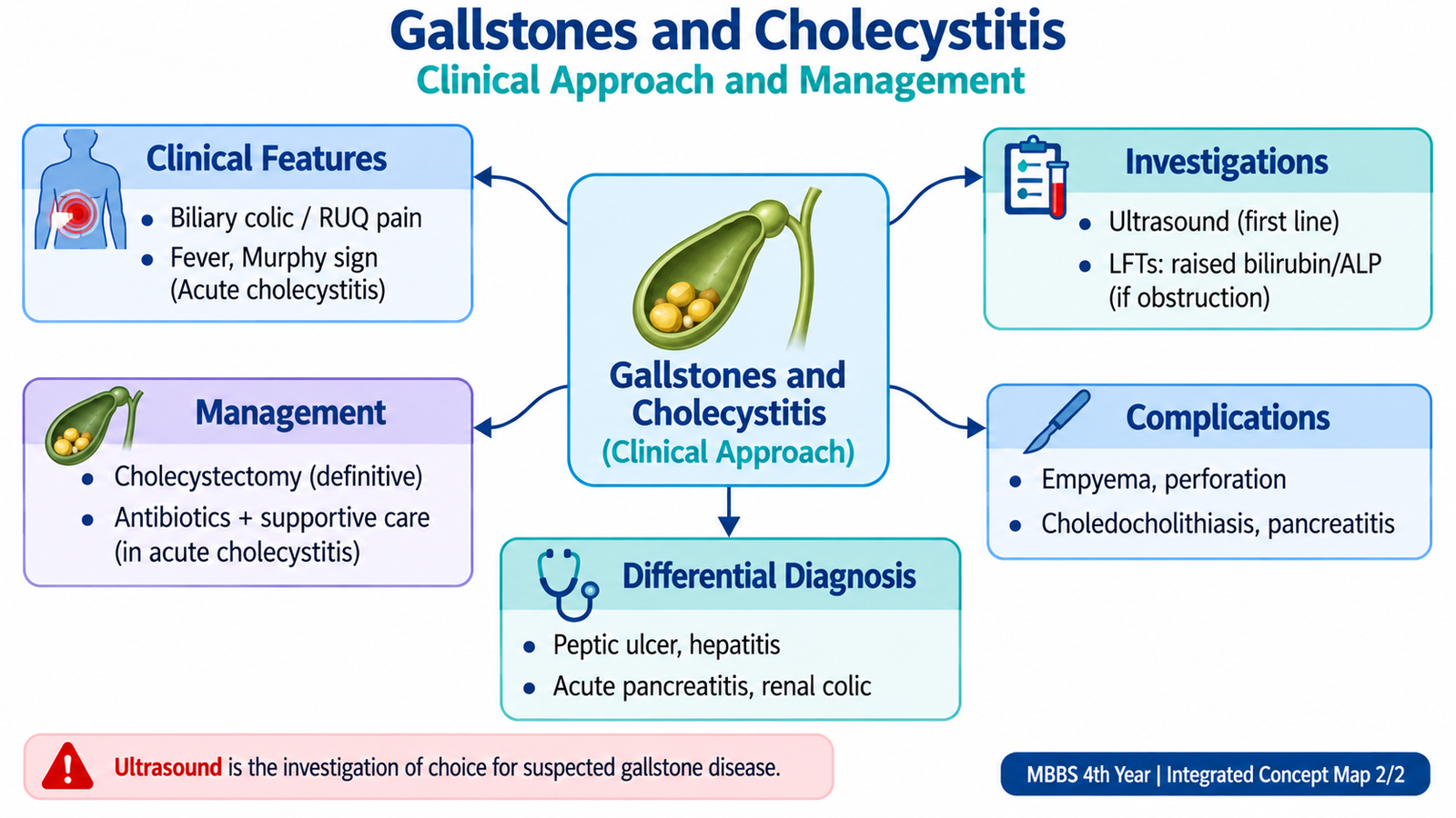
Many gallstones are found incidentally. When a stone transiently blocks the cystic duct, the gallbladder contracts against the obstruction and produces biliary colic. Despite the word “colic,” the pain is usually steady rather than rapidly fluctuating.
- Right upper-quadrant or epigastric pain.
- Pain may radiate toward the right scapular or shoulder region.
- Nausea and vomiting may accompany the pain.
- Symptoms may occur after a meal because gallbladder contraction increases against an obstructed outlet.
- Simple biliary colic does not usually produce persistent fever or marked inflammatory signs.
Investigations
Ultrasonography is the main initial imaging investigation because it can identify gallstones and assess the gallbladder and biliary tract without exposing the patient to radiation.
- Ultrasound: stones appear as echogenic structures and usually produce posterior acoustic shadowing.
- Liver-function tests: uncomplicated gallbladder stones may have normal results. A cholestatic pattern raises suspicion of common bile-duct obstruction.
- MRCP or endoscopic ultrasound: may be used when a common bile-duct stone is suspected but not adequately demonstrated by routine ultrasound.
- ERCP: is especially valuable when a duct stone needs therapeutic removal rather than simply diagnostic imaging.
Treatment principles
Treatment depends on whether stones are asymptomatic, symptomatic or complicated. The important surgical principle is that gallbladder stones causing recurrent symptoms are usually treated by removal of the gallbladder rather than repeated symptomatic treatment alone.
- Asymptomatic stones: most patients do not require routine cholecystectomy simply because an incidental stone is present.
- Symptomatic gallstones: laparoscopic cholecystectomy is the standard definitive treatment for suitable patients.
- Common bile-duct stones: require clearance of the duct, commonly by an endoscopic or surgical approach, together with management of the gallbladder source.
Complications
A stone becomes clinically dangerous when it obstructs, inflames, infects or erodes into surrounding structures.
- Acute cholecystitis: prolonged cystic-duct obstruction causes gallbladder inflammation.
- Choledocholithiasis: a stone enters the common bile duct.
- Obstructive jaundice: develops when bile flow through the extrahepatic biliary system is blocked.
- Ascending cholangitis: biliary obstruction permits infection of the biliary tree.
- Acute pancreatitis: a migrating stone may obstruct the distal biliary-pancreatic outflow region.
- Cholecystoenteric fistula and gallstone ileus: chronic inflammation may allow a large stone to erode into bowel and later produce intestinal obstruction.
C. Acute Cholecystitis: Etiology, Pathogenesis and Morphology
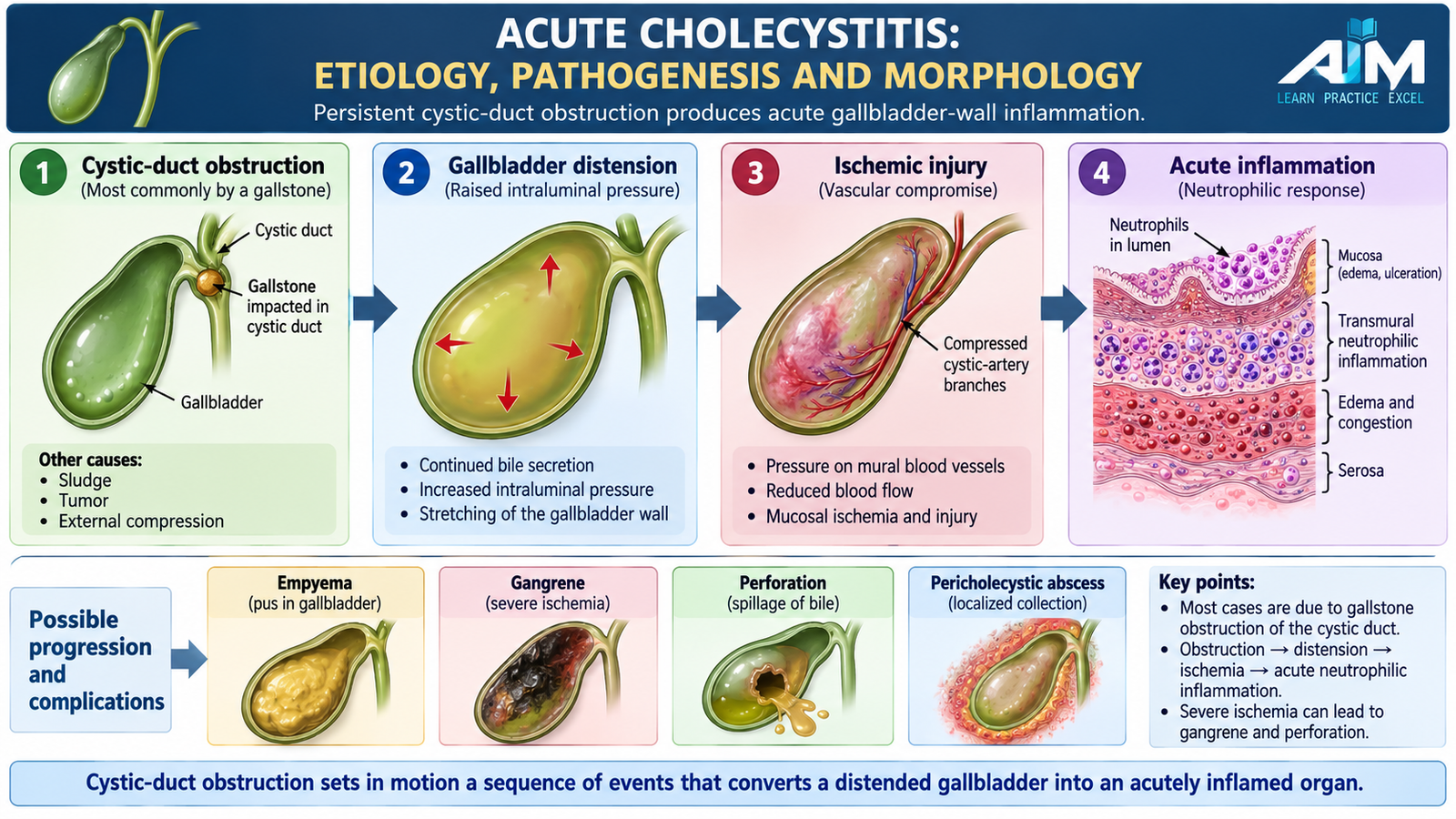
Acute cholecystitis is acute inflammation of the gallbladder. In most patients it is associated with a gallstone obstructing the cystic duct or gallbladder neck. Obstruction causes bile retention, distension and chemical injury to the gallbladder wall. The resulting inflammation may later be complicated by bacterial infection.
Calculous acute cholecystitis
This is the common form. A stone becomes impacted in the cystic duct or gallbladder neck. Continued secretion into an obstructed gallbladder increases intraluminal pressure and causes distension. The distended wall develops impaired venous and lymphatic drainage, followed by reduced blood flow and inflammatory injury.
Bacteria may be present in some cases, but the initial event is commonly mechanical and chemical rather than primary infection. This explains why obstruction itself can produce severe inflammation.
Acalculous acute cholecystitis
Acalculous cholecystitis occurs without gallstones and is particularly associated with severely ill patients. Gallbladder stasis, ischemia and systemic inflammation can damage the wall even when no stone is present.
Important settings include severe systemic illness, major trauma, burns, sepsis and prolonged critical care. Because these patients may already be very ill, the abdominal findings can be less obvious and the condition may progress rapidly.
Morphology
Gross morphology
- The gallbladder is usually enlarged and tense.
- The wall becomes thickened because of edema and inflammation.
- The serosal surface may appear congested and may be covered by fibrinous or fibrinopurulent exudate.
- Severe ischemic injury may produce dark areas of hemorrhage or gangrenous necrosis.
- The lumen may contain cloudy bile, pus or inflammatory debris.
Microscopic morphology
- Edema and vascular congestion of the gallbladder wall.
- Prominent acute inflammatory infiltration, particularly neutrophils.
- Mucosal ulceration or necrosis in more severe disease.
- Transmural necrosis may develop when vascular compromise is marked.
The morphological changes explain the clinical picture: wall inflammation causes localized tenderness, while tissue injury releases inflammatory mediators that produce fever and systemic inflammatory responses.
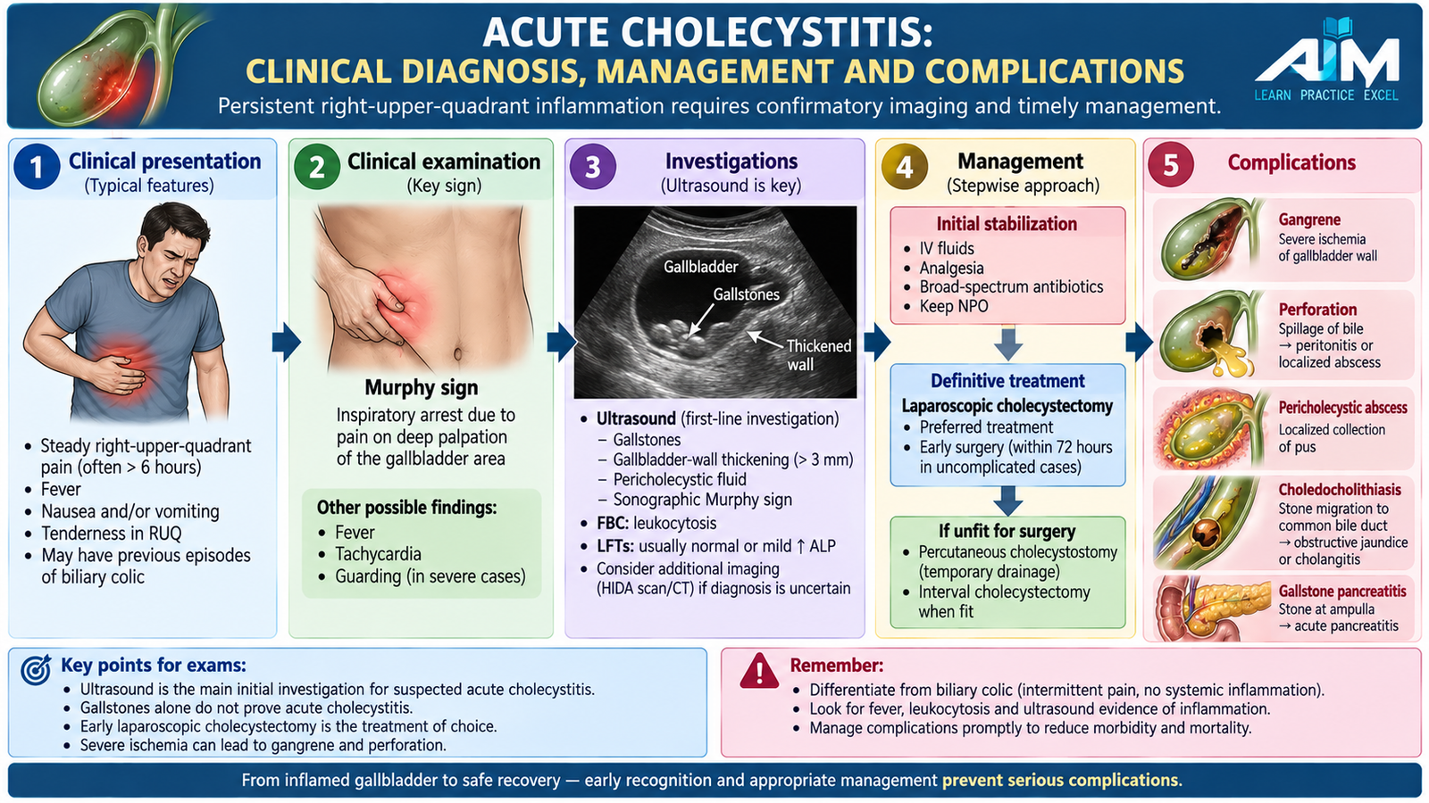
D. Acute Cholecystitis: Clinical Diagnosis, Management and Complications
Acute cholecystitis should be suspected when gallstone-type pain becomes persistent and is accompanied by localized inflammatory signs. The clinical diagnosis is strengthened by supportive laboratory findings and characteristic ultrasound appearances.
Clinical presentation
- Persistent right upper-quadrant or epigastric pain.
- Fever and malaise.
- Nausea and vomiting.
- Right upper-quadrant tenderness and guarding.
- Murphy sign: inspiration is suddenly interrupted because palpation beneath the right costal margin brings the inflamed gallbladder against the examiner’s hand and produces pain.
The key distinction from simple biliary colic is the presence of sustained inflammation. Fever, localized tenderness and inflammatory laboratory abnormalities therefore support acute cholecystitis rather than transient uncomplicated obstruction.
Investigations
Investigations confirm gallbladder inflammation, identify the obstructing stone and look for complications or associated bile-duct obstruction.
- Complete blood count: leukocytosis, often with neutrophilia, supports an acute inflammatory process.
- Liver-function tests: may be normal or mildly abnormal. Marked cholestatic abnormalities should raise suspicion of associated common bile-duct obstruction.
- Ultrasound: may demonstrate stones, gallbladder-wall thickening, distension, pericholecystic fluid and a sonographic Murphy sign.
- Hepatobiliary scintigraphy: may be useful when ultrasound is inconclusive and persistent cystic-duct obstruction remains suspected.
- CT: may help when complications such as perforation, abscess or another abdominal diagnosis are being considered.
Management
Initial management controls pain, maintains hydration and treats systemic illness while definitive gallbladder treatment is planned. The aim is not only to settle the current inflammatory episode but also to remove the diseased gallbladder when appropriate so recurrent obstruction does not continue.
- Temporary restriction of oral intake during the acute phase.
- Intravenous fluids when required.
- Analgesia and antiemetic treatment.
- Antibiotic therapy when bacterial infection or significant systemic inflammatory illness is suspected.
- Laparoscopic cholecystectomy is the definitive treatment for suitable patients once appropriately assessed and stabilized.
- Percutaneous gallbladder drainage may be considered in selected severely ill patients who cannot safely undergo definitive surgery initially.
Complications
- Empyema: infected purulent material accumulates in the gallbladder.
- Gangrene: severe ischemia causes necrosis of the gallbladder wall.
- Perforation: necrotic wall ruptures, producing localized abscess or generalized peritoneal contamination.
- Sepsis: severe infection may produce systemic deterioration.
- Fistula formation: persistent inflammation may cause adhesion and erosion into adjacent bowel.
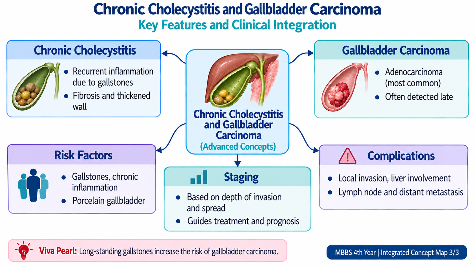
E. Chronic Cholecystitis: Pathology, Clinical Features and Management
Chronic cholecystitis is long-standing inflammation and fibrosis of the gallbladder, usually occurring in association with gallstones. It reflects repeated mechanical irritation, recurrent episodes of inflammation or persistent low-grade injury. Unlike acute cholecystitis, the dominant pathological process is fibrosis and chronic inflammatory change rather than neutrophil-rich acute inflammation.
Etiology and pathogenesis
Most affected gallbladders contain stones. Repeated obstruction and mucosal injury stimulate chronic inflammation and repair. Over time, collagen deposition causes thickening and fibrosis of the wall, while mucosal epithelium may become atrophic or grow downward into the wall.
Morphology
Gross morphology
- The gallbladder may be contracted or normal in size.
- The wall is commonly thickened and fibrotic.
- Gallstones are commonly present.
- The mucosa may appear flattened or irregular because of chronic injury.
Microscopic morphology
- Variable chronic inflammatory infiltrates, including lymphocytes and plasma cells.
- Fibrosis and thickening of the gallbladder wall.
- Mucosal atrophy may occur.
- Rokitansky–Aschoff sinuses: herniations of gallbladder mucosa extending into the muscular wall. They develop as mucosa pushes into areas of wall weakness during chronic injury and increased intraluminal pressure.
Clinical features
Patients commonly have recurrent episodes of biliary-type pain rather than the intense inflammatory illness of acute cholecystitis. Symptoms may be intermittent because obstruction can occur and then resolve.
- Recurrent right upper-quadrant or epigastric pain.
- Nausea or discomfort associated with meals in some patients.
- Previous episodes resembling biliary colic or acute cholecystitis.
- Persistent fever and marked systemic inflammation are not typical unless an acute episode is superimposed.
Investigations and treatment
Ultrasound commonly demonstrates gallstones and may show a contracted or thick-walled gallbladder. Laboratory tests may be normal between attacks because the disease is primarily localized to the gallbladder.
For symptomatic chronic gallstone disease, laparoscopic cholecystectomy provides definitive treatment by removing the gallbladder that contains the stones and has undergone chronic inflammatory change.
Complications
- Recurrent acute cholecystitis.
- Dense fibrosis and adhesions around the gallbladder.
- Cholecystoenteric fistula with possible gallstone ileus.
- Dystrophic calcification of the gallbladder wall, producing a porcelain gallbladder pattern.
- Long-standing gallstone-associated inflammation is an important background risk for gallbladder carcinoma.

F. Gallbladder Carcinoma: Risk Factors, Pathogenesis and Morphology
Gallbladder carcinoma is a malignant epithelial tumor arising from the gallbladder mucosa. Most cases are adenocarcinomas. The major pathological theme is long-standing mucosal injury and inflammation, which may promote progressive epithelial alteration and malignant transformation.
Risk factors and etiology
No single factor causes every case. However, chronic inflammatory injury is a common background feature, which explains the strong association with long-standing gallstone disease.
- Gallstones and chronic cholecystitis: repeated mechanical and inflammatory injury produces a long-standing abnormal mucosal environment.
- Porcelain gallbladder: chronic inflammation with calcification of the gallbladder wall is recognized as a risk-associated condition.
- Gallbladder epithelial polyps or adenomatous lesions: some may provide a pathway toward dysplasia and malignancy.
- Congenital abnormalities of the pancreatobiliary junction: abnormal reflux of pancreatic secretions may chronically damage biliary epithelium.
- Chronic biliary infection or carrier states: prolonged inflammation may contribute in susceptible individuals.
Pathogenesis
In many cases, repeated mucosal injury is followed by epithelial metaplasia and dysplasia. Dysplastic epithelium may then progress to invasive carcinoma. The process is therefore best understood as a sequence of chronic injury and abnormal epithelial repair rather than as a sudden event.
Gross morphology
Gallbladder carcinoma can grow either as a diffuse infiltrating lesion that thickens and stiffens the wall or as an exophytic mass projecting into the lumen. The infiltrating pattern can be difficult to distinguish grossly from severe chronic inflammatory thickening.
- Irregular thickening and induration of the gallbladder wall.
- Firm infiltrative areas or a polypoid/fungating mass.
- Ulceration of the mucosal surface may occur.
- Direct extension into the adjacent liver may occur relatively early because the gallbladder wall is thin and directly related to the hepatic bed.
Microscopic morphology
Most tumors are adenocarcinomas forming malignant glands within a fibrous, often desmoplastic stroma. The degree of gland formation varies with differentiation. Tumor may invade progressively through the wall into surrounding soft tissue, liver and adjacent structures.

G. Gallbladder Carcinoma: Clinical Features, Staging and Complications
Gallbladder carcinoma is often clinically silent during its early stages. Early symptoms may resemble ordinary gallstone disease, so the tumor may be discovered unexpectedly after cholecystectomy. Symptoms become clearer when the tumor invades the liver, obstructs the biliary system or spreads beyond the gallbladder.
Clinical features
- Persistent right upper-quadrant or upper abdominal pain.
- Loss of appetite and weight loss.
- Nausea or general constitutional symptoms.
- Jaundice when tumor involves or obstructs the biliary tract.
- Hepatomegaly or an upper abdominal mass may appear in advanced disease.
- Some tumors are detected incidentally during pathological examination of a removed gallbladder.
Staging principle
Gallbladder carcinoma is staged by the TNM system. The most important concept for undergraduate learning is that increasing depth of invasion through the gallbladder wall, involvement of regional lymph nodes and distant spread indicate progressively more advanced disease.
- Tis: carcinoma in situ, confined to the epithelium.
- T1: invasion remains within the inner layers of the gallbladder wall; T1a reaches the lamina propria and T1b reaches the muscle layer.
- T2: tumor extends into the perimuscular connective tissue but has not penetrated the serosal surface or directly invaded the liver.
- T3: tumor perforates the serosa and/or directly invades the liver or one adjacent organ or structure.
- T4: very advanced local disease involving major vascular structures or multiple adjacent extrahepatic organs.
- N category: describes regional lymph-node metastasis.
- M category: identifies distant metastasis.
The reason depth matters is anatomical: the gallbladder has a relatively thin wall and lacks a protective submucosal layer. Once carcinoma passes through the wall, it can gain access to adjacent liver tissue and lymphatic pathways.
Assessment of extent
Ultrasound may first demonstrate an abnormal mass, irregular wall thickening or associated biliary obstruction. Cross-sectional imaging such as CT or MRI is important for assessing local extension, liver involvement, lymph nodes and distant disease. Histopathology establishes the tumor type and depth of invasion when tissue is available.
Complications
- Liver invasion: occurs because of the close anatomical relationship between the gallbladder and hepatic bed.
- Biliary obstruction: produces obstructive jaundice when the tumor compromises major bile ducts.
- Regional lymph-node spread.
- Peritoneal dissemination.
- Distant metastasis in advanced disease.

Important Comparison: Biliary Colic, Acute Cholecystitis and Chronic Cholecystitis
| Feature | Biliary Colic | Acute Cholecystitis | Chronic Cholecystitis |
|---|---|---|---|
| Main process | Transient cystic-duct obstruction | Persistent obstruction with acute inflammation | Repeated injury with chronic inflammation and fibrosis |
| Pain | Episodic RUQ/epigastric pain | Persistent RUQ pain | Recurrent biliary-type pain |
| Fever | Usually absent | Common | Usually absent between attacks |
| Murphy sign | Usually absent | Often positive | Usually not prominent |
| Dominant pathology | No established wall inflammation | Edema, congestion and neutrophils | Fibrosis, chronic inflammatory cells and Rokitansky–Aschoff sinuses |
| Definitive principle | Cholecystectomy for recurrent symptomatic disease | Stabilization followed by definitive gallbladder treatment | Cholecystectomy for symptomatic disease |
Integrated Mechanism Flow
⭐ AIM High-Yield Review
Gallstones (Cholelithiasis)
Watch this visual explanation to reinforce gallstone formation, types, pathogenesis and important clinical concepts.