
This chapter follows the supplied KMU learning outcomes and builds the organisms, disease mechanisms, drug actions and prevention principles in a logical sequence. First understand how each parasite reaches and damages the host; then use the final high-yield review for revision.
18. Intestinal Protozoal and Helminthic Infections: Pathogenesis, Treatment and Control
GIT Module — Intestinal parasites, their life cycles and disease mechanisms, antiparasitic pharmacology, and epidemiological prevention and control.
Topic Introduction
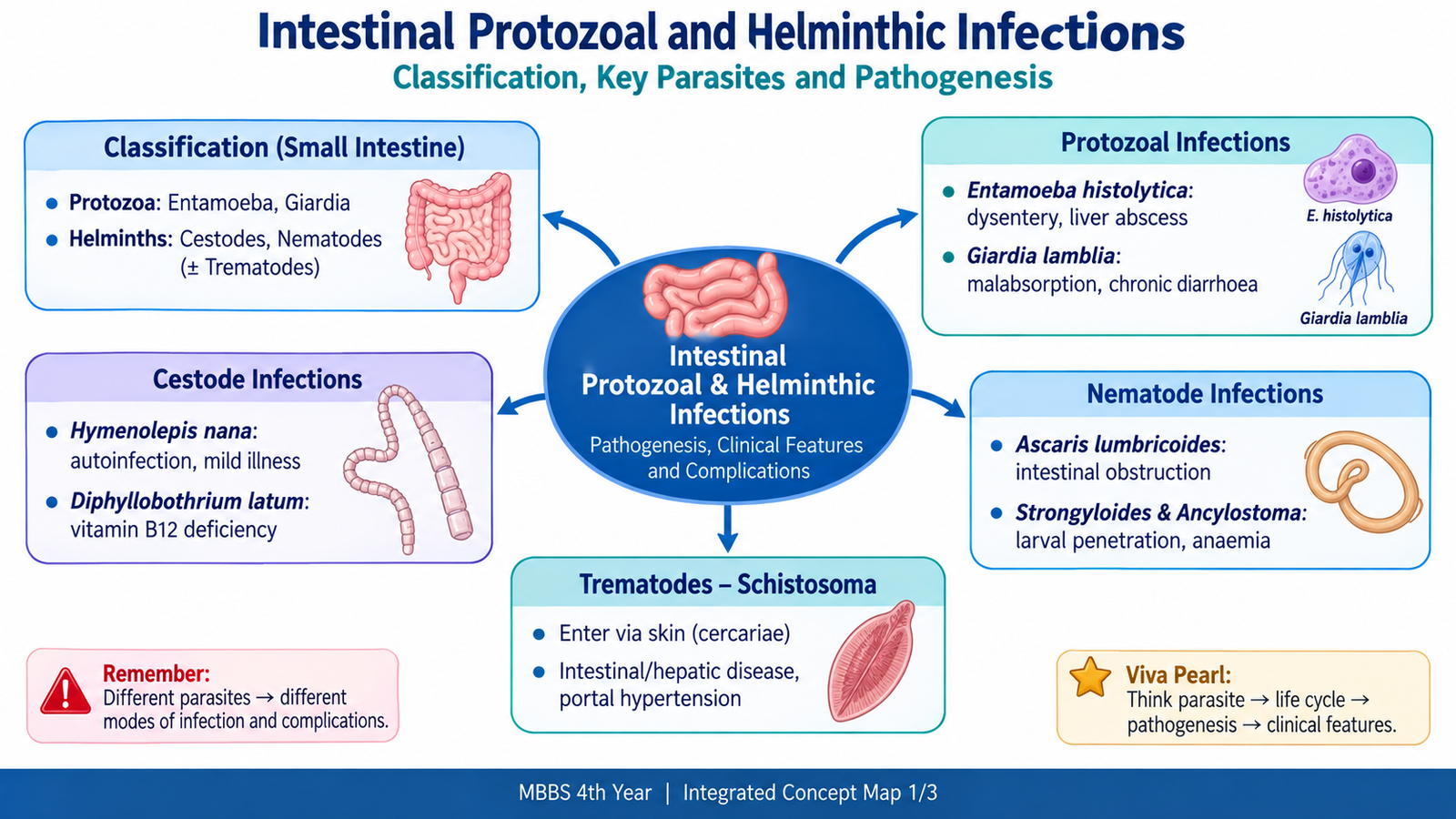
Intestinal parasitic infections are caused by protozoa and helminths that reach the gastrointestinal tract or related blood vessels through contaminated food or water, skin penetration, or infected food sources. Different parasites produce disease in different ways. Some invade intestinal mucosa, some attach to the surface and interfere with absorption, some remove blood or nutrients, and others cause inflammation when their eggs become trapped in tissues. This chapter explains the major intestinal protozoa and worms included in the curriculum, with emphasis on their life cycles, morphology, pathogenesis, clinical effects and complications. It then connects these mechanisms with anti-amoebic and anthelmintic drug actions and with practical community measures that interrupt transmission.
A. Intestinal Parasites: Core Classification and Transmission
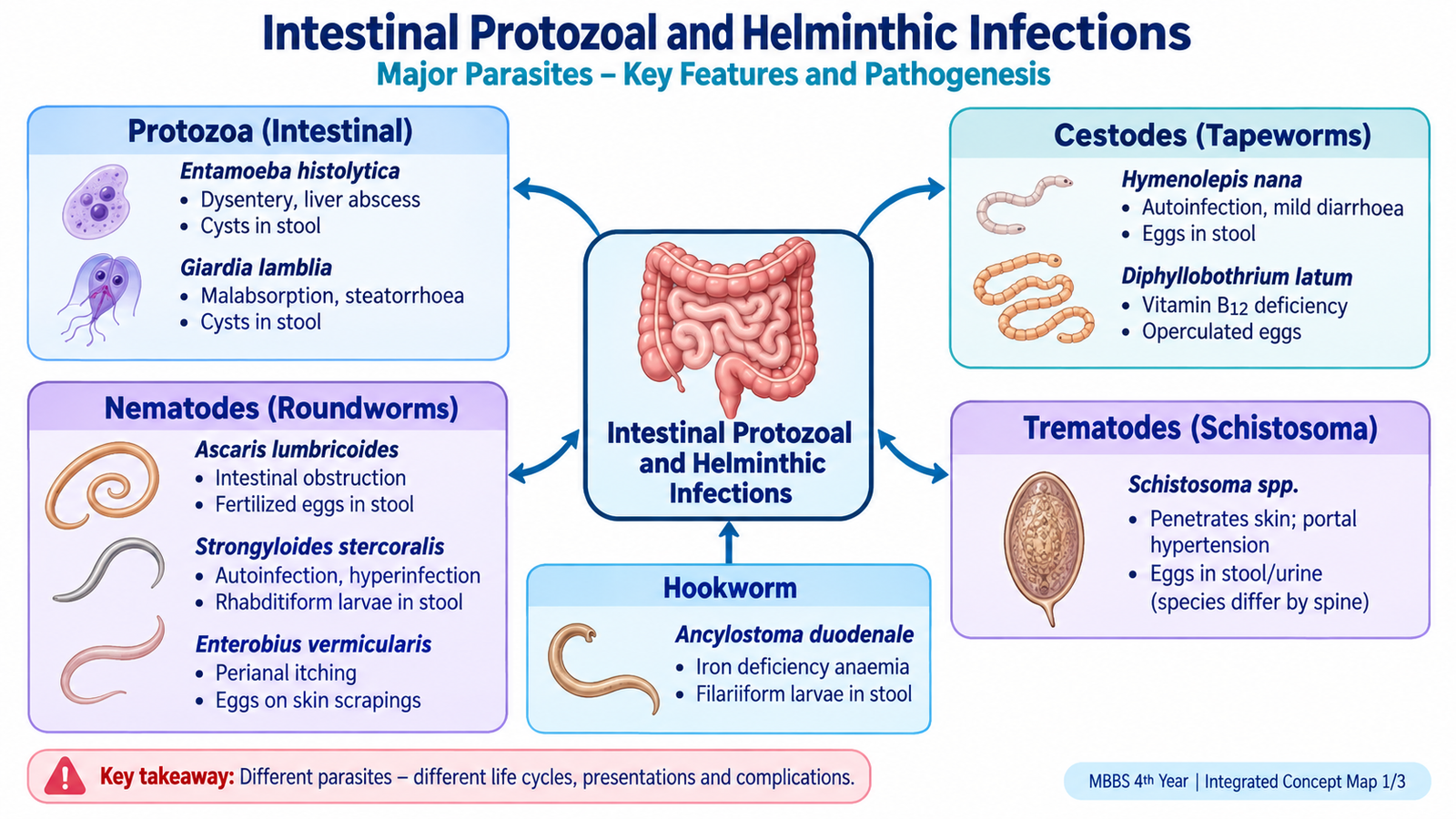
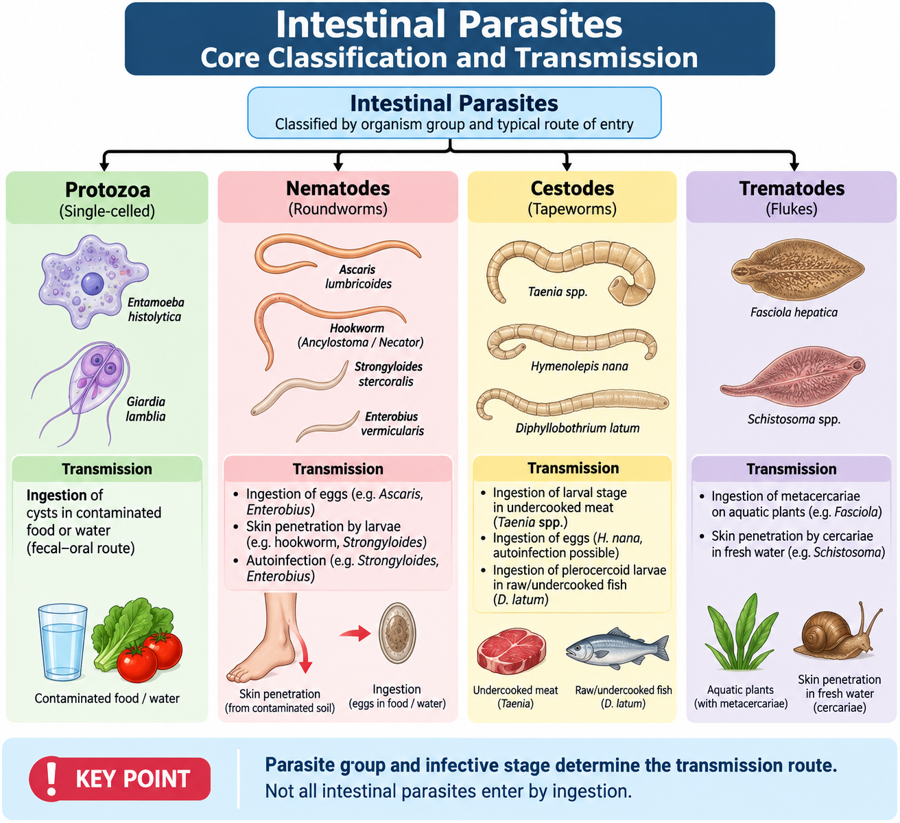
Intestinal parasites are broadly divided into protozoa, which are single-celled organisms, and helminths, which are multicellular worms. Helminths are further divided into nematodes, cestodes and trematodes. The site occupied by the parasite and the route by which it enters the body are important because they help explain the resulting clinical pattern.
Major groups relevant to this topic
- Protozoa: Entamoeba histolytica and Giardia lamblia.
- Nematodes: Ascaris lumbricoides, Strongyloides, Ancylostoma duodenale and Enterobius vermicularis.
- Cestodes: Hymenolepis nana and Diphyllobothrium latum.
- Trematodes: flukes, with Schistosoma being the major example in this curriculum.
The phrase “intestinal parasite” does not mean that every organism remains inside the small-intestinal lumen. Giardia, adult Ascaris, hookworms and intestinal tapeworms are closely associated with the small intestine. E. histolytica mainly colonizes and invades the large intestine, while adult Schistosoma worms live in blood vessels and their eggs produce intestinal or urinary tissue injury.
Important routes of acquisition
The infective stage differs between parasites, so transmission cannot be understood by memorizing “fecal–oral spread” for every organism.
- Ingestion of cysts: E. histolytica and Giardia.
- Ingestion of eggs: Ascaris, Enterobius and H. nana.
- Skin penetration by larvae: hookworms and Strongyloides.
- Skin penetration by cercariae in freshwater: Schistosoma.
- Eating infected freshwater fish: D. latum.
Key concept: The route of entry is linked to the life cycle. Parasites that penetrate skin may migrate through the lungs before reaching the intestine, whereas organisms acquired by ingestion usually begin their human phase in the gastrointestinal tract.
B. Amoebiasis and Giardiasis
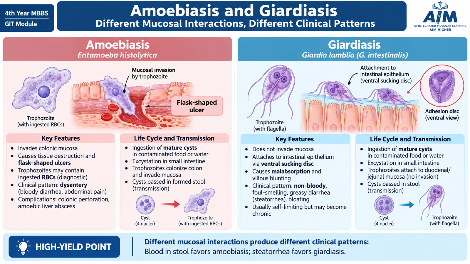
Entamoeba histolytica and Giardia lamblia are both intestinal protozoa acquired by ingestion of environmentally resistant cysts, but their disease mechanisms are very different. E. histolytica can invade intestinal tissue and produce ulceration, whereas Giardia remains mainly on the mucosal surface and interferes with absorption.
Entamoeba histolytica
E. histolytica causes amoebiasis. The infective form is the mature cyst. After cysts are swallowed, excystation occurs in the small intestine and releases trophozoites. These trophozoites subsequently reach the colon, multiply, and may either remain within the lumen or invade the colonic mucosa. Some organisms encyst again and cysts are passed in stool, allowing transmission to another person.
Morphology
- Trophozoite: motile form with a single nucleus, a small central karyosome and fine peripheral chromatin.
- Ingested red blood cells inside a trophozoite strongly support invasive E. histolytica.
- Cyst: rounded resistant form; the mature infective cyst contains four nuclei.
Pathogenesis
The trophozoite adheres to colonic epithelial cells and damages them through direct cytotoxic effects and proteolytic enzymes. Mucosal destruction then extends into the submucosa. The resulting ulcer classically has a relatively narrow mucosal opening with broader destruction beneath the surface, producing a flask-shaped ulcer.
Ingestion of cysts → excystation → colonic trophozoites → mucosal invasion → flask-shaped ulcers → dysentery or extraintestinal spread.
Clinical features and complications
Many infections remain asymptomatic. In invasive intestinal disease, mucosal ulceration produces abdominal pain, diarrhea, blood and mucus in stool and sometimes tenesmus. Trophozoites may enter portal circulation and reach the liver, where they can cause an amoebic liver abscess.
- Amoebic dysentery.
- Severe or fulminant colitis.
- Intestinal perforation in destructive disease.
- Amoeboma, an inflammatory mass that may mimic a colonic tumor.
- Amoebic liver abscess and, rarely, extension or rupture into nearby structures.
Giardia lamblia
Giardia lamblia causes giardiasis and mainly affects the upper small intestine. The infective cyst is swallowed in contaminated water or food. Excystation releases trophozoites, which attach to the duodenal and jejunal mucosa. They multiply but normally do not invade deeply into tissue. Trophozoites eventually encyst as they move distally, and cysts are passed in stool.
Morphology
- Trophozoite: pear-shaped and bilaterally symmetrical, with two nuclei, multiple flagella and a ventral adhesive disc.
- The two nuclei produce the characteristic “face-like” microscopic appearance.
- Cyst: oval and resistant; the mature cyst contains four nuclei.
Pathogenesis
Giardial trophozoites attach to the epithelial surface and disturb the normal brush-border environment. This reduces efficient digestion and absorption and may flatten or injure intestinal villi. Because there is usually no deep invasion, grossly bloody diarrhea is not typical.
Surface attachment → brush-border dysfunction → impaired absorption → steatorrhea, bloating and weight loss.
Clinical features and complications
- Watery or foul-smelling diarrhea.
- Greasy stool or steatorrhea due to fat malabsorption.
- Abdominal bloating, cramps and excessive gas.
- Weight loss when infection is prolonged.
- Persistent malabsorption may contribute to nutritional deficiencies.
- Temporary lactose intolerance may follow mucosal brush-border injury.
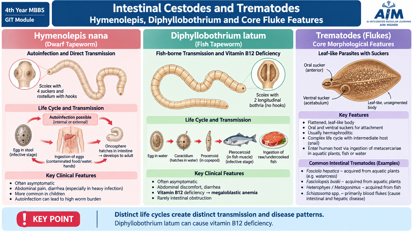
C. Intestinal Cestodes and Trematodes: Hymenolepis, Diphyllobothrium and Core Fluke Features
Cestodes are segmented flatworms commonly called tapeworms. They have a scolex for attachment and a chain of segments called proglottids. Two cestodes required in this topic are Hymenolepis nana and Diphyllobothrium latum. Trematodes, or flukes, are also flatworms but have a different body structure and life cycle.
Hymenolepis nana
H. nana, the dwarf tapeworm, is unusual because it can complete its life cycle in humans without requiring an obligatory intermediate host. Eggs are swallowed and release oncospheres in the small intestine. The oncosphere enters an intestinal villus and develops into a cysticercoid larva. It then returns to the lumen, attaches to the intestinal mucosa and matures into an adult worm.
Eggs released from the adult may leave the host in stool, but they may also hatch within the same host. This produces internal autoinfection and allows the parasite burden to increase without a new external exposure.
Morphology
- A small tapeworm with a scolex containing four suckers.
- The rostellum is armed with hooks.
- Eggs characteristically contain an oncosphere and polar filaments.
Pathogenesis, clinical features and complications
Light infections may cause no symptoms. A larger worm burden, especially when maintained by autoinfection, can irritate the intestinal mucosa and produce abdominal discomfort, anorexia and diarrhea. Heavy infection may lead to more persistent gastrointestinal symptoms and impaired nutrition, particularly in children.
Diphyllobothrium latum
D. latum, the broad fish tapeworm, is acquired by eating inadequately cooked infected freshwater fish containing the larval stage. The parasite attaches to the small intestine and develops into a long adult tapeworm.
The life cycle involves freshwater stages and more than one host. Eggs reach water, larvae are taken up by small aquatic crustaceans, and infected crustaceans are eaten by freshwater fish. Humans acquire infection when the infective fish stage is consumed.
Morphology
- The scolex has two longitudinal sucking grooves called bothria.
- The mature worm is long and composed of broad proglottids.
- Its eggs are operculated.
Pathogenesis and clinical effects
Many infected people are asymptomatic or develop mild abdominal discomfort and diarrhea. An important feature is the worm’s competition for vitamin B12. In susceptible individuals, prolonged depletion of vitamin B12 may produce megaloblastic anemia and, when severe, neurological consequences of vitamin B12 deficiency.
General physical characteristics of trematodes
Trematodes are dorsoventrally flattened, unsegmented worms. Most have an oral sucker and a ventral sucker that help them attach to host tissues. Their alimentary tract is incomplete. Most medically important trematodes are hermaphroditic, but Schistosoma is an important exception because male and female worms are separate. Most trematode eggs are operculated; schistosome eggs are another important exception.
D. Schistosomiasis: Life Cycle, Pathophysiology and Laboratory Recognition
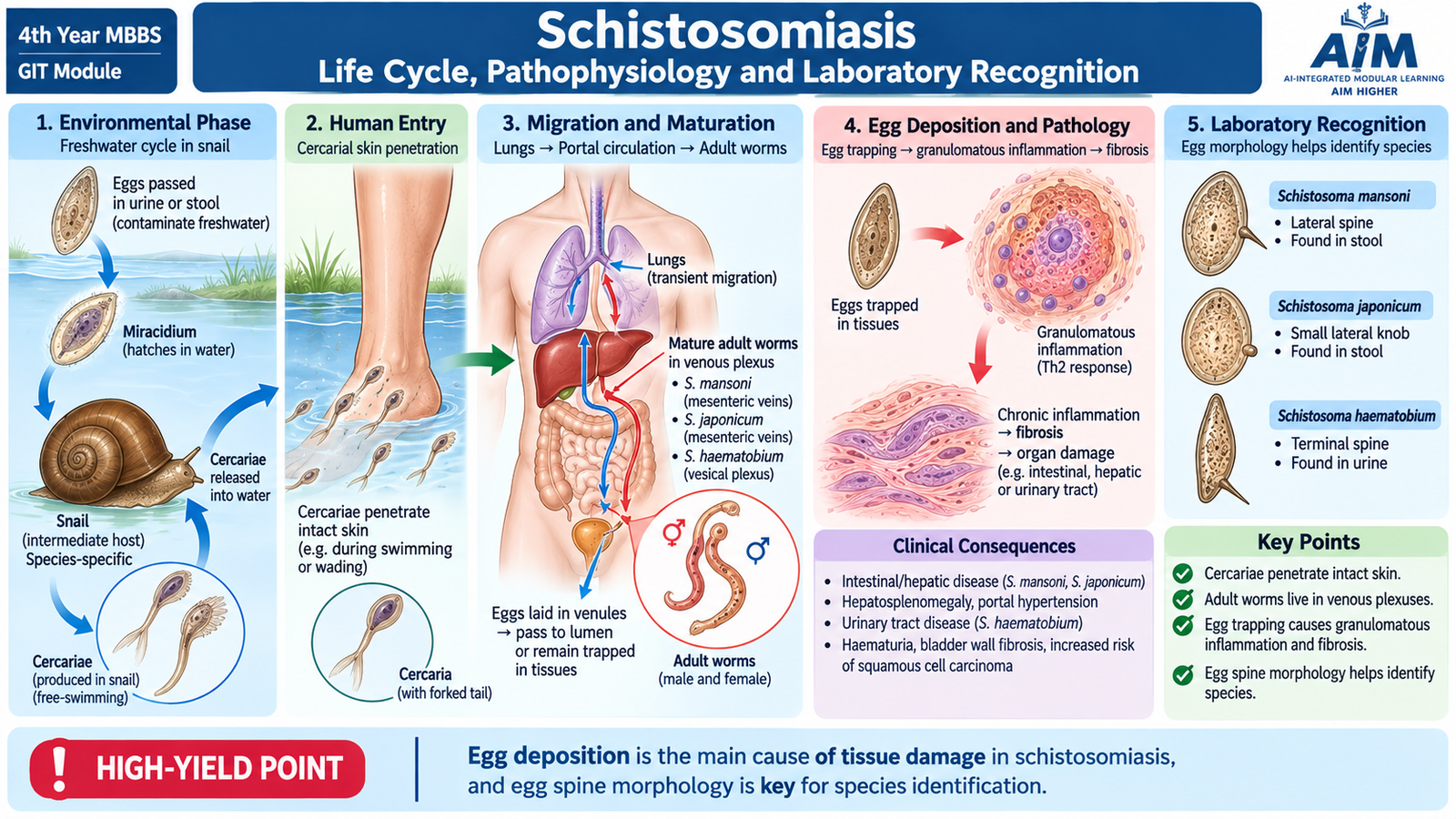
Schistosomiasis is caused by blood flukes of the genus Schistosoma. Unlike most other trematodes, adult schistosomes are separate male and female worms and live within the venous circulation. The major tissue damage is produced not by the adult worms themselves, but by the host inflammatory response to eggs trapped in tissues.
Major species and disease patterns
- Schistosoma mansoni: predominantly intestinal and hepatosplenic disease.
- Schistosoma japonicum: predominantly intestinal and hepatosplenic disease.
- Schistosoma haematobium: predominantly urinary disease.
Route of infection and life cycle
Humans acquire schistosomiasis when free-swimming cercariae released from infected freshwater snails penetrate intact skin. The cercaria loses its tail and becomes a schistosomulum. Schistosomula enter the circulation, migrate through the body and mature in the portal venous system. Male and female worms pair and then migrate to their preferred venous sites.
Female worms produce eggs. Some eggs cross the intestinal or urinary wall and leave the body in stool or urine. When eggs reach freshwater, they release miracidia that infect suitable snails. Development inside the snail eventually produces new cercariae, completing the cycle.
Pathophysiology
Eggs that fail to leave the body become trapped in intestinal, hepatic or urinary tissues. Their antigens stimulate a granulomatous inflammatory response. Repeated egg deposition leads to chronic inflammation and fibrosis.
In intestinal and hepatosplenic schistosomiasis, eggs may cause mucosal ulceration and chronic intestinal symptoms. Eggs carried to the liver can produce periportal fibrosis. Progressive fibrosis increases resistance to portal blood flow and may lead to portal hypertension and splenomegaly.
In urinary schistosomiasis, egg deposition in the bladder wall produces chronic inflammation, hematuria and fibrosis. Long-standing S. haematobium infection is associated with an increased risk of squamous cell carcinoma of the urinary bladder.
Clinical features
- Transient itchy dermatitis may occur at the site of cercarial penetration.
- An acute systemic illness may develop during early infection, with fever and eosinophilia.
- Intestinal disease may produce abdominal pain and diarrhea, sometimes with blood.
- Chronic hepatosplenic disease may produce hepatosplenic enlargement and features of portal hypertension.
- S. haematobium characteristically produces urinary symptoms and hematuria.
Laboratory diagnosis
Direct demonstration of characteristic eggs provides important evidence of active infection. Stool examination is used for intestinal species, while urine is the appropriate specimen for S. haematobium. Eosinophilia may support the diagnosis but is not specific.
Comparison of Schistosoma eggs
| Species | Key egg feature | Main specimen | Major disease pattern |
|---|---|---|---|
| S. mansoni | Prominent lateral spine | Stool | Intestinal/hepatosplenic |
| S. japonicum | Small lateral knob | Stool | Intestinal/hepatosplenic |
| S. haematobium | Terminal spine | Urine | Urinary |
E. Ascaris and Strongyloides Infections
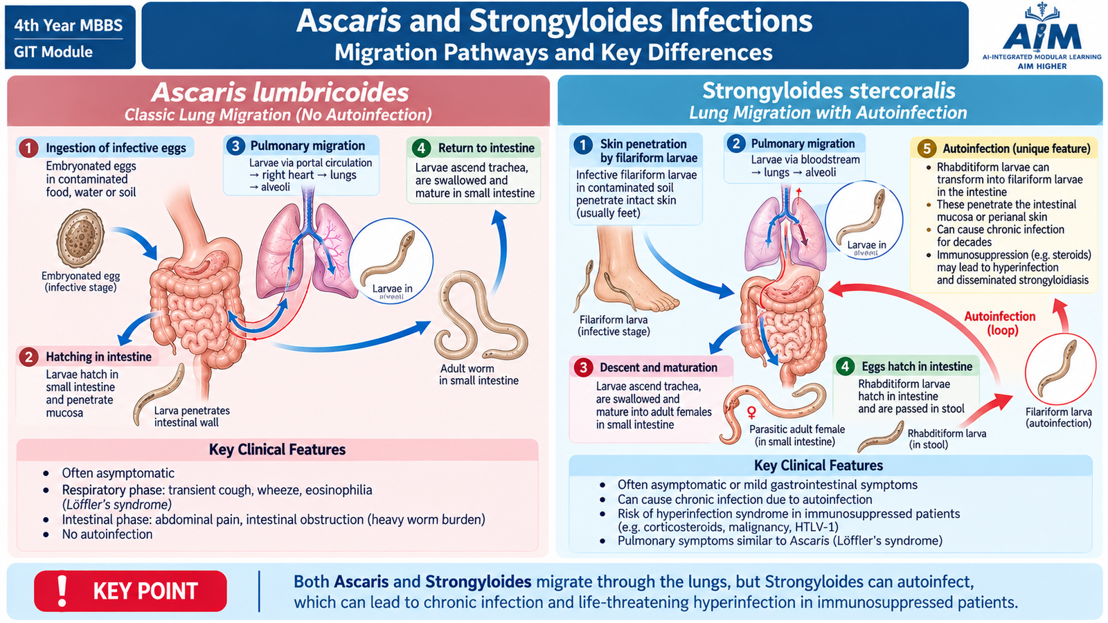
Ascaris lumbricoides and Strongyloides are intestinal nematodes whose larvae migrate through tissues before the mature parasite becomes established in the intestine. Their life cycles explain why gastrointestinal disease may be accompanied by pulmonary manifestations and eosinophilia.
Ascaris lumbricoides
Ascariasis begins when embryonated eggs are swallowed. Larvae hatch in the small intestine, penetrate the intestinal wall and enter the circulation. They migrate through the liver to the lungs, enter the alveoli, ascend the bronchial tree and are swallowed. On returning to the small intestine they develop into adult worms.
Ingested egg → intestinal larva → liver/lung migration → swallowed larva → adult worm in small intestine.
Morphology
- Large cylindrical roundworm with tapering ends.
- The male is smaller and has a curved posterior end.
- Fertilized eggs have a thick shell with a characteristic mammillated outer coat.
Pathogenesis and clinical features
Larval migration through the lungs can provoke eosinophilic inflammation, producing cough, wheeze and transient pulmonary infiltrates. This pulmonary hypersensitivity pattern is sometimes called Löffler syndrome. Adult intestinal worms may cause vague abdominal discomfort or remain asymptomatic.
A heavy worm burden can mechanically obstruct the bowel. Adult worms are also capable of abnormal migration into the biliary or pancreatic ducts, producing obstructive or inflammatory complications.
Major complications
- Intestinal obstruction.
- Biliary migration and biliary obstruction.
- Pancreatic duct migration with pancreatitis.
- Pulmonary hypersensitivity during larval migration.
Strongyloides
Strongyloidiasis usually begins when infective filariform larvae penetrate the skin. Larvae enter the circulation, reach the lungs, ascend the respiratory tract and are swallowed. In the small intestine, adult parthenogenetic female worms live within the mucosa and produce eggs that rapidly hatch into rhabditiform larvae.
Unlike many intestinal nematodes, larvae rather than typical eggs are usually found in stool. Some rhabditiform larvae transform into infective filariform larvae before leaving the host. These can penetrate intestinal mucosa or perianal skin and begin another cycle inside the same person. This is autoinfection.
Morphology and recognition
- Rhabditiform larvae are the usual diagnostic intestinal stage.
- Filariform larvae are the infective stage.
- Adult parasitic females are slender worms within the small-intestinal mucosa.
Pathogenesis and clinical features
Skin penetration may cause an itchy eruption. Larval migration through the lungs may cause cough or wheeze. Intestinal infection can produce abdominal pain, diarrhea and malabsorption. Rapidly moving serpiginous skin lesions around the buttocks or trunk are described as larva currens.
Hyperinfection
Autoinfection makes Strongyloides especially important in people who become immunosuppressed, particularly during corticosteroid exposure. Accelerated larval multiplication can produce hyperinfection or disseminated strongyloidiasis, with severe pulmonary and gastrointestinal disease. Larval migration may carry intestinal bacteria into the bloodstream, contributing to serious bacterial sepsis.
F. Hookworm and Enterobius Infections
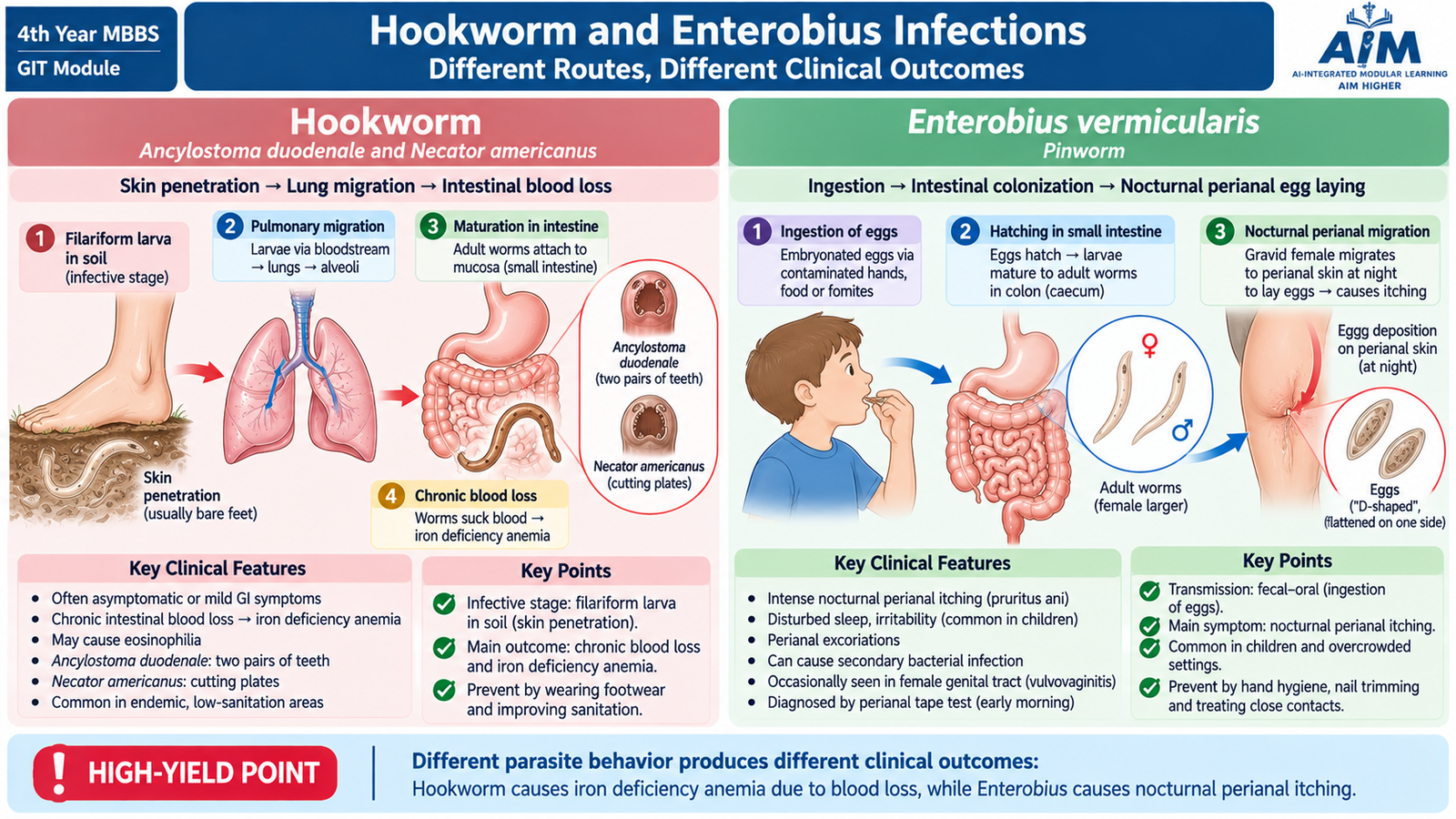
Ancylostoma duodenale and Enterobius vermicularis are nematodes, but their mechanisms are very different. Hookworms enter through skin and cause disease mainly by intestinal blood loss, whereas Enterobius is transmitted by eggs and causes characteristic nocturnal perianal itching when female worms deposit eggs around the anus.
Ancylostoma duodenale
Infective filariform hookworm larvae penetrate exposed skin and enter the circulation. They migrate to the lungs, ascend the tracheobronchial tree, are swallowed and reach the small intestine. Adult worms attach firmly to the mucosa using their buccal structures and feed on blood.
Morphology
- Adult A. duodenale has a curved anterior end.
- Its buccal capsule contains characteristic teeth used for attachment.
- Eggs are oval, thin-shelled and contain developing blastomeres when freshly passed.
Pathogenesis
Each attached worm damages small-intestinal mucosa and removes blood. Persistent blood loss gradually depletes iron stores. Therefore, the characteristic systemic consequence of substantial hookworm infection is iron-deficiency anemia.
Skin penetration → pulmonary migration → intestinal attachment → chronic blood loss → iron deficiency → microcytic hypochromic anemia.
Clinical features and complications
- Itching at the site of larval penetration.
- Transient respiratory symptoms during migration.
- Abdominal discomfort.
- Fatigue, pallor and reduced exercise tolerance from anemia.
- Severe or prolonged infection may impair nutrition, growth and functional capacity.
Enterobius vermicularis
Enterobius vermicularis, or pinworm, is acquired when infective eggs are swallowed. Larvae hatch in the intestine and adult worms live mainly around the cecum and adjacent colon. At night, the gravid female migrates onto the perianal skin and deposits large numbers of eggs.
The eggs rapidly become infective. Scratching contaminates fingers and fingernails, and hand-to-mouth transfer can produce repeated self-infection. Eggs may also contaminate bedding, clothing and household surfaces.
Morphology
- The female worm has a long, pointed posterior end, giving the name “pinworm.”
- Eggs are colorless and characteristically flattened on one side.
Clinical features and complications
The classic manifestation is nocturnal pruritus ani. Itching occurs because the female worm and its eggs irritate perianal skin. Repeated itching can disturb sleep and may produce excoriations or secondary irritation. Female worms may occasionally migrate into the female genital tract and produce vulvovaginal irritation.
Recognition clue: Because eggs are deposited around the anus rather than regularly released into stool, sampling the perianal region is more useful than routine stool examination for demonstrating the eggs.
G. Antiprotozoal and Anthelmintic Pharmacology
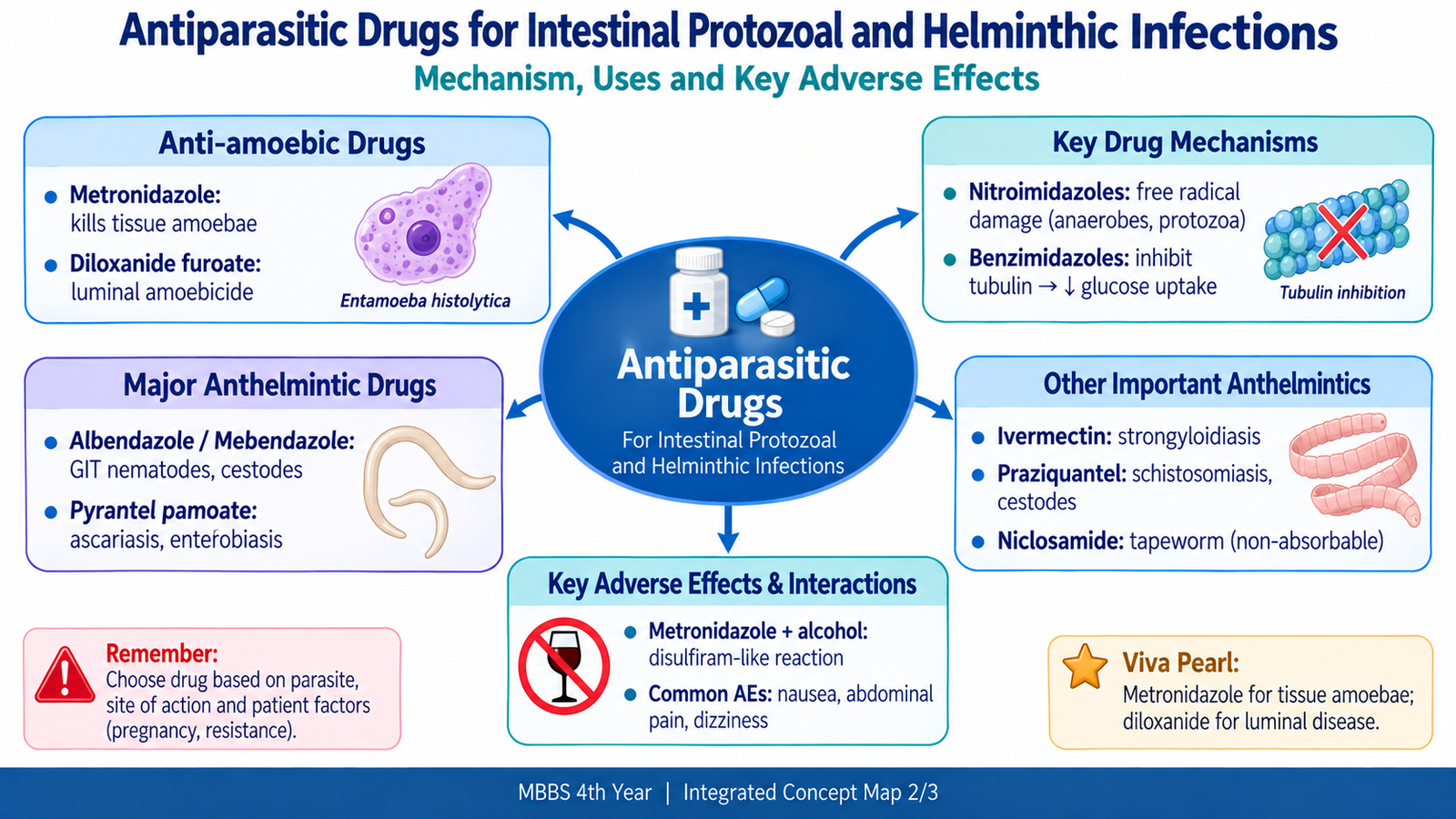
Antiparasitic treatment becomes easier to understand when the drug is matched to the location and biology of the parasite. Amoebiasis may require a drug that reaches invasive trophozoites as well as a second drug that clears organisms remaining in the intestinal lumen. Anthelmintic drugs act through several mechanisms, including disruption of microtubules, paralysis of worms and damage to parasite membranes.
Classification of anti-amoebic drugs
- Luminal amoebicides: act mainly on organisms in the intestinal lumen; important example: diloxanide furoate.
- Tissue/systemic amoebicides: act on invasive organisms in tissues.
- Nitroimidazoles: metronidazole is the major prototype used against invasive amoebiasis and is also effective in giardiasis.
Metronidazole
Mechanism: Metronidazole enters susceptible anaerobic organisms and protozoa, where its nitro group is reduced to reactive intermediates. These products damage DNA and inhibit nucleic-acid function, resulting in death of the organism.
Clinical uses in this topic: It is effective against invasive E. histolytica trophozoites and is also used for giardiasis. In invasive amoebiasis, metronidazole does not reliably eliminate all luminal cysts, so treatment is logically followed by an effective luminal amoebicide.
Adverse effects: nausea, abdominal discomfort, metallic taste and headache are common. Dark urine may occur. Prolonged exposure can rarely produce peripheral neuropathy.
Alcohol should be avoided during metronidazole treatment because a disulfiram-like reaction may occur, producing flushing, nausea, vomiting, headache and cardiovascular discomfort.
Important contraindication/caution: hypersensitivity to nitroimidazoles; caution is appropriate in significant neurological disease and severe hepatic dysfunction.
Diloxanide furoate
Diloxanide furoate is a luminal amoebicide. Its exact molecular mechanism is not as clearly defined as that of metronidazole, but its active form acts on amoebae within the intestinal lumen.
- Use: eradication of luminal E. histolytica, including asymptomatic intestinal carriage and residual luminal infection following treatment of invasive disease.
- Adverse effects: flatulence, nausea, abdominal discomfort and occasional allergic skin reactions.
- Caution: avoid in patients with hypersensitivity; use in pregnancy should follow appropriate clinical advice rather than routine self-medication.
Classification of important anthelmintic drugs
- Benzimidazoles: albendazole, mebendazole.
- Depolarizing neuromuscular agent: pyrantel pamoate.
- Macrocyclic lactone: ivermectin.
- Calcium-permeability enhancing drug: praziquantel.
- Intestinal cestocidal drug: niclosamide.
| Drug | Mechanism | Important uses in this topic | Important adverse effects | Major contraindication / caution |
|---|---|---|---|---|
| Albendazole | Binds parasite β-tubulin → inhibits microtubules and glucose uptake → energy depletion. | Broad intestinal nematode activity, including Ascaris, hookworm and Enterobius. | GI upset, headache; prolonged systemic use may cause hepatotoxicity or marrow suppression. | Benzimidazole hypersensitivity; avoid or use cautiously in pregnancy and significant liver disease. |
| Mebendazole | Inhibits β-tubulin-dependent microtubule formation and glucose utilization. | Ascaris, hookworm and Enterobius. | Abdominal pain, diarrhea; liver or blood abnormalities are uncommon with prolonged exposure. | Benzimidazole hypersensitivity; caution in pregnancy and hepatic disease. |
| Pyrantel pamoate | Activates nematode nicotinic receptors → persistent depolarization → spastic paralysis. | Enterobius, Ascaris and hookworm infections. | GI upset, dizziness and headache. | Hypersensitivity; caution in significant hepatic dysfunction and pregnancy. |
| Ivermectin | Activates parasite glutamate-gated chloride channels → hyperpolarization and paralysis. | Important drug for strongyloidiasis. | Dizziness, GI symptoms, pruritus and neurological toxicity in susceptible circumstances. | Hypersensitivity; pregnancy and situations increasing CNS toxicity require caution. |
| Praziquantel | Increases parasite membrane Ca²⁺ permeability → sustained contraction, paralysis and tegumental damage. | Schistosomiasis and important intestinal cestode infections including H. nana and D. latum. | Dizziness, headache, abdominal symptoms and transient inflammatory symptoms from parasite destruction. | Hypersensitivity; ocular cysticercosis is an important contraindication because inflammatory damage can threaten vision. |
| Niclosamide | Disrupts cestode energy production, including oxidative phosphorylation. | Selected intestinal tapeworm infections. | Usually mild GI upset, nausea or abdominal discomfort. | Hypersensitivity; it is not appropriate for tissue-stage cestode disease because systemic absorption is minimal. |
Therapeutic logic: Match the drug to the parasite and its location. Metronidazole targets invasive amoebic trophozoites, luminal therapy clears intestinal amoebae, ivermectin is particularly important for Strongyloides, benzimidazoles act broadly against intestinal nematodes, and praziquantel is important against schistosomes and several cestodes.

H. Epidemiological Determinants, Prevention and Control
Intestinal parasitic disease is strongly influenced by the environment and by human behavior. The most important community principle is that different parasites require different routes of transmission; therefore, effective control depends on identifying where the life cycle can be interrupted.
Important intestinal worm patterns in the local context
Where sanitation, safe water and hygiene are inadequate, fecal contamination favors transmission of worms such as Ascaris and H. nana. Barefoot contact with contaminated soil creates an opportunity for hookworm and Strongyloides larvae to penetrate skin. Enterobius spreads particularly efficiently where close household or institutional contact permits transfer of eggs between hands, clothes, bedding and surfaces.
Food and environmental exposures also matter. Consumption of infected inadequately cooked freshwater fish transmits D. latum, while skin contact with cercaria-contaminated freshwater is the key exposure for schistosomiasis in endemic settings.
Epidemiological determinants
- Unsafe disposal of feces: contaminates soil, food and water with cysts or eggs.
- Unsafe water: facilitates fecal–oral protozoal and helminthic transmission.
- Poor hand hygiene: increases ingestion of infective cysts and eggs.
- Walking barefoot on contaminated soil: permits skin penetration by hookworm and Strongyloides larvae.
- Crowding and close contact: assist Enterobius and other fecal–oral infections.
- Raw or inadequately cooked freshwater fish: permits transmission of D. latum.
- Freshwater exposure in endemic areas: creates risk of schistosomal cercarial penetration.
- Immunosuppression: does not initiate Strongyloides infection but can convert chronic infection into dangerous hyperinfection.
Prevention and control
Prevention works by breaking the transmission cycle before the parasite reaches a new host or by reducing the reservoir of infection.
- Safe sanitation: proper disposal of human feces reduces contamination with eggs, cysts and larvae.
- Safe drinking water: reduces ingestion of fecally transmitted parasites.
- Hand hygiene: wash hands after using the toilet and before preparing or eating food.
- Food hygiene: wash produce and adequately cook foods that may transmit parasites.
- Footwear: reduces skin contact with soil containing hookworm or Strongyloides larvae.
- Nail and household hygiene: short nails, handwashing and cleaning contaminated clothes or bedding help interrupt Enterobius transmission.
- Avoid inadequately cooked infected freshwater fish: prevents D. latum infection.
- Avoid unsafe freshwater exposure in schistosomiasis-endemic areas: prevents cercarial skin penetration.
- Early recognition and treatment: reduces morbidity and, for parasites transmitted from human feces, may reduce continuing environmental contamination.
- Population control measures: organized deworming or other community interventions should follow local public-health policy and epidemiological need rather than being applied indiscriminately.
Public-health principle: sanitation interrupts fecal contamination, footwear interrupts larval skin penetration, food safety interrupts food-borne infection, and avoidance of contaminated freshwater interrupts schistosomal transmission.

Integrated Mechanism Flow
Cysts, eggs, larvae, fish or freshwater
Ingestion or skin penetration
Intestinal lumen, mucosa or venous system
Invasion, malabsorption, blood loss, nutrient loss or egg granulomas
Dysentery, steatorrhea, anemia, pruritus or fibrosis
Targeted drug therapy plus transmission control
⭐ AIM High-Yield Review
- ⭐ E. histolytica invades colonic mucosa and produces characteristic flask-shaped ulcers; portal spread may cause a liver abscess.
- Giardia attaches to the upper small-intestinal surface without deep invasion, producing malabsorption, steatorrhea and bloating rather than typical bloody dysentery.
- H. nana can complete its life cycle in one human host and can cause internal autoinfection.
- D. latum is acquired from infected freshwater fish and may cause vitamin B12 deficiency and megaloblastic anemia.
- ⭐ Schistosomal tissue injury is mainly egg-mediated. Granulomatous inflammation and fibrosis, rather than direct adult-worm damage, produce chronic disease.
- S. mansoni: lateral spine; S. haematobium: terminal spine; S. japonicum: small lateral knob.
- Ascaris undergoes lung migration before returning to the intestine; heavy infection may cause intestinal obstruction or biliary migration.
- ⭐ Strongyloides can autoinfect the host. Immunosuppression, particularly corticosteroid exposure, can precipitate dangerous hyperinfection.
- Hookworm attaches to small-intestinal mucosa and causes chronic blood loss, producing iron-deficiency anemia.
- Enterobius causes nocturnal pruritus ani because gravid female worms deposit eggs on perianal skin at night.
- Metronidazole damages protozoal DNA after intracellular reduction; invasive amoebiasis also requires clearance of residual luminal organisms.
- Albendazole and mebendazole inhibit parasite microtubules; pyrantel causes spastic paralysis; ivermectin causes chloride-mediated paralysis.
- Praziquantel increases parasite calcium permeability and is important against schistosomes and several cestodes.
- Prevention follows transmission: sanitation for fecal contamination, footwear for soil-penetrating larvae, safe food preparation for food-borne parasites and freshwater precautions for schistosomiasis.