
21. Inflammatory Bowel Disease: Ulcerative Colitis and Crohn Disease
Topic Introduction
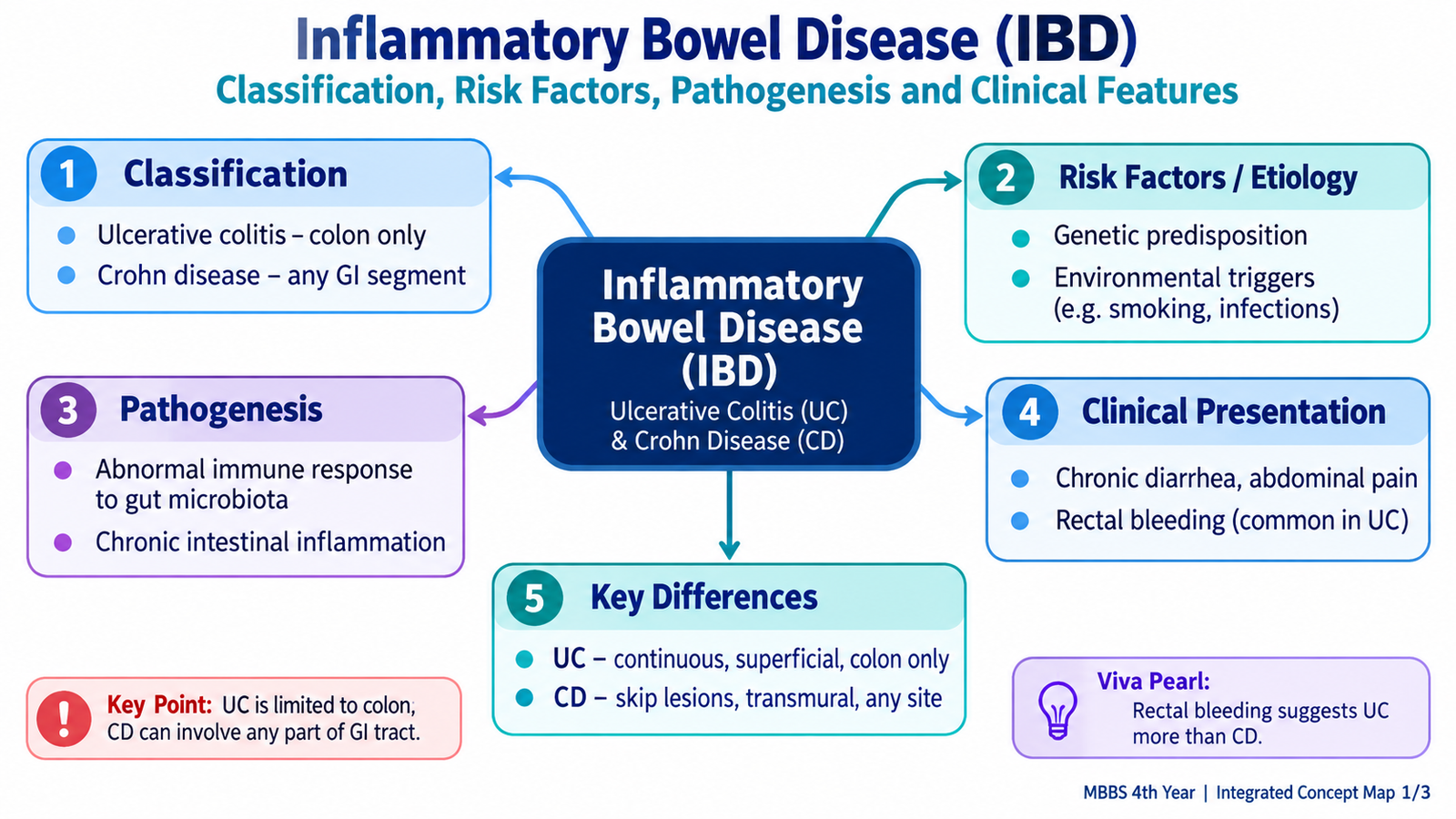
Inflammatory bowel disease, or IBD, refers mainly to ulcerative colitis and Crohn disease. Both are chronic inflammatory disorders of the gastrointestinal tract that usually follow a relapsing and remitting course. They arise when genetic susceptibility, environmental influences, intestinal microbes and an abnormal mucosal immune response interact to produce persistent intestinal inflammation. Although the two diseases share several features, they differ greatly in their site, depth and pattern of bowel involvement. These differences explain their characteristic symptoms, complications and surgical approaches. In this chapter, you will learn how IBD develops, how ulcerative colitis and Crohn disease are recognized and differentiated, how investigations establish the diagnosis, how the major drug groups work, when surgery becomes necessary and why long-term colonic surveillance is important.
A. IBD Classification, Etiology, Risk Factors and Pathogenesis
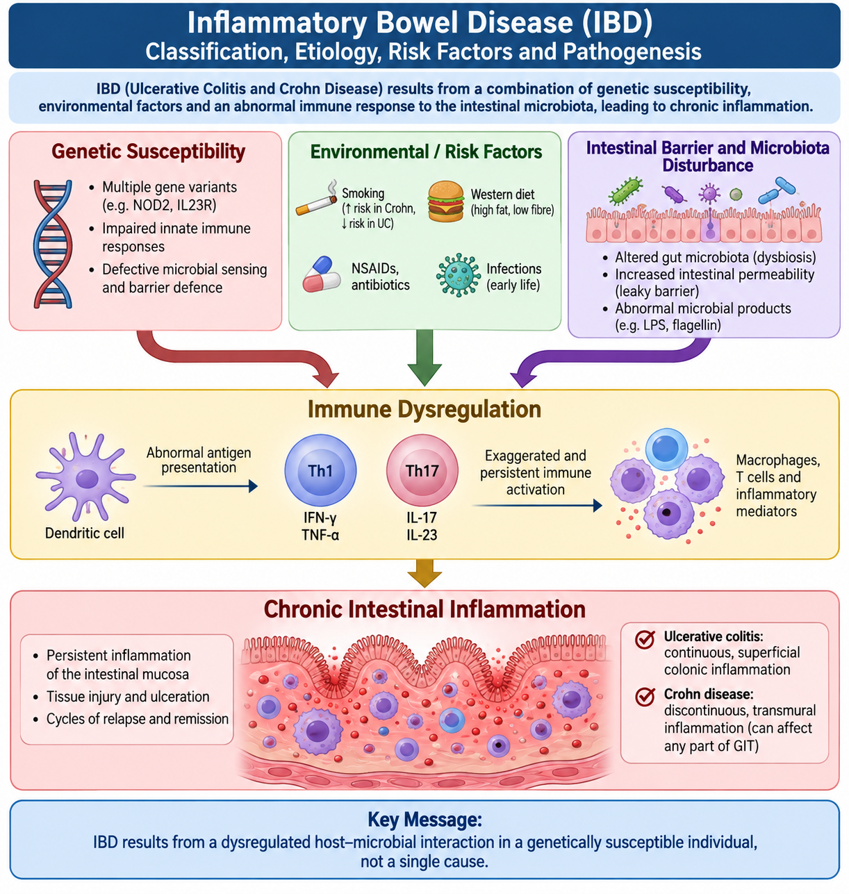
IBD is not simply an infection or a single-gene disorder. It is best understood as a state in which the intestinal immune system reacts abnormally to luminal contents and the normal intestinal microbiota in a genetically susceptible person. The resulting inflammation becomes persistent because the epithelial barrier, immune regulation and mechanisms that normally switch inflammation off do not function normally.
Classification
The two major forms are:
- Ulcerative colitis (UC): inflammatory disease limited to the colon, beginning in the rectum and extending proximally in a continuous pattern.
- Crohn disease (CD): inflammatory disease that can involve any part of the gastrointestinal tract and characteristically produces discontinuous, often transmural lesions.
Occasionally, colonic inflammatory disease cannot initially be classified confidently as either UC or Crohn disease. Such cases may be described as IBD-unclassified until the pattern becomes clearer.
Etiology and Risk Factors
No single cause explains IBD. Several factors interact:
- Genetic susceptibility: family clustering supports an inherited predisposition. Genetic abnormalities involved in microbial recognition and mucosal immune regulation are particularly important in Crohn disease.
- Abnormal immune regulation: intestinal immune responses become excessive or fail to terminate appropriately.
- Altered intestinal microbiota: the normal gut microbial community provides persistent antigenic stimulation in susceptible individuals.
- Epithelial barrier dysfunction: impaired mucosal barrier function allows greater contact between luminal antigens and mucosal immune cells.
- Environmental influences: environmental exposures modify risk and disease activity. Cigarette smoking is particularly associated with a worse course in Crohn disease.
Pathogenesis
The central mechanism is a failure of normal intestinal immune tolerance. In a susceptible host, epithelial barrier abnormalities increase exposure of mucosal immune cells to microbial and luminal antigens. Innate and adaptive immune responses become activated and release inflammatory cytokines. Instead of resolving normally, the inflammatory reaction continues and damages the bowel wall.
Genetic susceptibility + environmental influences → abnormal epithelial barrier and microbial interaction → dysregulated mucosal immunity → persistent cytokine-driven inflammation → intestinal injury and complications.
In Crohn disease, immune responses involving Th1 and Th17 pathways are prominent. In UC, the immune pattern is different and is largely restricted to the colonic mucosa. These immunological differences help explain why modern therapies can control IBD by blocking selected inflammatory cytokines or preventing inflammatory lymphocytes from reaching the intestinal mucosa.
B. Ulcerative Colitis: Pathology and Clinical Presentation
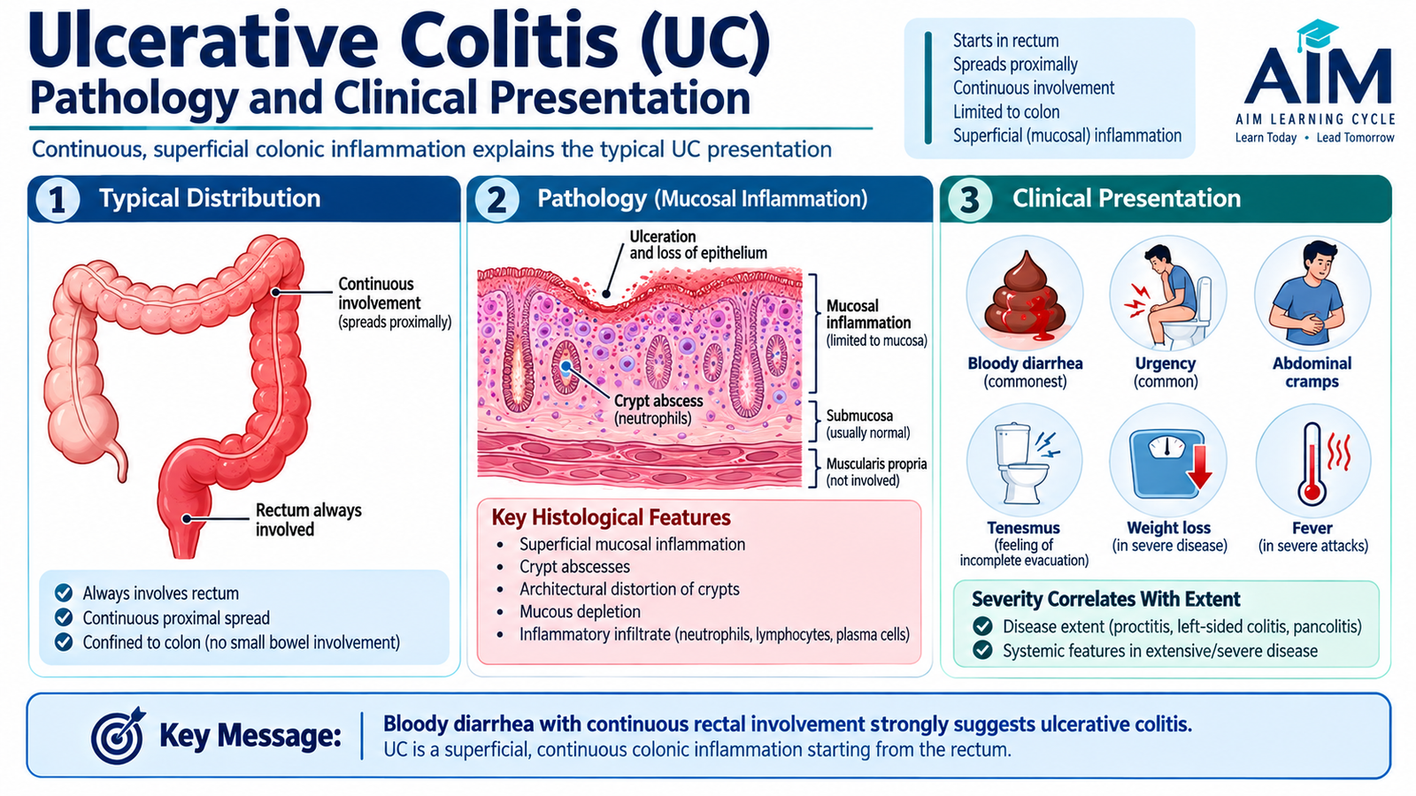
Ulcerative colitis is a chronic inflammatory disease of the colon in which inflammation starts in the rectum and extends proximally in a continuous manner. The inflammatory process is mainly limited to the mucosa and superficial submucosa. This superficial but diffuse mucosal injury explains why rectal bleeding and bloody diarrhea are such characteristic features.
Distribution
- The rectum is typically involved.
- Disease spreads proximally without normal intervening segments.
- It may remain limited to the rectum, involve the left colon or extend throughout the colon.
- The small intestine is generally not affected, although minimal terminal ileal inflammation may occasionally occur in extensive colitis.
Gross Morphology
Repeated mucosal inflammation produces a red, granular and friable mucosal surface. Superficial ulceration develops, and surviving islands of regenerating mucosa may project between ulcers as inflammatory pseudopolyps. Because the inflammation is not usually transmural, deep fissures, fistulas and marked bowel-wall thickening are not characteristic.
Microscopic Morphology
Active disease shows mucosal inflammation with neutrophils entering colonic crypts. When neutrophils accumulate within crypt lumina, they form crypt abscesses. Chronic disease produces architectural distortion of the crypts together with chronic inflammatory cells in the lamina propria.
Clinical Features
Because the inflamed colonic mucosa ulcerates easily and loses its normal ability to absorb water, patients commonly develop frequent loose stools containing blood and mucus.
- Bloody diarrhea is the classic presentation.
- Rectal urgency and tenesmus are common because rectal inflammation is typical.
- Crampy lower abdominal pain may accompany bowel movements.
- More extensive or severe disease can produce fever, anemia, fatigue and weight loss.
Inflammation and superficial ulceration directly damage the highly vascular colonic mucosa, so blood readily enters the intestinal lumen.
C. Crohn Disease: Pathology and Clinical Presentation
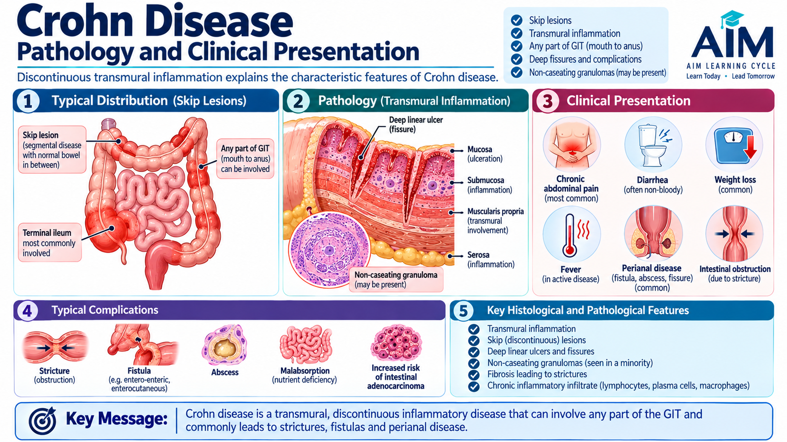
Crohn disease is a chronic inflammatory disorder that can affect any part of the gastrointestinal tract from the mouth to the anus. The terminal ileum and colon are common sites. Unlike UC, Crohn disease is characteristically discontinuous and transmural, meaning inflammation may extend through the full thickness of the bowel wall. These two features explain many of its distinctive complications.
Distribution and Gross Morphology
- Diseased segments are separated by relatively normal bowel, creating skip lesions.
- Early mucosal lesions may develop into deeper linear or serpiginous ulcers.
- Intersecting ulcers and residual swollen mucosa can produce a cobblestone appearance.
- Transmural inflammation causes bowel-wall thickening and fibrosis.
- Mesenteric fat may extend around the serosal surface, producing creeping fat.
- Deep fissures may penetrate the bowel wall and form fistulas or sinus tracts.
Microscopic Morphology
Microscopy shows chronic active inflammation that may extend through the bowel wall. Non-caseating granulomas may be found and strongly support Crohn disease when present, but they are not present in every patient and therefore are not required for diagnosis.
Clinical Features
Symptoms depend on the site and severity of disease. Ileal inflammation commonly produces abdominal pain and diarrhea, while chronic inflammation may interfere with nutrient absorption.
- Chronic or recurrent diarrhea, which may be non-bloody.
- Abdominal pain, commonly involving the right lower abdomen when the terminal ileum is affected.
- Weight loss and reduced appetite.
- Fever during active inflammation or septic complications.
- Perianal disease, including fissures, fistulas or abscesses.
- Features of malabsorption when substantial small-bowel disease is present.
Why Transmural Inflammation Matters
When inflammation extends through the full thickness of the bowel wall, healing is accompanied by fibrosis and scarring. This narrows the intestinal lumen and can produce strictures and obstruction. Deep inflammatory tracks may also penetrate into adjacent bowel loops, skin or organs and form fistulas.
Transmural inflammation → deep ulceration → fibrosis or penetrating disease → stricture, fistula or abscess.
D. Diagnostic Approach and Investigations in IBD
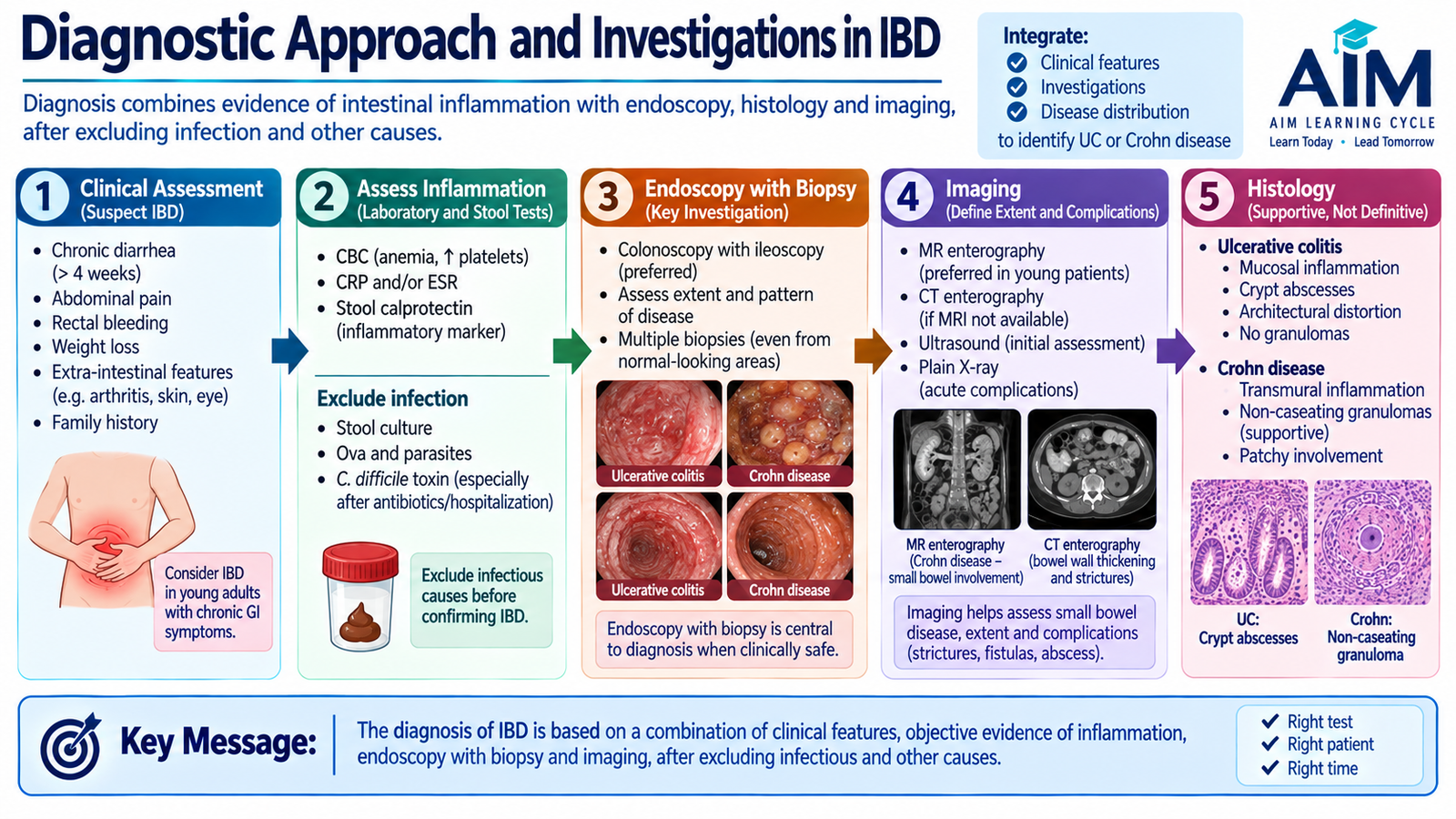
IBD is diagnosed by combining the clinical pattern with laboratory evidence of inflammation, stool testing, endoscopic appearance, histology and imaging when needed. No single blood test establishes the diagnosis. The diagnostic approach must also exclude infection and identify the extent and complications of disease.
Initial Laboratory Assessment
- Complete blood count: may show anemia from chronic blood loss, inflammation or nutritional deficiency, and leukocytosis during active inflammation.
- CRP and ESR: support the presence and activity of systemic inflammation but are not specific for IBD.
- Serum albumin: may fall in severe inflammation, protein loss or poor nutritional status.
- Electrolytes and renal function: are useful when significant diarrhea or dehydration is present.
Stool Assessment
Infection can mimic or worsen IBD, so stool studies are important during the diagnostic evaluation and during an apparent flare when infection is suspected. Fecal calprotectin reflects neutrophil activity within the intestine. A raised level supports intestinal inflammation and can help distinguish inflammatory disease from non-inflammatory bowel symptoms, but it does not by itself distinguish UC from Crohn disease.
Endoscopy and Biopsy
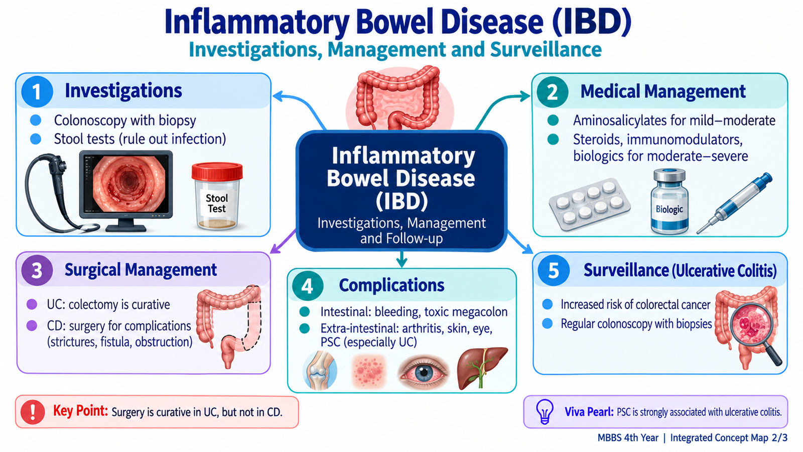
Colonoscopy with biopsies is central to diagnosis when clinically safe. In UC, it demonstrates continuous inflammation beginning in the rectum. In Crohn disease, it may show skip lesions, aphthous or deep ulceration, cobblestoning and terminal ileal involvement. Biopsies confirm chronic inflammatory changes and help distinguish the two patterns.
| Investigation | Main Role |
|---|---|
| Blood tests | Assess inflammation, anemia, nutritional status and systemic effects. |
| Stool testing | Exclude infectious causes and assess intestinal inflammation. |
| Colonoscopy + biopsy | Define distribution, directly inspect mucosa and obtain histology. |
| Cross-sectional imaging | Assess small bowel, bowel wall and complications such as abscess, fistula or obstruction. |
Imaging
Cross-sectional imaging is especially useful in Crohn disease because a significant part of the small bowel cannot be assessed fully by routine colonoscopy. CT or MR enteric imaging can demonstrate bowel-wall thickening, strictures, penetrating disease, fistulas and abscesses. In severe colitis, abdominal imaging may also identify colonic dilatation, perforation or other complications.
E. Intestinal and Extra-Intestinal Manifestations and Complications
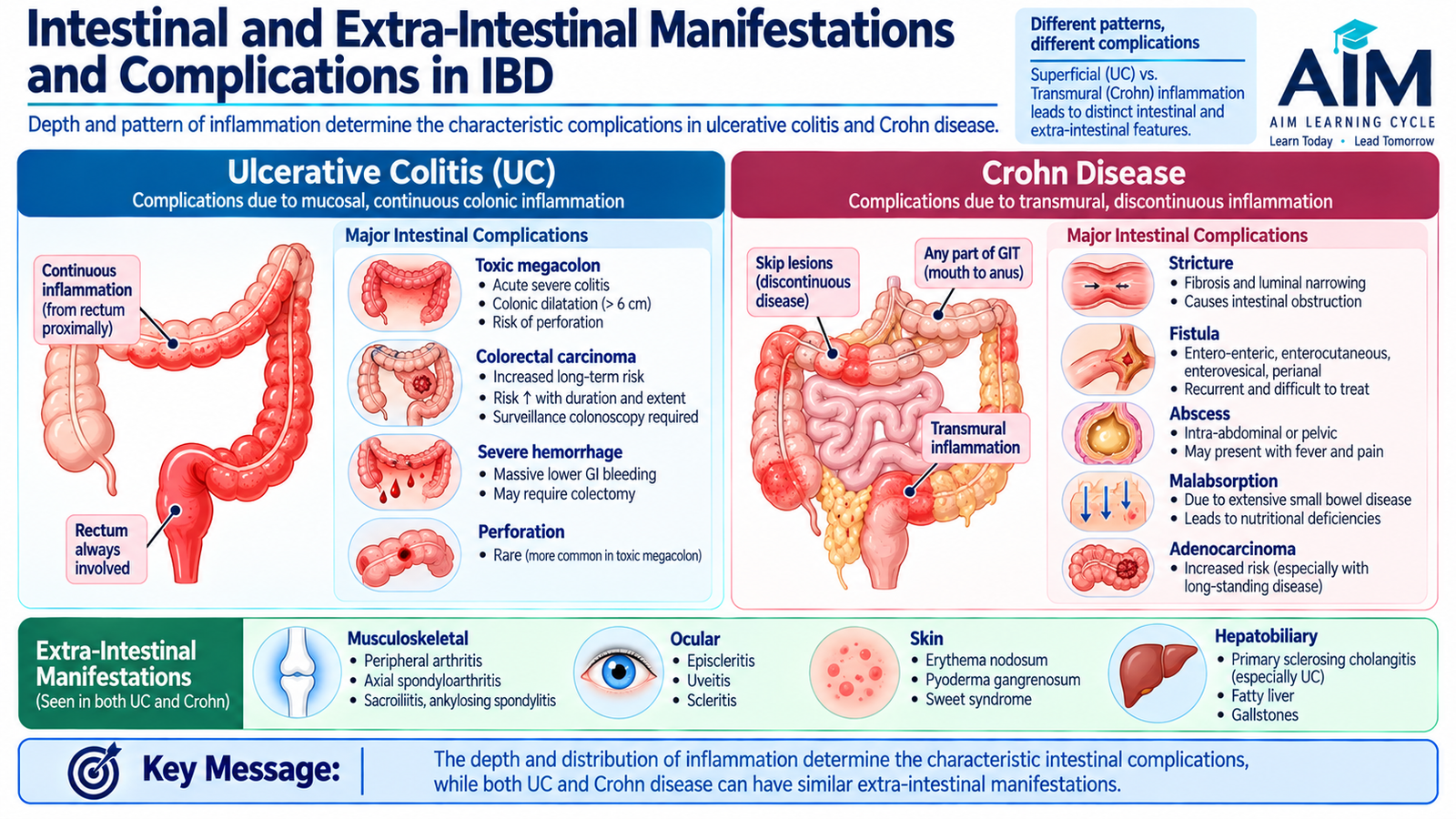
IBD is primarily a gastrointestinal disease, but persistent immune activation can affect organs outside the bowel. Complications also differ according to the depth and location of intestinal inflammation. Recognizing this distinction helps explain why toxic megacolon is strongly associated with severe colitis, whereas fistulas and strictures are characteristic of transmural Crohn disease.
Ulcerative Colitis Complications
- Severe hemorrhage: extensive mucosal ulceration can produce significant bleeding.
- Toxic megacolon: severe inflammation impairs colonic neuromuscular function, resulting in marked dilatation with systemic toxicity.
- Perforation: severe inflammation and dilatation may weaken the bowel wall.
- Colorectal neoplasia: longstanding colonic inflammation increases the risk of dysplasia and colorectal carcinoma.
Crohn Disease Complications
- Strictures and intestinal obstruction from repeated transmural inflammation and fibrosis.
- Fistulas connecting bowel to bowel, skin or nearby organs.
- Abscess formation following penetrating inflammation.
- Perianal disease including fissures, fistulas and abscesses.
- Malabsorption and nutritional deficiency when substantial small bowel is involved or resected.
- Longstanding colonic involvement also increases colorectal cancer risk.
Extra-Intestinal Manifestations
Several manifestations reflect systemic immune dysregulation rather than direct spread of bowel inflammation.
- Musculoskeletal: peripheral arthritis and axial inflammatory disease.
- Skin: erythema nodosum and pyoderma gangrenosum.
- Eye: episcleritis or uveitis.
- Hepatobiliary: primary sclerosing cholangitis, particularly associated with UC.
- Thromboembolic disease: active inflammation increases the tendency toward venous thrombosis.
Some extra-intestinal manifestations tend to improve as intestinal inflammation is controlled, while others may follow a course that is partly independent of bowel activity.
F. Pharmacotherapy of IBD: Drug Classes, Mechanisms and Adverse Effects
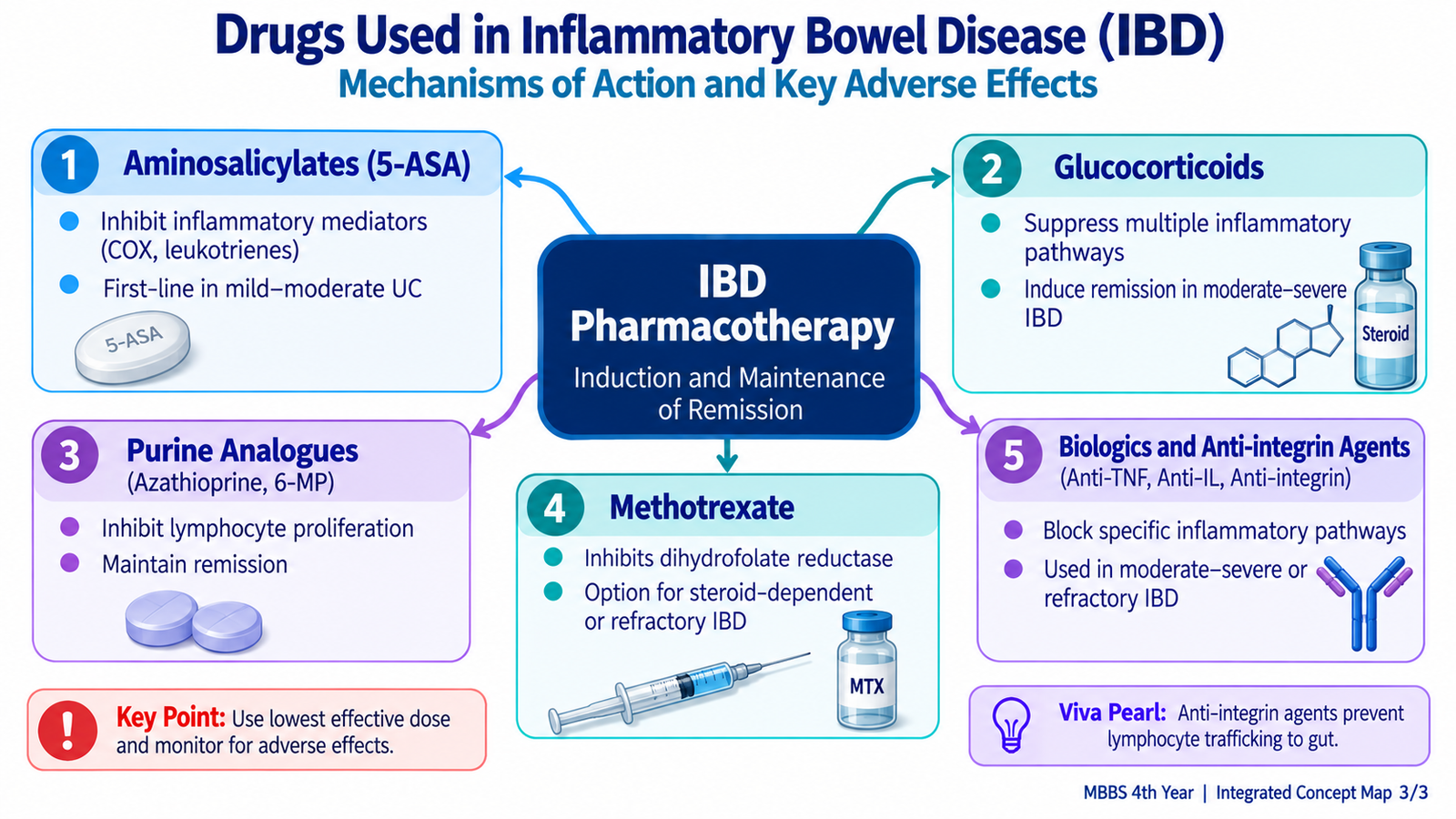
Drug treatment in IBD aims to suppress active inflammation, achieve remission and then maintain disease control. The appropriate class depends on the type of IBD, disease severity, distribution and previous response. Some drugs act locally in the intestinal mucosa, whereas others suppress systemic immune responses or selectively block inflammatory pathways.
1. Aminosalicylates
Mesalamine and sulfasalazine deliver 5-aminosalicylic acid activity to the inflamed bowel. They reduce production and action of inflammatory mediators within the intestinal mucosa. Their role is greatest in mild-to-moderate ulcerative colitis.
Adverse effects: nausea, abdominal discomfort, headache and hypersensitivity may occur. Renal injury is uncommon but important. Sulfasalazine can additionally produce folate deficiency, reversible impairment of male fertility and adverse reactions related to its sulfapyridine component.
2. Glucocorticoids
Glucocorticoids enter cells and alter transcription of genes involved in inflammation. They reduce cytokine production, leukocyte activation and inflammatory tissue injury. Because they act rapidly, they are valuable for induction of remission during active disease, but their toxicity makes them unsuitable as routine long-term maintenance therapy.
Important adverse effects: increased infection risk, hyperglycemia, hypertension, osteoporosis, mood changes, cataract formation and suppression of the hypothalamic-pituitary-adrenal axis.
3. Purine Analogues
Azathioprine and 6-mercaptopurine interfere with purine metabolism and reduce proliferation and activity of immune cells. Their onset is slow, so they are not drugs for immediate control of an acute severe flare. Their main role is as steroid-sparing immunomodulators for maintaining disease control in selected patients.
Important adverse effects: bone-marrow suppression, hepatotoxicity, pancreatitis and increased susceptibility to infection.
4. Methotrexate
Methotrexate interferes with folate-dependent cellular pathways and has immunomodulatory effects. In IBD, its established role is greater in Crohn disease than in UC. It may be used as a steroid-sparing agent in selected patients.
Important adverse effects: bone-marrow suppression, hepatotoxicity, oral ulceration, pulmonary toxicity and teratogenicity.
5. Monoclonal Antibodies
Biological drugs target specific inflammatory mediators rather than producing broad nonspecific immunosuppression. Important examples are anti-TNF monoclonal antibodies such as infliximab and adalimumab.
These agents can be particularly valuable in disease that does not respond adequately to conventional therapy and in selected fistulizing Crohn disease.
Important adverse effects: serious infection, reactivation of latent infections, infusion or injection reactions and other immune-mediated complications. Patients require appropriate clinical assessment before and during therapy.
6. Anti-Integrin Agents
Integrins are adhesion molecules that help lymphocytes move from blood into tissues. Vedolizumab blocks gut-directed lymphocyte trafficking by interfering with the α4β7 integrin pathway. Fewer inflammatory lymphocytes therefore reach the intestinal mucosa.
Potential adverse effects include infusion-related reactions and infection. Integrin-directed drugs differ in their tissue selectivity; this is clinically important because broader integrin blockade can produce more serious neurological risk than gut-selective therapy.
| Drug Class | Main Mechanistic Idea | Major Clinical Role | Important Toxicity |
|---|---|---|---|
| Aminosalicylates | Reduce mucosal inflammatory mediator activity | Especially UC | GI effects, hypersensitivity, renal injury |
| Glucocorticoids | Suppress inflammatory gene transcription | Induction during active disease | Infection, metabolic and bone effects |
| Purine analogues | Reduce lymphocyte proliferation | Steroid-sparing maintenance | Myelosuppression, hepatotoxicity, pancreatitis |
| Methotrexate | Folate-related immunomodulation | Selected Crohn disease | Hepatic and marrow toxicity, teratogenicity |
| Anti-TNF antibodies | Neutralize TNF signalling | Moderate-to-severe/refractory IBD | Serious infection/reactivation |
| Anti-integrin agents | Reduce lymphocyte trafficking into gut | Moderate-to-severe IBD | Infection and infusion reactions |

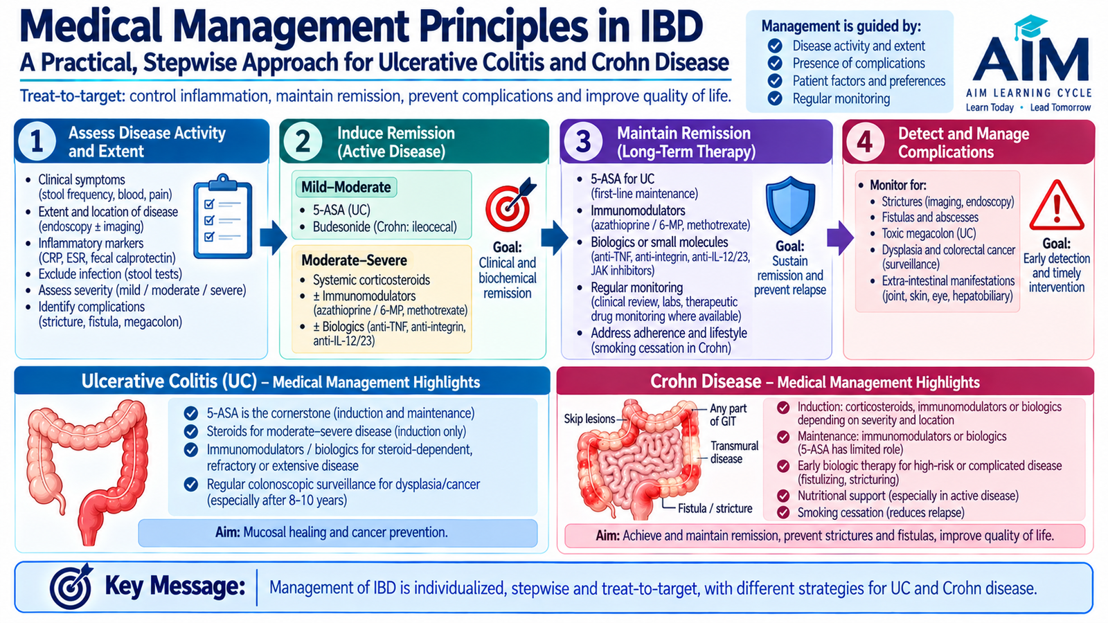
G. Medical Management Principles in Ulcerative Colitis and Crohn Disease
Management is planned according to the diagnosis, site, severity, complications and previous treatment response. A useful distinction is between induction of remission, in which active inflammation is brought under control, and maintenance of remission, in which treatment is continued to reduce relapse. The management plan also includes correction of nutritional problems and identification of complications rather than focusing on drug therapy alone.
Ulcerative Colitis
Mild-to-moderate UC is commonly managed with an aminosalicylate because the disease is predominantly mucosal and colonic. More active disease may require a glucocorticoid for induction. Patients who have persistent, steroid-dependent or more severe disease may require an immunomodulator or biologic agent.
A patient with severe colitis, systemic illness, major bleeding or suspected toxic megacolon requires hospital assessment because rapid deterioration, perforation and the need for surgery are possible.
Crohn Disease
Crohn disease treatment must account for the site and behaviour of disease. Corticosteroids can control active inflammatory disease, while immunomodulators and biological agents are used when longer-term immune control is required. Aminosalicylates have a much less important role than in UC.
Medical treatment alone cannot correct a fixed fibrotic stricture or drain a significant abscess. This is why distinguishing active inflammation from a structural complication is clinically important.
Supportive Principles
- Correct dehydration and electrolyte disturbances when significant diarrhea is present.
- Assess and manage anemia and nutritional deficiencies.
- Address weight loss and inadequate nutritional intake.
- Avoid cigarette smoking in Crohn disease because it is associated with worse disease behaviour.
- Investigate new deterioration for infection and complications rather than automatically assuming that symptoms represent uncomplicated inflammatory activity.
Active inflammatory disease → anti-inflammatory or immunomodulatory therapy.
Fixed obstruction, perforation, abscess or other structural complication → procedural or surgical assessment.
H. Surgical Management of Ulcerative Colitis and Crohn Disease
Surgery has fundamentally different implications in UC and Crohn disease. In UC, the inflammatory disease is restricted to the colon and rectum, so removal of the affected large bowel can eliminate intestinal UC. In Crohn disease, inflammation may occur at multiple gastrointestinal sites and can recur after resection. Therefore, Crohn surgery treats complications or localized refractory disease rather than curing the underlying tendency to inflammation.
Surgery in Ulcerative Colitis
Important reasons for surgical intervention include:
- Acute severe colitis that fails appropriate medical treatment.
- Toxic megacolon.
- Perforation.
- Severe uncontrolled hemorrhage.
- Dysplasia or colorectal carcinoma.
- Chronic disease that remains unacceptable despite medical management.
The broad surgical principle is removal of the colon and rectum. Depending on clinical circumstances, reconstruction may permit intestinal continuity using an ileal pouch connected to the anus, or an ileostomy may be required. Detailed operative technique is not necessary for understanding the undergraduate principle.
Surgery in Crohn Disease
Common indications include:
- Persistent obstruction from a fibrotic stricture.
- Abscess or penetrating septic complication.
- Complex fistula requiring intervention.
- Perforation.
- Significant bleeding.
- Localized disease that remains uncontrolled despite appropriate medical therapy.
- Malignancy or strong suspicion of malignancy.
Because Crohn disease can recur in remaining bowel, the surgeon tries to preserve as much healthy intestine as practical. Limited resection or bowel-preserving approaches may therefore be used when appropriate. Repeated extensive resection can eventually reduce absorptive bowel length and contribute to nutritional problems.
UC: surgery can remove the intestinal disease.
Crohn disease: surgery treats complications but recurrence can occur elsewhere.

I. Surveillance Colonoscopy and Long-Term Cancer Risk in Ulcerative Colitis
Longstanding inflammation of the colonic mucosa can promote a sequence of epithelial injury, regeneration and accumulation of neoplastic changes. For this reason, patients with longstanding colonic UC have an increased risk of colorectal dysplasia and carcinoma. Surveillance colonoscopy is therefore performed not because the patient necessarily has symptoms of cancer, but because precancerous mucosal change may develop before an invasive cancer becomes clinically obvious.
Purpose of Surveillance
- Examine the colon for visible suspicious mucosal lesions.
- Detect dysplasia before progression to invasive colorectal cancer.
- Obtain targeted or appropriately selected biopsies according to the surveillance technique being used.
- Identify patients who may need closer specialist follow-up or surgical assessment.
Who Requires Greater Attention?
Cancer risk is influenced by factors such as the duration and anatomical extent of colonic inflammation, persistent inflammatory activity, associated primary sclerosing cholangitis, family history and previous dysplasia. Therefore, surveillance is individualized rather than being identical for every patient.
The exact starting point and interval of surveillance depend on the patient’s risk profile and the guideline being followed. The important undergraduate principle is that longstanding colonic UC requires planned surveillance for dysplasia and colorectal cancer.
Chronic colonic inflammation → repeated epithelial injury and regeneration → dysplasia risk → colorectal carcinoma risk → surveillance aims to detect precancerous change early.

Integrated Mechanism Flow
Genetic and environmental influences
Abnormal interaction with luminal antigens
Persistent inflammatory signalling
UC: mucosal; Crohn: transmural
Diarrhea, bleeding, pain and systemic features
Megacolon versus stricture/fistula
Important Comparison: Ulcerative Colitis versus Crohn Disease
| Feature | Ulcerative Colitis | Crohn Disease |
|---|---|---|
| Site | Colon only | Any GI segment; terminal ileum/colon common |
| Rectum | Typically involved | May be spared |
| Distribution | Continuous | Skip lesions |
| Depth | Mainly mucosal | Transmural |
| Gross pattern | Friable mucosa, superficial ulcers, pseudopolyps | Deep fissures, cobblestoning, wall thickening, creeping fat |
| Histology clue | Crypt architectural distortion and crypt abscesses in active disease | Transmural inflammation; non-caseating granulomas may occur |
| Typical stool pattern | Bloody diarrhea common | Chronic diarrhea; may be non-bloody |
| Characteristic complication | Toxic megacolon | Stricture, fistula, abscess |
| Smoking | Different epidemiological relationship; smoking is not recommended as therapy | Associated with worse disease course |
| Surgical principle | Removal of colon/rectum can eliminate intestinal UC | Not curative; recurrence may occur |
⭐ AIM High-Yield Review
Inflammatory Bowel Disease (IBD): Ulcerative Colitis & Crohn Disease
Video support for pathophysiology, clinical features, complications, diagnosis and management of inflammatory bowel disease.