
This chapter follows the KMU learning outcomes and connects pathology with the surgical approach in a logical sequence. First understand how gastric polyps and cancers develop, then connect morphology with diagnosis and management. Finish with the AIM High-Yield Review for focused revision.
Gastric Polyps, Gastric Cancer and Gastric Outlet Obstruction
A structured study of gastric polyps and tumors, the development and morphology of gastric carcinoma, its diagnostic and surgical management principles, and the causes, diagnosis and management of gastric outlet obstruction.
Topic Introduction
Gastric polyps are localized projections of the gastric mucosa, but they are not all true tumors. Some develop as a response to chronic mucosal injury, whereas others are neoplastic and may carry a risk of malignant transformation. Gastric cancer, particularly gastric adenocarcinoma, develops through several pathological pathways and may present as a localized mass, an ulcerative lesion or diffuse thickening of the stomach wall. Understanding its risk factors, pathogenesis and morphology helps explain how it is diagnosed and treated. Gastric outlet obstruction is an important complication of benign or malignant disease near the pylorus and produces characteristic vomiting, dehydration and biochemical abnormalities. This chapter connects these pathological changes with practical diagnosis and surgical management.
A. Gastric Polyps: Classification, Pathogenesis and Morphology
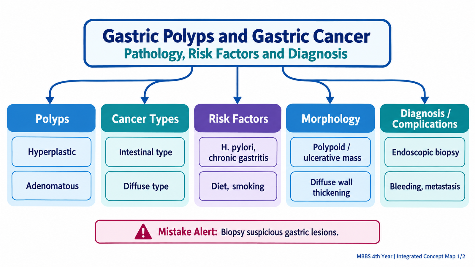
A gastric polyp is a localized lesion that projects above the surrounding gastric mucosa. The word “polyp” describes its shape rather than its biological behaviour. Some gastric polyps are produced by reactive or regenerative mucosal changes, while others represent true neoplasms. This distinction is important because their risk of progression to carcinoma is different.
Classification
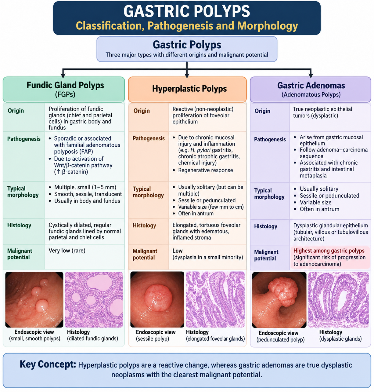
The common gastric epithelial polyps can be organized into three clinically important groups:
- Fundic gland polyps — composed of cystically dilated glands in the body and fundus.
- Hyperplastic polyps — regenerative lesions that usually arise in chronically injured or inflamed mucosa.
- Gastric adenomas — true dysplastic epithelial neoplasms with definite malignant potential.
Fundic gland polyps
Fundic gland polyps arise from the acid-secreting mucosa of the body and fundus. They consist of glands that become cystically dilated. They may occur sporadically, may be associated with prolonged acid-suppressive therapy, or may occur in association with inherited polyposis syndromes. Most sporadic lesions are small and have very low malignant potential.
Morphology: They are usually small, smooth, well-circumscribed mucosal elevations. Microscopically, the characteristic finding is cystic dilatation of fundic glands lined by relatively bland epithelial cells.
Hyperplastic polyps
Hyperplastic polyps usually represent an exaggerated regenerative response to chronic mucosal injury. Repeated epithelial damage causes increased regeneration of gastric foveolar cells, producing elongated and tortuous surface glands.
Morphology: These lesions are often smooth or lobulated. Microscopically, elongated, branching or cystically dilated foveolar glands are present in an inflamed and edematous stroma. The surrounding mucosa often shows evidence of chronic gastritis.
Most hyperplastic polyps are non-neoplastic. However, dysplasia may occasionally develop, particularly in larger lesions, and the abnormal background mucosa may itself carry an increased cancer risk.
Gastric adenomas
Gastric adenomas are true neoplastic polyps composed of dysplastic epithelium. They frequently arise in a background of chronic atrophic gastritis and intestinal metaplasia. Because dysplasia is already present, adenomas have a much greater relationship with gastric adenocarcinoma than ordinary hyperplastic or sporadic fundic gland polyps.
Morphology: Adenomas may appear elevated, velvety or lobulated. Microscopically, the glands show epithelial dysplasia, including nuclear enlargement, hyperchromasia, crowding and loss of normal maturation.
Diagnosis and complications
Endoscopy identifies the polyp and permits biopsy or removal for histopathological examination. Histology is important because the external appearance alone cannot reliably distinguish a benign regenerative lesion from a dysplastic adenoma.
- Polyps may cause chronic blood loss and anemia if their surface becomes eroded.
- Large lesions near the pylorus may rarely contribute to obstruction.
- The most important complication of an adenoma is progression to adenocarcinoma.
B. Gastric Tumors: Classification and Gastric Carcinoma Types
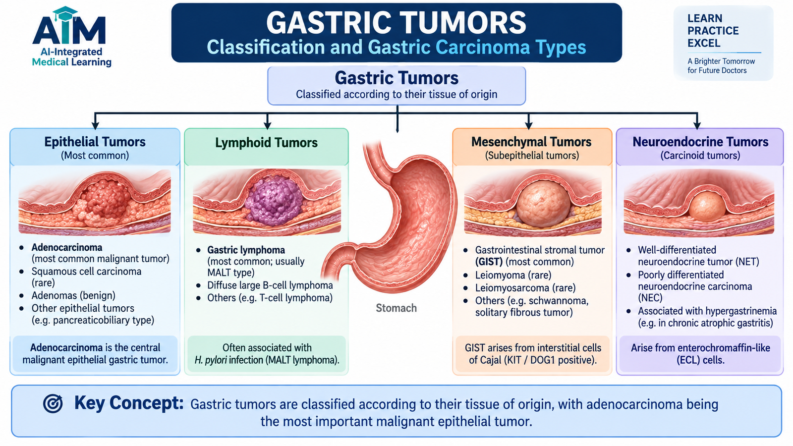
The stomach may develop tumors from several different tissues. The major classification is based on the cell or tissue from which the tumor originates. For this topic, gastric adenocarcinoma is the central malignant tumor because it is the principal gastric carcinoma encountered in surgical practice.
| Tissue of origin | Important gastric tumor | Core concept |
|---|---|---|
| Glandular epithelium | Adenocarcinoma | Main malignant epithelial tumor of stomach |
| Lymphoid tissue | Gastric lymphoma | Malignancy of gastric lymphoid cells |
| Mesenchymal tissue | Gastrointestinal stromal tumor | Major mesenchymal gastric tumor |
| Neuroendocrine cells | Neuroendocrine tumor | Tumor showing neuroendocrine differentiation |
Main histological types of gastric adenocarcinoma
For undergraduate pathology, the most useful division is the intestinal type and the diffuse type. These differ in their pathogenesis and microscopic appearance.
- Intestinal type: malignant cells form gland-like structures and usually develop through a sequence of chronic mucosal injury, atrophy, intestinal metaplasia and dysplasia.
- Diffuse type: poorly cohesive malignant cells infiltrate the stomach wall individually or in small groups. Signet-ring cells are characteristic.
Early and advanced gastric carcinoma
Early gastric carcinoma is confined to the mucosa and submucosa, irrespective of whether regional lymph nodes are involved. Once the tumor extends deeper through the gastric wall, it is considered advanced. This distinction matters because very superficial cancers may be suitable for less extensive local treatment, whereas deeper invasive cancers generally require formal oncological surgery and additional staging.
C. Gastric Adenocarcinoma: Etiology, Risk Factors and Pathogenesis
Gastric adenocarcinoma develops when gastric epithelial cells accumulate changes that allow uncontrolled growth, survival and invasion. However, the pathway is not identical in every patient. The intestinal type commonly develops on a background of long-standing mucosal injury and metaplastic change, whereas the diffuse type is more strongly associated with loss of cell-to-cell adhesion.
Important risk factors
Risk factors are important because they help explain why chronic epithelial injury and abnormal regeneration can eventually produce dysplasia and carcinoma.
- Helicobacter pylori-associated chronic gastritis, particularly when prolonged inflammation progresses to mucosal atrophy and intestinal metaplasia.
- Chronic atrophic gastritis, including autoimmune atrophic gastritis.
- Intestinal metaplasia and gastric epithelial dysplasia.
- Gastric adenomas, because they already contain dysplastic epithelium.
- Smoking.
- A diet repeatedly rich in heavily salted, preserved or smoked foods.
- Previous gastric surgery with long-standing mucosal alteration in the gastric remnant.
- Inherited predisposition in a small proportion of patients, particularly abnormalities affecting CDH1/E-cadherin in hereditary diffuse gastric cancer.
Intestinal-type pathway
Long-standing inflammation damages the normal gastric glands. Repeated injury produces glandular loss or atrophy. The mucosa may then replace its normal gastric epithelial pattern with intestinal-type epithelium, called intestinal metaplasia. Further accumulation of abnormalities produces dysplasia, followed by invasive carcinoma.
This pathway explains why the intestinal type often arises in a stomach showing pre-existing chronic gastritis, atrophy and metaplastic change.
Diffuse-type pathway
The diffuse type does not depend on formation of intestinal-type glands. Instead, impaired function of E-cadherin, an important cell-adhesion molecule, reduces adhesion between epithelial cells. The malignant cells therefore become discohesive and spread individually through the stomach wall.
Some tumor cells contain abundant intracellular mucin that pushes the nucleus to one side, creating the classic signet-ring cell appearance.

D. Gastric Cancer: Morphology, Spread and Complications
Morphology describes how gastric cancer appears to the naked eye and under the microscope. These structural changes are clinically important because they determine whether a tumor causes ulceration, bleeding, obstruction or diffuse loss of gastric distensibility, and they influence how the disease spreads.
Gross morphology
Advanced gastric cancers may have several gross patterns:
- Exophytic or fungating: a mass projects into the gastric lumen.
- Ulcerative: the tumor forms an irregular ulcer with raised or infiltrated margins.
- Infiltrative: malignant cells spread widely through the wall and produce marked thickening and rigidity.
Diffuse infiltration may involve a large part of the stomach. The wall becomes thick, firm and poorly distensible, producing the classic appearance called linitis plastica or a “leather-bottle” stomach.
Microscopic morphology
In intestinal-type adenocarcinoma, malignant epithelial cells retain enough cohesion to form abnormal glandular structures. In the diffuse type, cells are poorly cohesive and infiltrate singly or in small clusters. Signet-ring cells may be prominent because intracellular mucin displaces the nucleus toward the cell edge.
Routes of spread
As the carcinoma penetrates deeper layers of the gastric wall, it gains access to lymphatic vessels, blood vessels and the peritoneal surface. This explains the major metastatic patterns.
- Direct extension into surrounding tissues and adjacent organs.
- Lymphatic spread to regional gastric nodes and, in advanced disease, more distant lymph nodes.
- Hematogenous spread, particularly to the liver.
- Transcoelomic spread across the peritoneal cavity, producing peritoneal deposits and sometimes ovarian metastases.
Complications
Complications follow directly from ulceration, progressive local growth and metastatic spread.
- Chronic blood loss and anemia: caused by ulceration and bleeding from the tumor surface.
- Gastric outlet obstruction: especially when a tumor narrows the distal stomach or pyloric region.
- Reduced food intake and weight loss: due to impaired gastric function and the systemic effects of malignancy.
- Perforation: uncommon, but possible when tumor destruction extends through the gastric wall.
- Metastatic disease: involving lymph nodes, liver, peritoneum or other sites.

E. Gastric Cancer: Diagnostic Approach and Laboratory Confirmation
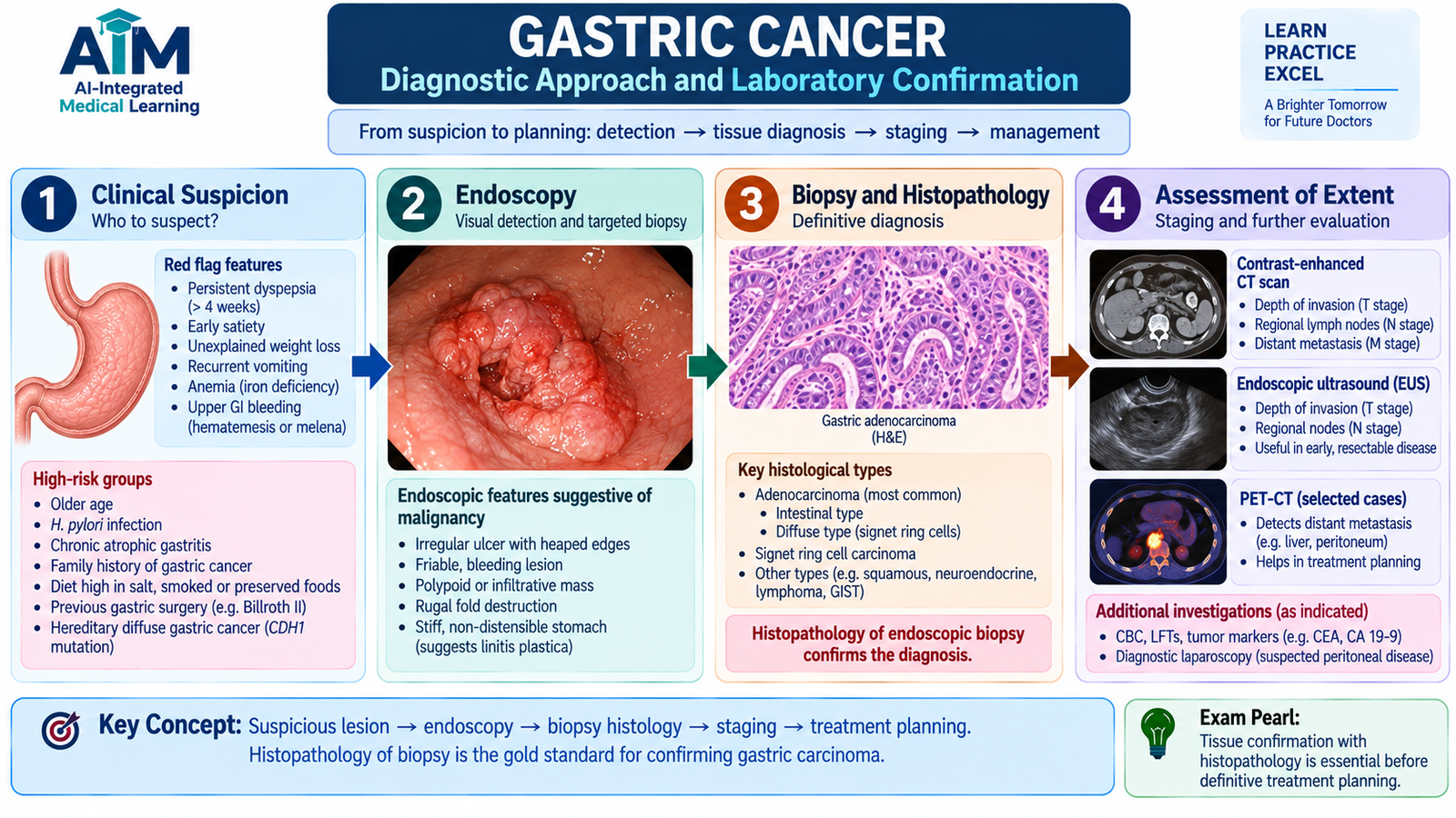
The diagnosis of gastric cancer requires direct visualization of the lesion and histopathological confirmation. Routine blood tests may show consequences of the disease, but they cannot establish the diagnosis by themselves. After cancer is confirmed, imaging is used to determine its extent and guide treatment planning.
Clinical suspicion
Gastric malignancy may be suspected when persistent upper gastrointestinal symptoms are accompanied by concerning features such as progressive weight loss, reduced appetite, chronic gastrointestinal blood loss, anemia, persistent vomiting or evidence of gastric outlet obstruction. These findings are not specific, so investigation is required.
Laboratory assessment
Laboratory tests mainly identify the physiological effects or complications of the cancer.
- Complete blood count: may demonstrate anemia, particularly when there has been chronic occult bleeding.
- Iron studies: may support iron deficiency when chronic gastrointestinal blood loss has occurred.
- Renal and electrolyte assessment: is important when vomiting and dehydration are present.
- Liver biochemical tests: may become abnormal if there is significant hepatic involvement, but they are not specific for gastric cancer.
Serum tumor markers may sometimes be used as additional information in patients with established malignancy, but they are not sufficiently specific to replace endoscopy and tissue diagnosis.
Upper gastrointestinal endoscopy and biopsy
Upper gastrointestinal endoscopy allows direct inspection of the gastric mucosa. A suspicious mass, ulcerated lesion or infiltrated area can be sampled. Histological examination of endoscopic biopsy is the definitive method for confirming gastric carcinoma.
The pathologist assesses the tissue architecture and cytological features to identify malignancy and determine the histological pattern. Intestinal-type carcinoma shows malignant gland formation, whereas diffuse carcinoma shows poorly cohesive cells and may contain signet-ring cells.
Assessment of disease extent
Once carcinoma has been confirmed, cross-sectional imaging is used to look for local extension, lymph-node disease and distant metastases. Endoscopic ultrasonography may provide additional information about the depth of invasion and regional nodes in selected patients. The purpose is not merely to describe the tumor, but to determine whether potentially curative treatment is technically and oncologically appropriate.
F. Gastric Cancer: Management Principles
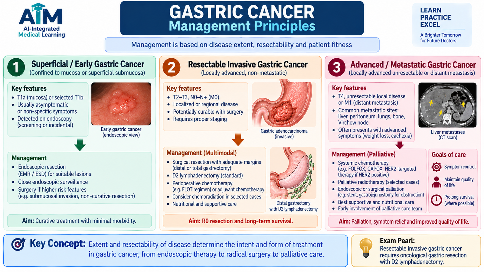
Management depends mainly on the depth and extent of the tumor, whether complete removal is possible, the presence of metastatic disease and the patient’s overall condition. Treatment should therefore follow proper staging rather than using the same operation for every patient. Surgery is the central potentially curative treatment for resectable invasive gastric carcinoma.
Very early superficial disease
Selected cancers confined to the superficial mucosa and meeting appropriate pathological criteria may be removed endoscopically. The purpose is to remove the complete superficial lesion while avoiding major gastric surgery when the probability of deeper or nodal disease is sufficiently low.
Resectable invasive gastric cancer
For a localized invasive carcinoma that can be completely removed, the principle is oncological gastric resection together with appropriate regional lymph-node removal.
- A distal lesion may be treated by an oncologically adequate distal or subtotal gastrectomy when suitable.
- More extensive or proximally located disease may require total gastrectomy.
- The operative objective is complete removal of the primary tumor with clear margins while addressing regional lymphatic spread.
The exact operation depends on tumor location and extent. Detailed operative technique is a specialist subject and is not required for understanding the undergraduate management principle.
Role of systemic oncological treatment
Many patients with invasive gastric cancer require systemic anticancer therapy in addition to local treatment. Depending on stage and resectability, systemic therapy may be incorporated around surgery or used for advanced disease. The important undergraduate concept is that treatment is multidisciplinary: surgery controls removable local disease, while oncological treatment addresses the risk of disease beyond the primary tumor.
Unresectable or metastatic disease
When cure by complete resection is not possible, treatment focuses on controlling tumor burden and relieving major symptoms. Important goals include maintaining nutrition, treating anemia or bleeding, controlling pain and relieving obstruction.
- Systemic therapy may be used where appropriate.
- Endoscopic stenting may restore passage through a malignant obstruction in selected patients.
- A surgical bypass may be considered when obstruction requires durable palliation and the patient is suitable.
- Supportive nutritional and symptom-directed care remains important throughout treatment.
G. Gastric Outlet Obstruction: Etiology, Diagnosis and Management
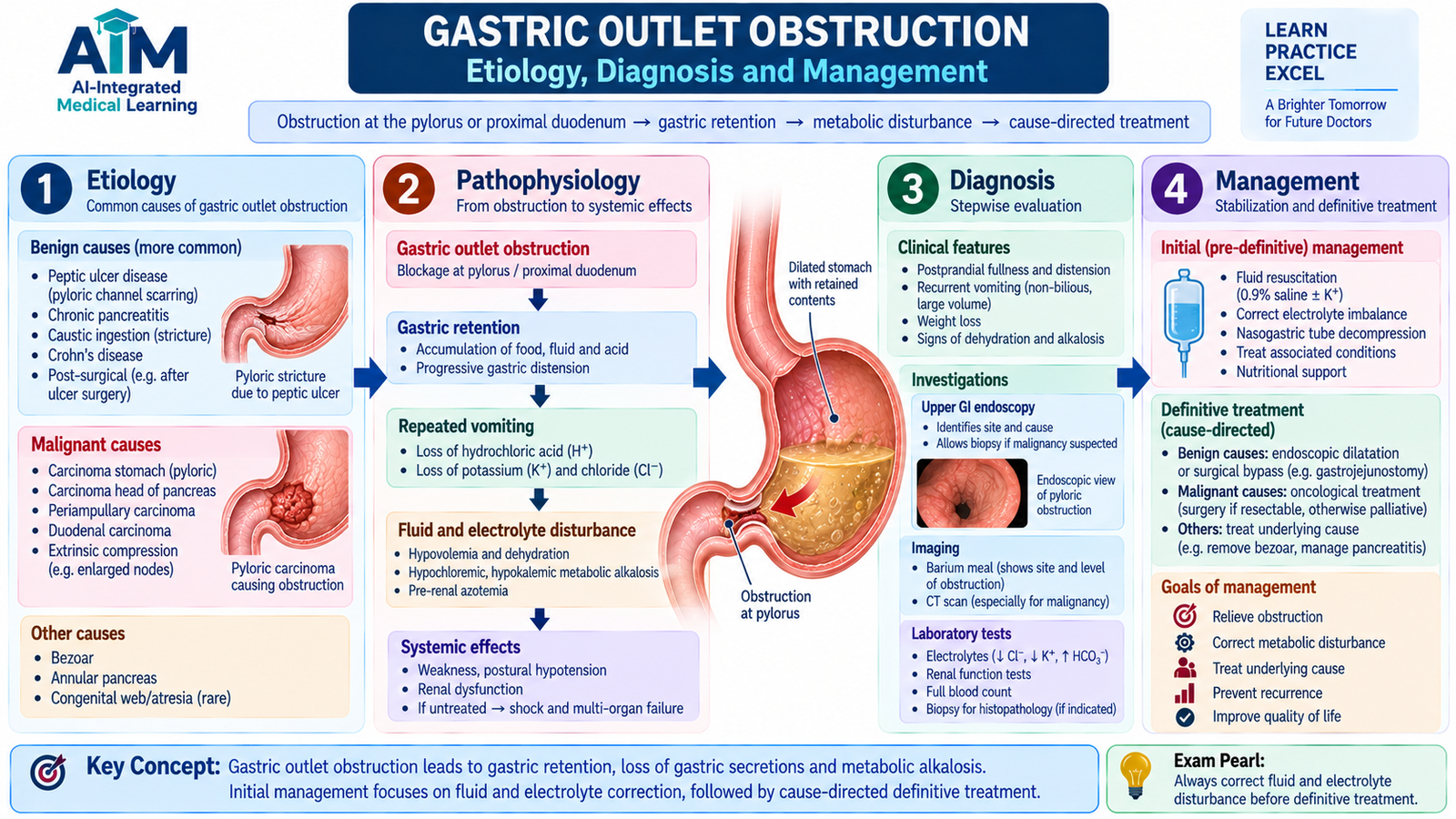
Gastric outlet obstruction (GOO) is a mechanical or functional blockage that prevents normal emptying of gastric contents through the distal stomach and proximal duodenum. Food and gastric secretions therefore accumulate in the stomach. Persistent obstruction causes repeated vomiting, dehydration, electrolyte loss and progressive nutritional deterioration.
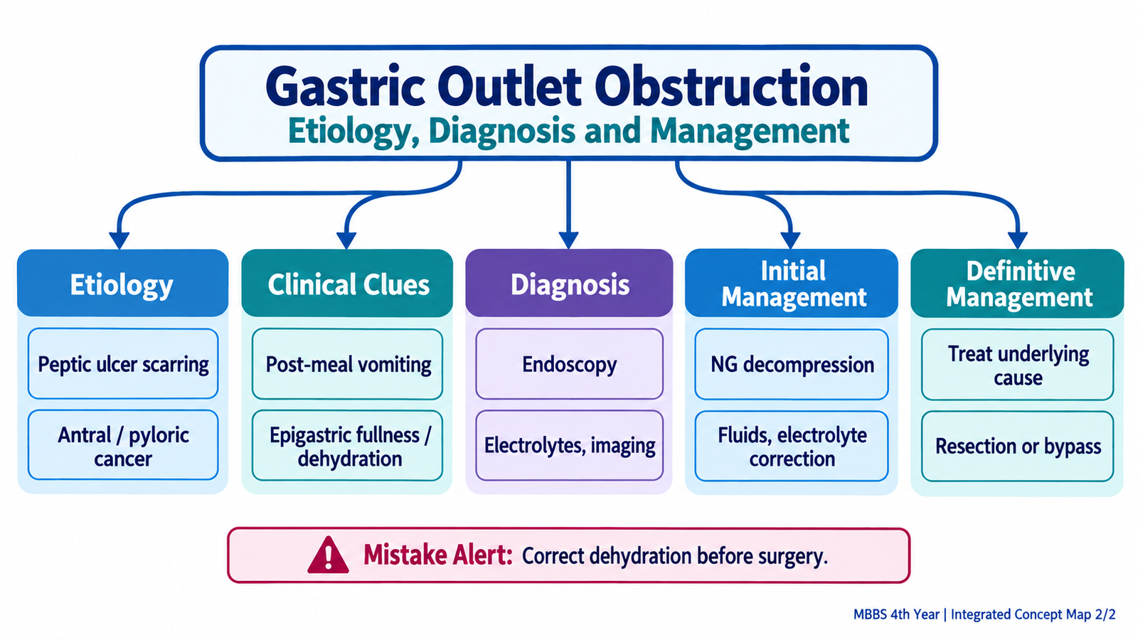
Etiology
The causes can broadly be divided into benign and malignant obstruction.
- Peptic ulcer-related obstruction: acute inflammation and edema may narrow the pyloroduodenal region, while repeated ulceration may later produce fibrotic scarring and fixed stenosis.
- Distal gastric carcinoma: progressive tumor growth can narrow the pylorus or distal stomach.
- Other malignant lesions: tumors arising around the proximal duodenum or pancreas may compress or invade the gastric outlet.
- Less common benign strictures may follow severe inflammatory or corrosive injury.
Clinical and biochemical consequences
Because gastric contents cannot pass normally into the duodenum, patients develop recurrent vomiting of retained gastric contents. Vomiting is commonly non-bilious when the obstruction lies proximal to entry of bile into the duodenum.
- Early satiety and postprandial fullness may occur because the stomach remains distended.
- Repeated vomiting leads to dehydration and weight loss.
- Loss of gastric hydrochloric acid removes hydrogen and chloride ions from the body.
- Volume depletion activates renal mechanisms that further promote potassium and hydrogen ion loss.
On examination, the upper abdomen may be distended. Visible gastric peristalsis may occur as the stomach contracts forcefully against an obstruction. A succussion splash after an appropriate period of fasting suggests retained fluid and gas in the stomach.
Diagnosis
Diagnosis begins by confirming that gastric emptying is impaired and then identifying the cause.
- Blood tests: assess dehydration, renal function and electrolyte/acid-base disturbance.
- Upper gastrointestinal endoscopy: directly identifies pyloric or distal gastric narrowing and permits biopsy when malignancy is suspected.
- Cross-sectional imaging: helps define a gastric mass, extrinsic compression, regional disease and other causes of obstruction.
Endoscopic biopsy is particularly important when obstruction is associated with an abnormal gastric mass because management differs greatly between benign scar-related stenosis and malignant obstruction.
Initial management
Before definitive treatment, the physiological consequences of obstruction must be corrected. Surgery or endoscopic intervention is less safe in a severely dehydrated patient with major electrolyte disturbance.
- Temporarily stop oral intake when significant obstruction is present.
- Restore circulating volume with intravenous fluid.
- Correct electrolyte and acid-base abnormalities.
- Use nasogastric decompression when substantial gastric retention or repeated vomiting requires relief.
- Assess and support nutritional status.
Definitive management
Definitive treatment depends on the underlying cause.
Benign obstruction: Active inflammatory narrowing from peptic ulcer disease may improve as the underlying ulcer disease is treated. A persistent fibrotic stricture may be suitable for endoscopic balloon dilatation, while refractory or unsuitable benign obstruction may require surgery.
Malignant obstruction: If the gastric cancer is resectable and the patient is suitable, treatment follows oncological resection principles. If the disease is not curable by resection, passage may be restored by an endoscopic stent or by surgical bypass in appropriately selected patients.
Integrated Mechanism Flow
This sequence explains the common intestinal-type progression. Diffuse gastric carcinoma follows a different pathway in which reduced cell adhesion produces widespread infiltration by poorly cohesive cells.
Important Comparison: Intestinal vs Diffuse Gastric Adenocarcinoma
| Feature | Intestinal type | Diffuse type |
|---|---|---|
| Typical pathway | Chronic gastritis → atrophy → metaplasia → dysplasia | Loss of effective cell adhesion |
| Cell cohesion | Relatively cohesive | Poorly cohesive |
| Microscopy | Malignant gland formation | Dispersed cells; signet-ring cells may occur |
| Gross tendency | Mass-forming or ulcerative lesion | Diffuse wall infiltration |
| Characteristic concept | Precursor mucosal changes are important | May produce linitis plastica |
⭐ AIM High-Yield Review
🎥 Video Learning: Gastric Polyps and Gastric Tumors
Watch this video after completing the learning material to reinforce the classification, pathology and morphology of gastric polyps and gastric tumors.