
8. Corrosive, Acid, Alkali and Cyanide Poisoning
Topic Introduction
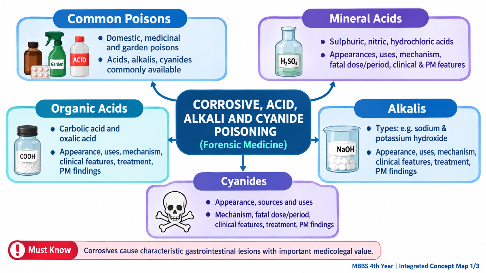
Corrosive poisons cause direct chemical destruction of tissues with which they come into contact. Strong mineral acids, alkalis and some organic acids therefore mainly damage the mouth, pharynx, esophagus and stomach after ingestion, although systemic toxicity may also occur. Different corrosives produce different types of tissue necrosis, and these mechanisms explain many of their characteristic clinical and postmortem appearances. Cyanide is different: its major action is not corrosion but rapid inhibition of cellular oxygen utilization. In this chapter, you will learn the important sources, appearances, mechanisms, fatal-dose concepts, clinical features, treatment principles, postmortem findings and forensic importance of the poisons specified in the KMU curriculum.
A. Orientation: Common Poisons and Toxicological Classification
A poison is a substance capable of causing illness or death when it enters the body in a sufficient amount. In forensic practice, the source of exposure often provides an important clue. Poisons may be encountered in homes, medicines, workplaces, gardens and agricultural environments. The purpose of recognizing these groups is not to memorize every possible poison, but to understand where clinically important exposures may arise.
Common Sources
- Domestic poisons: household cleaning acids and alkalis, bleach-containing products, phenolic disinfectants, kerosene and some rodenticides or insecticides.
- Medicinal poisons: analgesics, sedative-hypnotics and other drugs when taken in excessive amounts.
- Garden and agricultural poisons: pesticides, herbicides, rodenticides and toxic plant products.
In the present topic, the major groups are mineral acids, alkalis, organic acids and cyanides. The first three can produce important local tissue injury. Cyanide primarily produces rapid systemic cellular toxicity.

B. Mineral Acids: Classification and Core Corrosive Mechanism
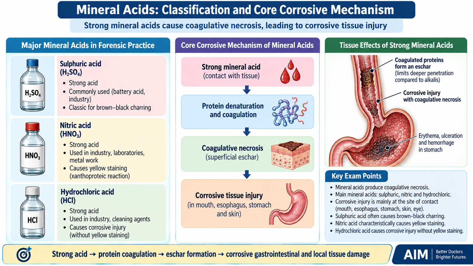
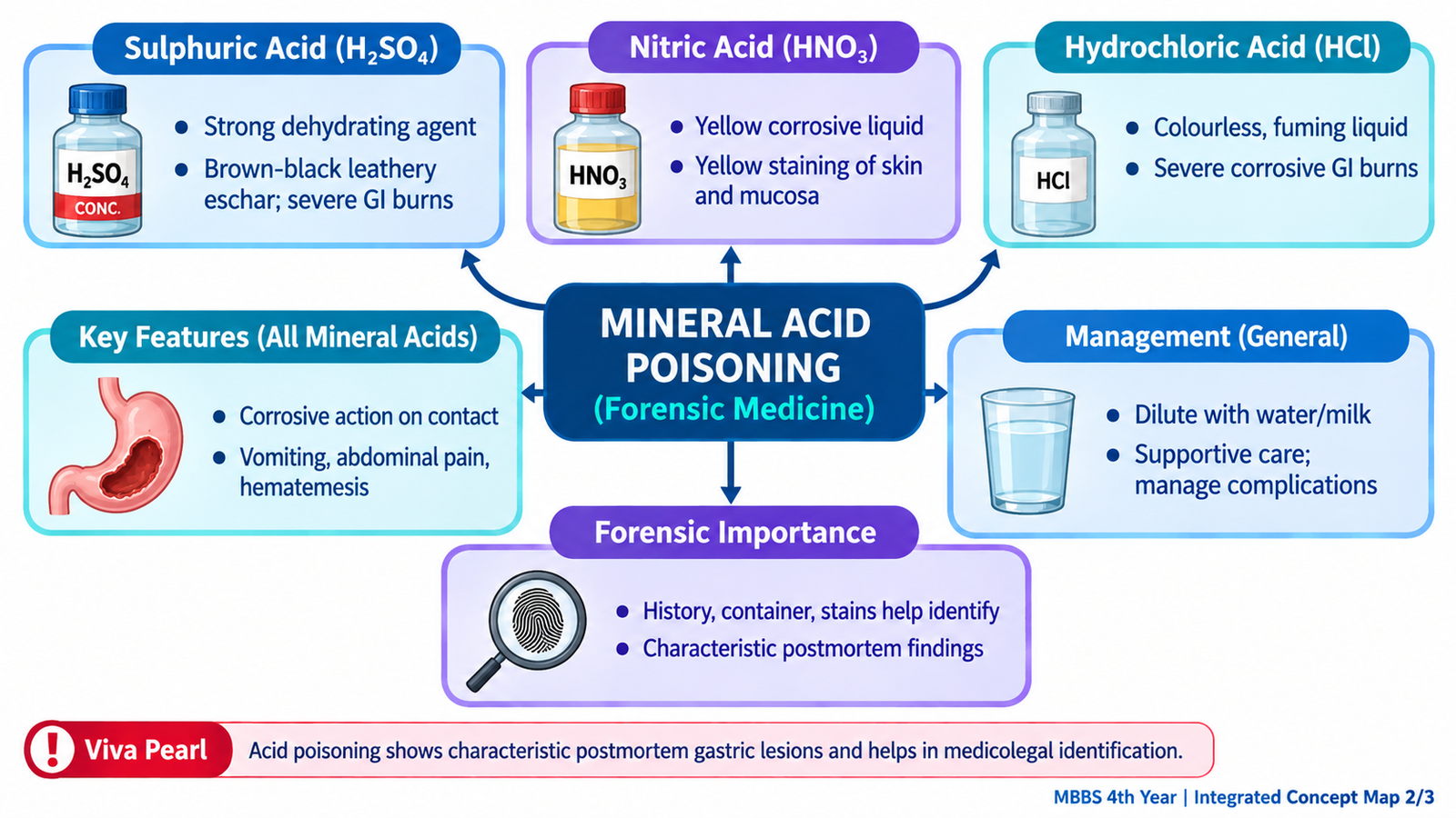
Mineral acids are strong inorganic acids capable of producing chemical burns. The important mineral acids in forensic toxicology are sulphuric acid, nitric acid and hydrochloric acid. Their common action is release of hydrogen ions and chemical destruction of proteins and tissues. Concentrated acids also remove water from tissues and produce protein coagulation.
Classification
- Sulphuric acid — H₂SO₄
- Nitric acid — HNO₃
- Hydrochloric acid — HCl
How Strong Acids Damage Tissue
Coagulated proteins may form an eschar, a layer of chemically damaged tissue. This can partially limit deeper penetration compared with the liquefactive necrosis produced by strong alkalis, although concentrated acids can still cause devastating full-thickness injury.
Acid ingestion typically produces immediate burning pain, salivation, painful swallowing, vomiting and upper gastrointestinal injury. Vomitus may contain blood when mucosal vessels are damaged. Airway edema may occur when the mouth, pharynx or larynx is involved.
C. Sulphuric Acid Poisoning
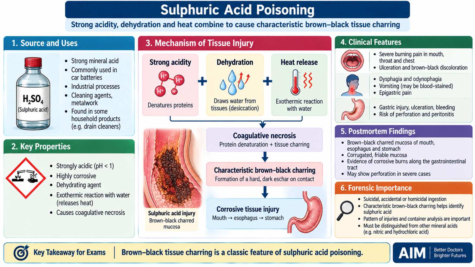
Sulphuric acid is one of the most powerful mineral acid corrosives. Concentrated sulphuric acid produces tissue damage through its strong acidity, marked dehydrating action and heat generation when it mixes with water. These combined effects account for its characteristic dark, charred appearance in severely affected tissues.
Appearance and Uses
Concentrated sulphuric acid is typically a colorless, heavy, oily and highly corrosive liquid. Commercial preparations may appear darker because of impurities. Important uses include industrial chemical manufacture, fertilizers, metal processing and lead-acid batteries.
Mechanism of Injury
Sulphuric acid causes severe protein coagulation and removes water from tissues. Contact with water is strongly exothermic, meaning heat is released. The combination of chemical corrosion, dehydration and heat produces deep burns and may result in dark brown or black charring.
Fatal Dose and Fatal Period
Traditional undergraduate forensic teaching gives an approximate fatal dose of 5–10 mL of concentrated sulphuric acid, with a commonly quoted fatal period of approximately 12–24 hours. These values are not absolute. Outcome depends on concentration, amount actually swallowed, aspiration, site of injury and the speed of medical treatment.
Clinical Features
- Immediate burning pain in the mouth, throat, retrosternal region and epigastrium.
- Burns around the lips and oral cavity.
- Drooling and painful or difficult swallowing because of mucosal injury.
- Vomiting, sometimes with altered or fresh blood.
- Abdominal tenderness due to gastric injury.
- Hoarseness, stridor or respiratory difficulty if the upper airway is involved.
- Severe exposure may cause hypovolemic shock, perforation and peritonitis.
- Healing of severe injury may later produce fibrotic narrowing, particularly gastrointestinal strictures.
Treatment Principles
There is no specific antidote. Management is based on preventing further exposure and supporting vital functions while the extent of corrosive injury is assessed.
- Assess and protect the airway, breathing and circulation.
- Remove contaminated clothing and irrigate exposed skin or eyes thoroughly with water.
- Do not induce vomiting, because the corrosive is re-exposed to the upper gastrointestinal tract.
- Do not attempt chemical neutralization with an alkali; neutralization may release heat and worsen tissue injury.
- Gastric lavage is generally avoided in significant corrosive ingestion because of the risk of additional injury or perforation.
- Provide analgesia, intravenous fluid support and treatment of shock when required.
- Specialist assessment of gastrointestinal injury is required after significant ingestion.
- Urgent surgical assessment is necessary when perforation or peritonitis is suspected.
Postmortem Appearance
Corrosive burns may be visible around the mouth and lips. The esophagus and stomach may show extensive brown to black discoloration, necrosis and ulceration. The stomach wall may be softened or perforated, allowing corrosive material to damage adjacent tissues. Hemorrhage and evidence of shock may accompany the local lesions.
Forensic Importance
Exposure may be accidental, suicidal, occupational or associated with deliberate corrosive assault. Sulphuric acid has historically been associated with acid attacks because it can cause severe burns and permanent disfigurement. The presence of characteristic corrosive lesions may support exposure to a strong acid, but the injuries alone cannot establish whether the manner was accidental, suicidal or homicidal.
D. Nitric Acid and Hydrochloric Acid Poisoning
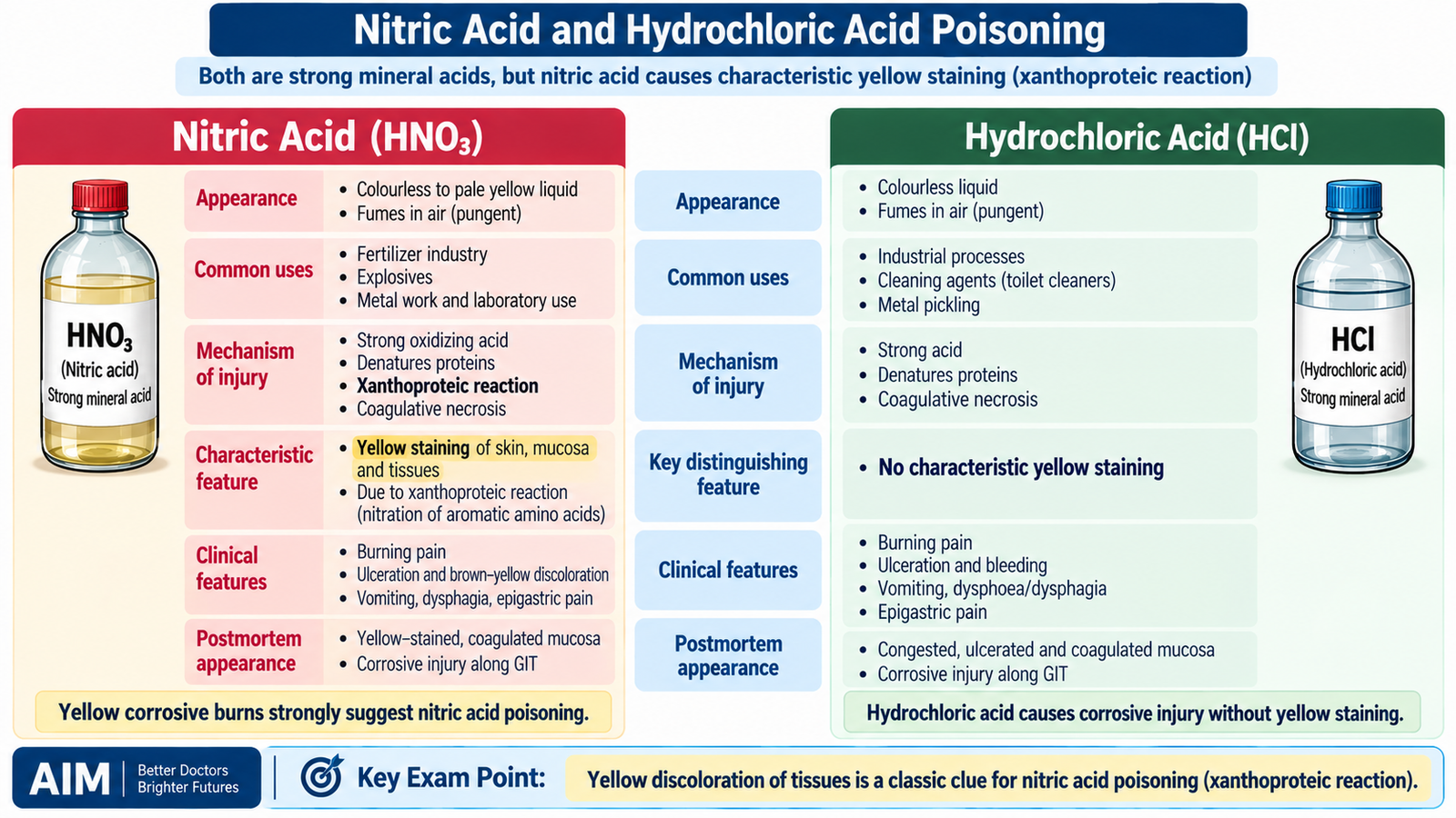
Nitric acid and hydrochloric acid share the general corrosive action of strong mineral acids, but their appearances and tissue effects provide useful distinguishing clues. Nitric acid is particularly memorable because it can produce yellow staining, whereas hydrochloric acid usually lacks such a striking color reaction.
Nitric Acid
Pure nitric acid is a colorless, highly corrosive liquid, but it may acquire a yellow color on standing because of dissolved nitrogen oxides. It has a sharp, irritating character and is used in chemical manufacture, metal work, fertilizers and laboratory processes.
Mechanism and Characteristic Yellow Stain
Nitric acid produces coagulative necrosis like other strong acids. It also reacts with aromatic amino acids in tissue proteins. This xanthoproteic reaction produces characteristic yellow staining of affected skin and mucosa.
Fatal Dose and Fatal Period
A traditionally quoted approximate fatal dose is 10–15 mL of concentrated nitric acid, with a fatal period commonly taught as approximately 12–24 hours. As with all corrosives, these figures vary considerably according to concentration, amount absorbed, complications and treatment.
Clinical Features
- Severe burning pain in the mouth, throat, chest and epigastrium.
- Yellow discoloration of exposed skin or mucosa.
- Salivation, dysphagia and vomiting.
- Hematemesis when gastrointestinal mucosal damage is severe.
- Airway irritation and respiratory symptoms following exposure to fumes.
- Shock and gastrointestinal perforation in severe poisoning.
Postmortem Appearance
Yellow staining may be seen around the mouth and on the gastrointestinal mucosa. The esophagus and stomach can show corrosive necrosis, ulceration, hemorrhage and perforation. The yellow discoloration is an important clue but must be interpreted with the complete circumstances and toxicological evidence.
Hydrochloric Acid
Hydrochloric acid is a colorless, strongly corrosive liquid with irritating fumes. It is widely used in laboratories and industrial processes and may also be present in strong cleaning preparations.
Clinical Features
Ingestion produces the typical manifestations of strong acid corrosion: burning pain, oral and pharyngeal injury, salivation, dysphagia, vomiting, abdominal pain and possible hematemesis. Severe exposure may lead to airway edema, circulatory shock, perforation and subsequent scarring.
Postmortem Appearance
The mouth, esophagus and stomach may show gray-white, brownish or dark areas of corrosive mucosal damage, together with erosions, ulceration and hemorrhage. Deep injury may cause gastric or esophageal perforation. Unlike nitric acid, hydrochloric acid does not produce the characteristic xanthoproteic yellow staining.
E. Alkali Poisoning
Strong alkalis are important corrosive poisons found in household and industrial cleaning products. Their tissue injury differs fundamentally from that of strong acids. Instead of primarily producing coagulative necrosis, alkalis dissolve proteins and saponify fats, producing liquefactive necrosis. This allows the chemical to penetrate deeply into tissues.
Types, Appearance and Uses
- Sodium hydroxide — caustic soda: usually a white solid or concentrated solution; used in drain and industrial cleaning products and soap manufacture.
- Potassium hydroxide — caustic potash: a strong white alkaline solid or solution used in chemical and cleaning processes.
- Ammonium hydroxide / ammonia solutions: colorless preparations with a characteristically pungent irritating odor; used in cleaning and industry.
Mechanism of Injury
Alkalis tend to cause particularly severe injury in the esophagus, although the stomach and airway can also be affected. Deep penetration continues as long as active alkali remains in contact with tissue.
Clinical Features
- Burning pain in the mouth, throat and chest.
- Drooling and inability or difficulty swallowing.
- Vomiting and upper abdominal pain.
- Hoarseness or stridor if laryngeal edema develops.
- Deep esophageal injury may occur even when external oral burns appear limited.
- Severe poisoning may produce mediastinal or gastrointestinal perforation, infection and shock.
- Healing may lead to fibrotic esophageal stricture and persistent dysphagia.
Treatment Principles
The main objective is to stop further exposure, protect vital functions and identify deep gastrointestinal or airway injury. There is no specific antidote.
- Assess airway, breathing and circulation immediately.
- Remove contaminated clothing and irrigate skin or eyes with abundant water.
- Do not induce vomiting.
- Do not attempt neutralization with an acid because the reaction may release heat.
- Avoid traumatic gastric procedures in significant corrosive ingestion.
- Provide analgesia, intravenous fluids and supportive care.
- Arrange specialist assessment for significant esophageal or gastric injury.
- Suspected perforation requires urgent surgical evaluation.
Postmortem Appearance
Affected mucosa may appear soft, swollen, slimy and extensively necrotic because tissue has undergone liquefaction. The esophagus may show deep ulceration or perforation. Severe gastrointestinal injury may be accompanied by hemorrhage, mediastinal inflammation, peritonitis or evidence of shock.
Forensic Importance
Strong alkalis are readily encountered in domestic and occupational environments. Poisoning may therefore be accidental, particularly when corrosive products are stored in inappropriate containers, or intentional. The pattern of tissue injury helps identify a corrosive mechanism, but the manner of exposure must be determined from the circumstances rather than from tissue appearance alone.

F. Carbolic Acid (Phenol) Poisoning
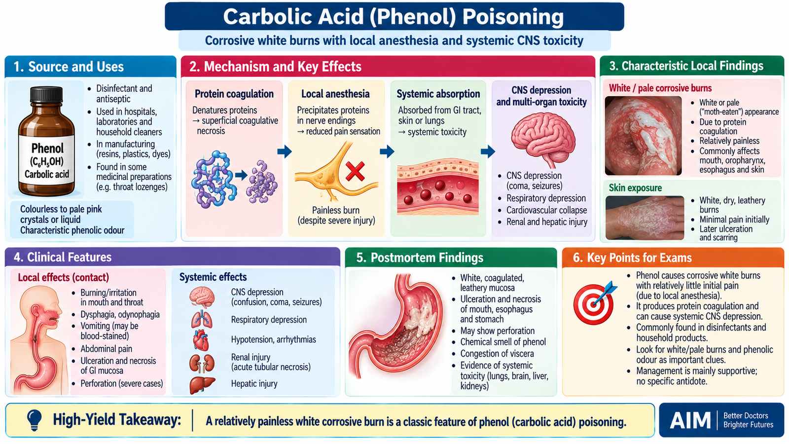
Carbolic acid is the traditional name for phenol. Although called an acid, phenol differs from strong mineral acids because it produces both local corrosive injury and important systemic toxicity after absorption. It also has a local anesthetic effect, so serious phenol burns may initially be less painful than expected.
Organic Acids: Classification
Important organic corrosive poisons traditionally discussed in forensic toxicology include carbolic acid (phenol), oxalic acid and acetic acid. The KMU outcomes emphasize phenol and oxalic acid because they have distinctive mechanisms and examination findings.
Appearance and Uses
Pure phenol forms colorless or white crystalline material and may become pinkish on exposure to air and light. It has a characteristic phenolic odor. Phenol has been used as a disinfectant and antiseptic and remains important in chemical and industrial processes.
Mechanism
Phenol denatures and coagulates proteins at the site of contact, producing corrosive burns. Its local anesthetic action can reduce pain sensation. When absorbed, phenol can depress the central nervous system and impair cardiovascular, renal and other organ functions.
Fatal Dose and Fatal Period
A traditional approximate fatal dose is about 10–15 g, and the fatal period in severe poisoning is commonly taught as approximately 3–4 hours. These are approximate forensic teaching values rather than fixed biological thresholds.
Clinical Features
- Characteristic phenolic odor.
- White or pale burns of the lips, mouth or skin, later becoming brownish.
- Burned areas may initially be relatively painless because of local anesthetic action.
- Nausea, vomiting and abdominal pain after ingestion.
- Dizziness, confusion, CNS depression, seizures or coma in severe systemic poisoning.
- Hypotension and cardiovascular collapse may occur.
- Renal injury can cause oliguria; urine may become unusually dark or greenish in significant phenol exposure.
Treatment Principles
- Protect the airway and support breathing and circulation.
- Remove contaminated clothing rapidly.
- Decontaminate exposed skin promptly; polyethylene glycol can assist removal of phenol when available, while copious irrigation is important when alternative decontamination is not immediately available.
- Do not induce vomiting after ingestion.
- Provide supportive management for seizures, hypotension and respiratory depression.
- Monitor renal function and manage acute kidney injury when it develops.
- There is no specific antidote for phenol poisoning.
Postmortem Appearance
The lips, oral mucosa, esophagus and stomach may show white, gray or brown corrosive changes. A phenolic odor may be appreciable. Internal organs may be congested, and evidence of systemic circulatory or renal toxicity may accompany the local lesions.
Forensic Importance
Phenol may cause accidental, occupational or intentional poisoning. Its characteristic odor and pale corrosive lesions can provide useful clues, but neither is individually diagnostic. Circumstantial history and toxicological confirmation remain important.
G. Oxalic Acid Poisoning
Oxalic acid is an organic acid with two important actions. First, it can directly corrode the gastrointestinal tract. Second, absorbed oxalate combines with calcium to form insoluble calcium oxalate. This lowers biologically available calcium and promotes crystal deposition in the kidneys, explaining the characteristic combination of hypocalcemia and renal injury.
Appearance and Uses
Oxalic acid is usually encountered as a colorless or white crystalline solid that dissolves in water. It has been used in cleaning and bleaching processes, particularly for removal of rust and stains, and in industrial applications.
Mechanism
Fatal Dose and Fatal Period
A traditionally quoted approximate fatal dose is 15–20 g. Severe poisoning may cause death within approximately 1–2 hours, although survival time varies depending on the amount absorbed, metabolic disturbance, renal injury and treatment.
Clinical Features
- Burning pain in the mouth, throat and epigastrium.
- Nausea, vomiting and gastrointestinal bleeding from corrosive injury.
- Hypocalcemia, which may produce muscle cramps, tetany, paresthesia or seizures.
- Cardiac electrical instability and arrhythmias may occur when hypocalcemia is severe.
- Renal deposition of calcium oxalate may produce oliguria and acute kidney injury.
- Severe poisoning may progress to shock, convulsions, coma or death.
Treatment Principles
Treatment addresses both corrosive injury and the specific calcium-binding effect of oxalate.
- Stabilize airway, breathing and circulation.
- Do not induce vomiting after significant corrosive ingestion.
- Correct clinically important hypocalcemia with calcium replacement.
- Provide fluid and supportive management while monitoring renal function.
- Manage seizures or cardiac complications when present.
- Severe acute kidney injury may require specialist renal support.
Postmortem Appearance
The upper gastrointestinal tract may show pale or whitish corrosive lesions, congestion, erosions and hemorrhage. An important systemic finding is renal injury. Calcium oxalate crystals may be demonstrated in renal tubules, supporting the mechanism of oxalate-related nephrotoxicity.
Forensic Importance
Poisoning may follow accidental or intentional ingestion of oxalic-acid-containing materials. The combination of gastrointestinal corrosion, hypocalcemic manifestations and renal calcium oxalate deposition is particularly useful for understanding and investigating suspected exposure.

H. Cyanide Poisoning
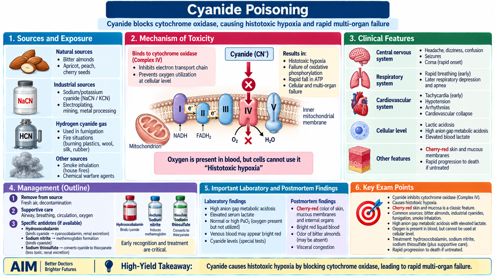
Cyanide is a rapidly acting systemic poison. Unlike corrosive acids and alkalis, its major toxic effect occurs after absorption. Cyanide prevents cells from utilizing oxygen by blocking mitochondrial oxidative phosphorylation. Oxygen may therefore be present in the blood but cannot be used effectively by tissues—a state called histotoxic hypoxia.
Appearance, Sources and Uses
- Hydrogen cyanide: a highly volatile, colorless liquid or gas.
- Sodium and potassium cyanide: generally white crystalline salts.
- A bitter-almond odor may be present, but many people cannot detect it; therefore its absence does not exclude cyanide.
- Industrial sources include metal extraction, electroplating and chemical manufacture.
- Cyanide exposure may also occur during combustion of certain nitrogen-containing materials in enclosed fires.
- Cyanogenic compounds occur naturally in some plants and seeds.
Mechanism
Cyanide binds to the ferric iron of mitochondrial cytochrome c oxidase (Complex IV). This blocks transfer of electrons to oxygen at the end of the respiratory chain. Oxidative phosphorylation stops, ATP production falls sharply and cells are forced toward anaerobic metabolism.
The brain and heart are affected rapidly because they have high oxygen and energy requirements. Because peripheral tissues extract less oxygen, venous blood may remain unusually oxygenated and appear bright red.
Fatal Dose and Fatal Period
Traditional forensic teaching quotes an approximate fatal dose of 50–60 mg for hydrogen cyanide and approximately 200–300 mg for potassium cyanide. A large absorbed dose can cause death within minutes; a frequently taught fatal period for severe hydrogen cyanide exposure is approximately 2–10 minutes. Actual outcome depends strongly on chemical form, route, dose, absorption and rapidity of antidotal treatment.
Clinical Features
The onset may be extremely rapid. Early symptoms reflect cellular hypoxia in the nervous and cardiovascular systems.
- Headache, dizziness, anxiety and confusion.
- Nausea and vomiting.
- Rapid breathing initially, followed by respiratory failure in severe poisoning.
- Altered consciousness, seizures and coma.
- Hypotension, cardiac arrhythmias and cardiovascular collapse.
- Severe metabolic, particularly lactic, acidosis.
- Skin or venous blood may appear unusually pink or bright red, but this is not consistently present.
Treatment
Cyanide poisoning requires immediate resuscitation and antidotal treatment. Treatment should not be delayed in a severely ill patient when exposure is strongly suspected.
- Remove the patient safely from the source of exposure.
- Support airway, ventilation and circulation and provide high-concentration oxygen.
- Treat seizures and cardiovascular instability.
- Hydroxocobalamin binds cyanide to form cyanocobalamin and is an important cyanide antidote.
- Sodium thiosulfate provides sulfur that promotes conversion of cyanide to the less toxic thiocyanate.
- Nitrite-based antidotal therapy produces methemoglobin, which can bind cyanide, but it requires particular caution when associated carbon monoxide or smoke-related hypoxia is possible.
Why the Antidotes Work
Sodium thiosulfate: provides sulfur → facilitates conversion of cyanide to thiocyanate → less toxic compound available for elimination.
Postmortem Appearance
Findings are not invariably diagnostic. A bitter-almond odor may be noticed, although not everyone can perceive it. Postmortem lividity and blood may appear bright red or pink because tissues have failed to extract oxygen normally. Internal organs may be congested, and the gastric mucosa may show irritation after ingestion of cyanide salts.
Forensic Importance
Cyanide exposure may be occupational, industrial, accidental, suicidal or, less commonly, homicidal. It is also relevant in deaths associated with enclosed-space fires because combustion products can contain hydrogen cyanide. Neither an almond odor nor bright-red lividity is sufficiently specific to establish the diagnosis by itself; circumstances and toxicological evidence remain important.

Integrated Mechanism Flow
Acid: coagulation
Alkali: liquefaction
Cyanide: mitochondrial block
Bleeding / perforation
Hypocalcemia / AKI
Cellular hypoxia
Shock, organ failure, death or late stricture
Important Comparison: Strong Acids versus Strong Alkalis
| Feature | Strong Acids | Strong Alkalis |
|---|---|---|
| Main necrosis | Coagulative necrosis | Liquefactive necrosis |
| Main chemical effect | Protein denaturation and dehydration | Protein dissolution and fat saponification |
| Tissue penetration | Eschar may partly limit penetration | Often deeper because liquefied tissue does not form a protective eschar |
| Typical GI tendency | Severe gastric injury is common | Severe esophageal injury is especially important |
| Characteristic examples | H₂SO₄, HNO₃, HCl | NaOH, KOH, ammonia solutions |
| Important late effect | Scarring and gastrointestinal narrowing may occur | Esophageal fibrosis and stricture are particularly important |
⭐ AIM High-Yield Review
🎥 AIM Video Learning
Watch these videos after completing the learning material to reinforce the mechanisms, clinical features and forensic importance of corrosive and cyanide poisoning.