
This chapter follows the supplied KMU learning outcomes and explains the forensic toxicology and abdominal-injury concepts in a logical sequence. First understand the mechanisms and characteristic patterns; then use the final AIM High-Yield Review for rapid revision.
20. Gastrointestinal & Abdominal Forensic Medicine: Irritant Poisons and Abdominal Injuries
Module / Theme: GIT
A forensic-medicine approach to irritant poisons, envenomation, characteristic postmortem findings and the interpretation of traumatic injuries involving the abdominal wall and gastrointestinal and solid abdominal organs.
1. Topic Introduction
Forensic gastrointestinal and abdominal medicine brings together two important areas: poisoning by substances that irritate or damage tissues, and traumatic injury to abdominal organs. Irritant poisons may act locally on the skin, gastrointestinal tract or respiratory mucosa and may also produce serious systemic toxicity. Their forensic assessment depends on recognizing the poison, understanding its mechanism, clinical effects, treatment principles, postmortem appearances and medico-legal significance. Abdominal trauma requires a similar cause-and-effect approach: the type of force influences which tissues are damaged, how internal bleeding or perforation develops and how the findings should be interpreted. This chapter covers the specific poisons and abdominal injuries included in the KMU curriculum at an appropriate undergraduate level.
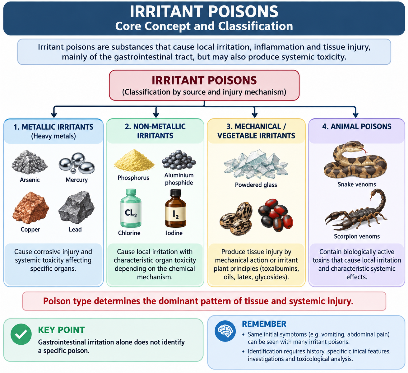
A. Irritant Poisons: Core Concept and Classification
An irritant poison is a substance that produces inflammation, tissue injury or both at the site where it comes into contact with the body. When swallowed, the gastrointestinal tract is commonly affected, producing burning pain, vomiting and diarrhea. Some irritants remain mainly local, while others are absorbed and produce important systemic effects such as hepatic failure, renal injury, neurological dysfunction, cardiovascular collapse or hemolysis.
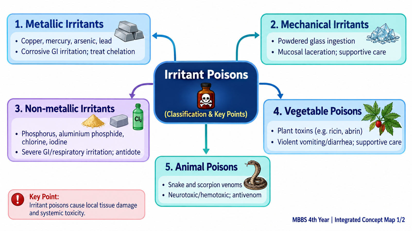
Classification
The classification is based mainly on the nature and origin of the irritant. This helps the student organize a large number of poisons without assuming that all members behave identically.
- Metallic irritants: arsenic, mercury, copper and lead.
- Non-metallic irritants: phosphorus, aluminium phosphide, chlorine and iodine.
- Mechanical irritants: classically powdered glass.
- Vegetable irritants: toxic plants or seeds containing irritant oils, toxalbumins, glycosides or vesicant substances.
- Animal poisons: particularly snake and scorpion venom in the present curriculum.
How Irritant Poisoning Produces Disease
Local gastrointestinal irritation explains abdominal pain, vomiting and diarrhea. Repeated vomiting and diarrhea can cause dehydration and electrolyte disturbance. When the poison is absorbed, its particular biochemical action determines the additional pattern of toxicity. For example, lead interferes with heme synthesis, aluminium phosphide produces profound cellular and cardiovascular toxicity, and some plant toxalbumins inhibit protein synthesis.
Forensic Interpretation
Gastrointestinal congestion or erosion alone does not identify a particular poison. Interpretation requires correlation with the history, scene findings, clinical course, external and internal examination and toxicological analysis. Cause of death should therefore not be assigned from a single nonspecific postmortem appearance.

B. Metallic Irritant Poisons: Arsenic, Mercury, Copper and Lead
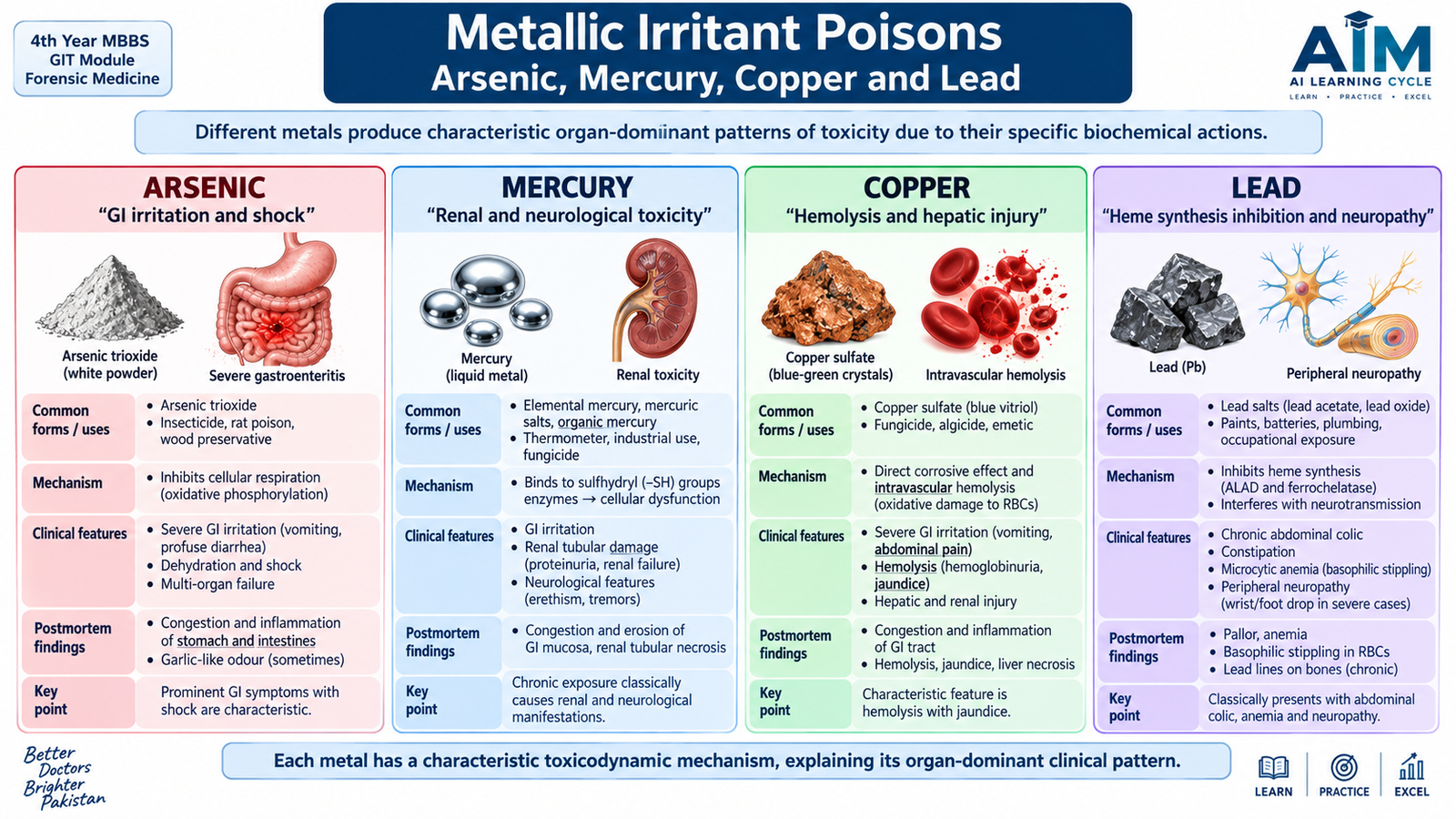
Metallic poisons can produce gastrointestinal irritation, but their major systemic effects differ according to the metal and its chemical form. In forensic practice, the pattern of exposure is important: a single large exposure may cause an acute illness, while repeated occupational or environmental exposure may produce chronic toxicity.
Arsenic
Arsenic trioxide is the classic forensic arsenical poison. It is a white powder and historically gained medico-legal importance because it could be mixed with food without producing a striking colour. Arsenic compounds have also had industrial and pesticide-related uses.
Mechanism
Arsenic binds to sulfhydryl-containing enzymes and disturbs cellular energy metabolism. Acute poisoning causes severe capillary and gastrointestinal injury, while prolonged exposure can affect the skin, peripheral nerves, bone marrow and other organs.
Clinical Features
- Severe burning abdominal pain, nausea and repeated vomiting.
- Profuse watery diarrhea, sometimes described as resembling severe gastroenteritis.
- Dehydration, hypotension and circulatory collapse in severe poisoning.
- Cardiac rhythm disturbances may occur.
- Neuropathy may appear during recovery or chronic exposure.
- Chronic exposure may produce pigmentation changes, hyperkeratosis and peripheral neuropathy.
Treatment Principle
Management begins with airway, breathing and circulation, replacement of fluid and electrolyte losses and treatment of cardiovascular complications. Chelation may be indicated; agents used in arsenic poisoning include dimercaprol and oral sulfhydryl chelators such as succimer, depending on the clinical situation and specialist guidance.
Postmortem and Medico-Legal Importance
The stomach and intestinal mucosa may be congested, inflamed and hemorrhagic. Fatty change and visceral congestion may accompany systemic toxicity. Historically arsenic was important in homicidal poisoning, but modern cases may also arise from occupational, environmental or accidental exposure. Toxicological confirmation is essential because the gross findings are not specific.
Mercury
Mercury occurs as elemental mercury, inorganic salts and organic mercury compounds. Elemental mercury is the familiar silvery liquid metal. Toxicity depends strongly on its chemical form: inhaled mercury vapor mainly produces systemic toxicity, whereas corrosive inorganic salts can cause marked gastrointestinal injury.
Mechanism and Clinical Features
Mercury binds sulfhydryl groups in cellular proteins and enzymes. Acute ingestion of a corrosive mercury salt may cause burning pain in the mouth and abdomen, vomiting, diarrhea, gastrointestinal bleeding and renal injury. Chronic exposure may produce tremor, neuropsychiatric changes, gingivostomatitis and renal dysfunction.
Treatment
Further exposure is stopped and supportive treatment is provided. Chelation with agents such as succimer or DMPS may be used for appropriate forms of mercury poisoning under toxicology guidance. Choice of chelator depends on the mercury compound and clinical presentation.
Postmortem and Medico-Legal Importance
Corrosive inorganic mercury compounds may produce erosive or necrotic gastrointestinal lesions together with renal damage. Chronic poisoning is more likely to be occupational or environmental than homicidal. Interpretation requires identification of the chemical form and exposure route.
Copper
Copper sulfate is a blue crystalline copper salt that has been used in agriculture and industry. Its colour may provide an exposure clue, although colour alone is never sufficient proof.
Mechanism and Clinical Features
Copper salts cause direct gastrointestinal irritation and, after absorption, can produce oxidative injury to red blood cells. Severe poisoning may therefore combine vomiting and abdominal pain with intravascular hemolysis, jaundice, hemoglobinuria, methemoglobinemia and acute kidney injury.
Treatment
Management is supportive, with attention to shock, hemolysis, methemoglobinemia and renal complications. Chelation, commonly with penicillamine in appropriate cases, may be considered with specialist advice.
Postmortem and Medico-Legal Importance
Gastrointestinal congestion and erosion may be present, together with evidence of hemolysis, jaundice and renal tubular injury. Exposure may be accidental, occupational or intentional.
Lead
Lead is a bluish-gray heavy metal encountered particularly in batteries, smelting, pigments and contaminated occupational environments. Unlike strongly corrosive metallic irritants, lead is especially important because of its cumulative chronic toxicity.
Mechanism
Clinical Features
- Abdominal colic and constipation.
- Anemia; basophilic stippling may be seen in red cells.
- Peripheral motor neuropathy, classically producing wrist or foot weakness in advanced disease.
- Encephalopathy may occur in severe poisoning, particularly in children.
- A blue-black gingival line may occur in chronic exposure but is not present in every case.
- Chronic renal injury and hypertension may develop.
Treatment
The source of exposure must be identified and removed. Chelating agents used according to severity include succimer, calcium disodium EDTA and, in severe encephalopathic poisoning, regimens incorporating dimercaprol under specialist management.
Postmortem and Medico-Legal Importance
Postmortem findings of chronic lead poisoning are often nonspecific. Renal damage and cerebral edema may occur in severe disease. Medico-legally, lead poisoning is particularly important in occupational and environmental exposure, especially when several people share the same source.
C. Mechanical Irritant: Powdered Glass
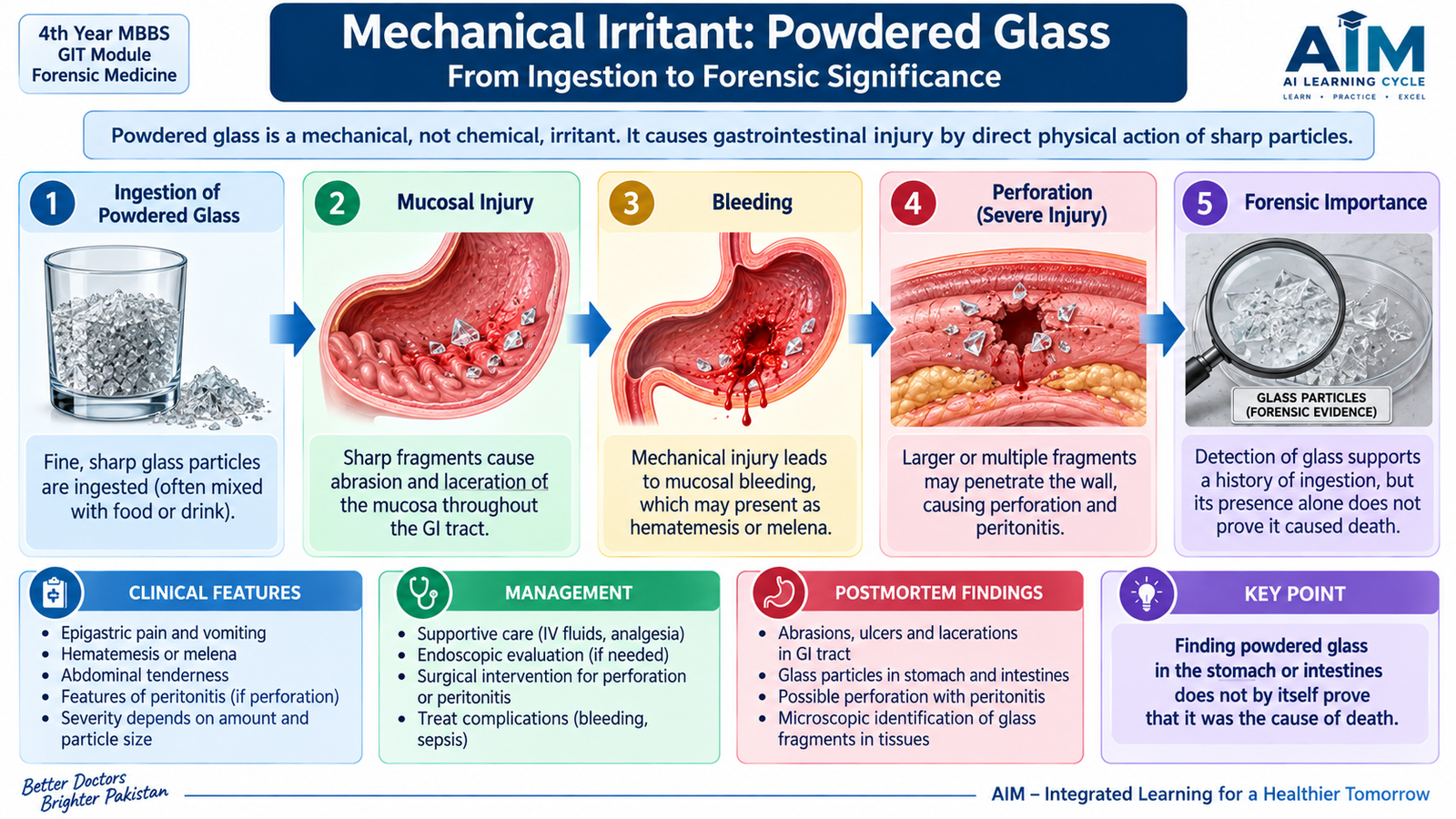
Powdered or fragmented glass is traditionally described as a mechanical irritant because injury, when it occurs, results from physical abrasion or penetration rather than a chemical toxin. An important forensic point is that finely powdered glass does not automatically cause severe or fatal gastrointestinal injury. Many traditional claims regarding its lethality are exaggerated.
Mechanism and Symptoms
Sharp fragments may damage the lips, oral cavity, pharynx or gastrointestinal mucosa. Larger or irregular fragments carry greater potential for injury than very fine powder.
- Pain in the mouth, throat or abdomen.
- Nausea and vomiting.
- Hematemesis or blood in stool if significant mucosal injury occurs.
- Perforation and peritonitis are possible with sharp fragments but are not inevitable.
Treatment
There is no antidote. Management is based on assessment for mechanical injury, bleeding or perforation. Inducing vomiting is inappropriate because it may cause additional trauma. Significant symptoms require clinical evaluation and appropriate imaging or endoscopic/surgical assessment according to the suspected site of injury.
Postmortem Appearance
If injury has occurred, abrasions, cuts, hemorrhagic areas or perforations may be seen in the upper gastrointestinal tract or bowel. Particles may be present in gastrointestinal contents, but merely finding glass does not prove that it caused death.
Forensic Importance
Alleged administration of glass may arise in suspected homicidal poisoning or fabricated accounts. The forensic examiner must demonstrate actual traumatic lesions and correlate them with recovered material. The historical reputation of powdered glass as a reliable poison should not replace objective evidence.
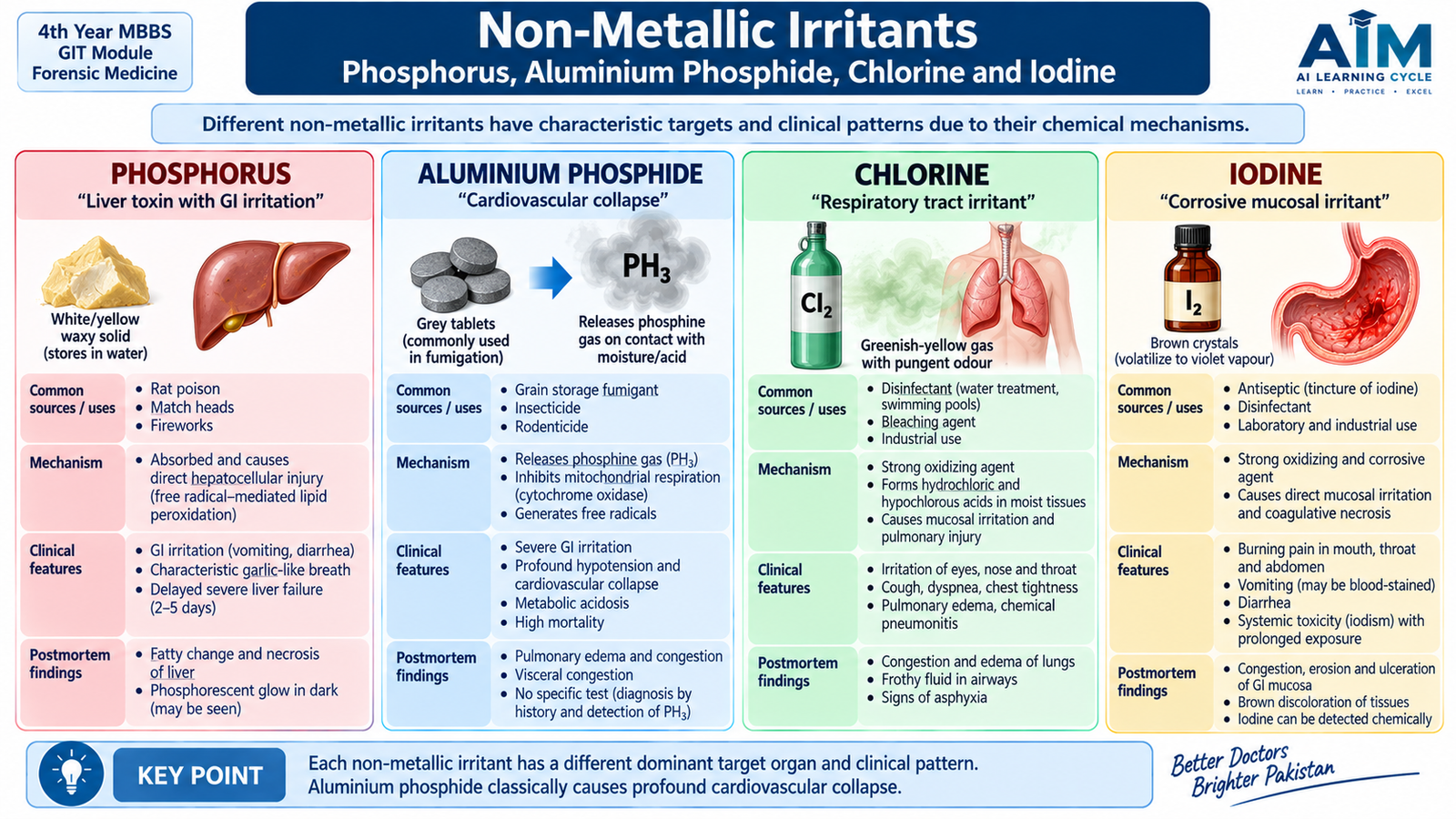
D. Non-Metallic Irritants: Phosphorus, Aluminium Phosphide, Chlorine and Iodine
These poisons are grouped together as non-metallic irritants, but they have very different mechanisms. White phosphorus is an important hepatotoxic poison, aluminium phosphide releases the highly toxic gas phosphine, chlorine primarily damages moist respiratory surfaces, and iodine acts as a local irritant and corrosive when sufficiently concentrated.
White Phosphorus
White or yellow phosphorus is a waxy substance that may have a garlic-like odor and can glow in the dark because of slow oxidation. Historically it has been used in products such as certain rodenticides and pyrotechnic materials.
Mechanism
Phosphorus causes severe gastrointestinal irritation and, after absorption, profound cellular toxicity. The liver is especially vulnerable, and severe poisoning may progress to hepatic necrosis and failure.
Clinical Features
- Burning abdominal pain, nausea and vomiting.
- Garlic-like odor may be noticed.
- Severe cases develop jaundice, coagulopathy, hypoglycemia, hepatic encephalopathy and cardiovascular collapse.
- Renal and myocardial injury may accompany hepatic failure.
Fatal Dose and Period
Traditional forensic teaching commonly quotes roughly 60–120 mg of white phosphorus as potentially fatal in an adult, with death often occurring after several days from hepatic or multisystem failure. These historical figures are approximate; actual toxicity varies considerably with preparation and exposure.
Treatment and Postmortem Appearance
There is no specific antidote. Management is aggressive supportive care with close monitoring for hepatic failure, hypoglycemia, coagulopathy, renal injury and cardiovascular instability. Obsolete toxic emetics should not be used. At postmortem examination, the stomach may show inflammation and hemorrhage; a garlic-like odor or luminescent material may occasionally be noted. The liver commonly shows severe fatty change and toxic injury.
Medico-Legal Importance
Cases may be accidental or intentional. Historical use in rodenticides and fireworks explains its traditional forensic importance.
Aluminium Phosphide
Aluminium phosphide is a solid fumigant used to protect stored grain. When exposed to moisture or gastric acid, it releases phosphine gas, which is responsible for most of its toxicity.
Mechanism
Clinical Features
- Nausea, vomiting and abdominal discomfort.
- Profound hypotension and shock.
- Cardiac arrhythmias and myocardial dysfunction.
- Metabolic acidosis.
- Pulmonary edema and respiratory distress.
- Hepatic and renal dysfunction in severe poisoning.
Fatal Dose and Period
Aluminium phosphide has a narrow margin of safety; even part of a commercial fumigant tablet may cause severe or fatal poisoning. Traditional forensic texts give approximate lethal quantities rather than a dependable threshold. Fatal deterioration is often rapid, commonly within the first day, although survival for several days may occur. These figures must not be used to predict outcome in an individual patient.
Treatment, Postmortem and Forensic Importance
There is no established specific antidote. Treatment is aggressive supportive management of airway, circulation, shock, arrhythmias, acidosis and organ failure. Postmortem findings may include visceral congestion, pulmonary edema and hemorrhagic or congested gastrointestinal mucosa; the findings are not specific. The poison is important medico-legally because of agricultural availability and both accidental and intentional exposures.
Chlorine
Chlorine is a yellow-green, pungent gas used in industrial processes and disinfection. Clinically important exposure is usually by inhalation rather than ingestion.
Mechanism
When chlorine contacts moist mucosal surfaces, reactive acids and oxidants are formed. These damage the eyes and respiratory epithelium, leading to inflammation, bronchospasm and increased pulmonary capillary permeability.
Clinical Features and Treatment
- Burning of the eyes, nose and throat.
- Cough, choking sensation and chest tightness.
- Wheezing or bronchospasm.
- Severe exposure may produce chemical pneumonitis and pulmonary edema.
The person is removed from exposure and given fresh air, oxygen and supportive respiratory treatment. Bronchodilators are used when bronchospasm is present. There is no specific antidote.
Postmortem and Medico-Legal Importance
Severe fatal exposure may produce congested, heavy, edematous lungs and inflamed respiratory mucosa. Most cases are accidental or occupational. Dangerous chlorine exposure can also occur when incompatible household cleaning chemicals are mixed.
Iodine
Iodine is a dark bluish-black crystalline substance that produces violet vapor on heating. Iodine compounds are widely associated with antiseptic and laboratory use.
Mechanism and Clinical Features
Concentrated iodine acts as a local irritant and corrosive. It may cause burning in the mouth and throat, vomiting, abdominal pain, diarrhea, gastrointestinal bleeding, dehydration and shock. Renal injury may occur in severe systemic toxicity.
Fatal Dose and Period
Older forensic texts traditionally quote approximately 2–4 g of elemental iodine as a potentially fatal oral quantity, with severe fatal poisoning often evolving within about a day. Such figures are approximate because toxicity depends on formulation, concentration and clinical circumstances.
Treatment and Postmortem Appearance
Treatment is supportive, with attention to corrosive gastrointestinal injury, fluid loss and shock. Chemical neutralization should not be improvised. Postmortem examination may show brownish staining, congestion, erosion and hemorrhage of the upper gastrointestinal mucosa. Toxicological confirmation remains necessary.
E. Vegetable Irritant Poisons
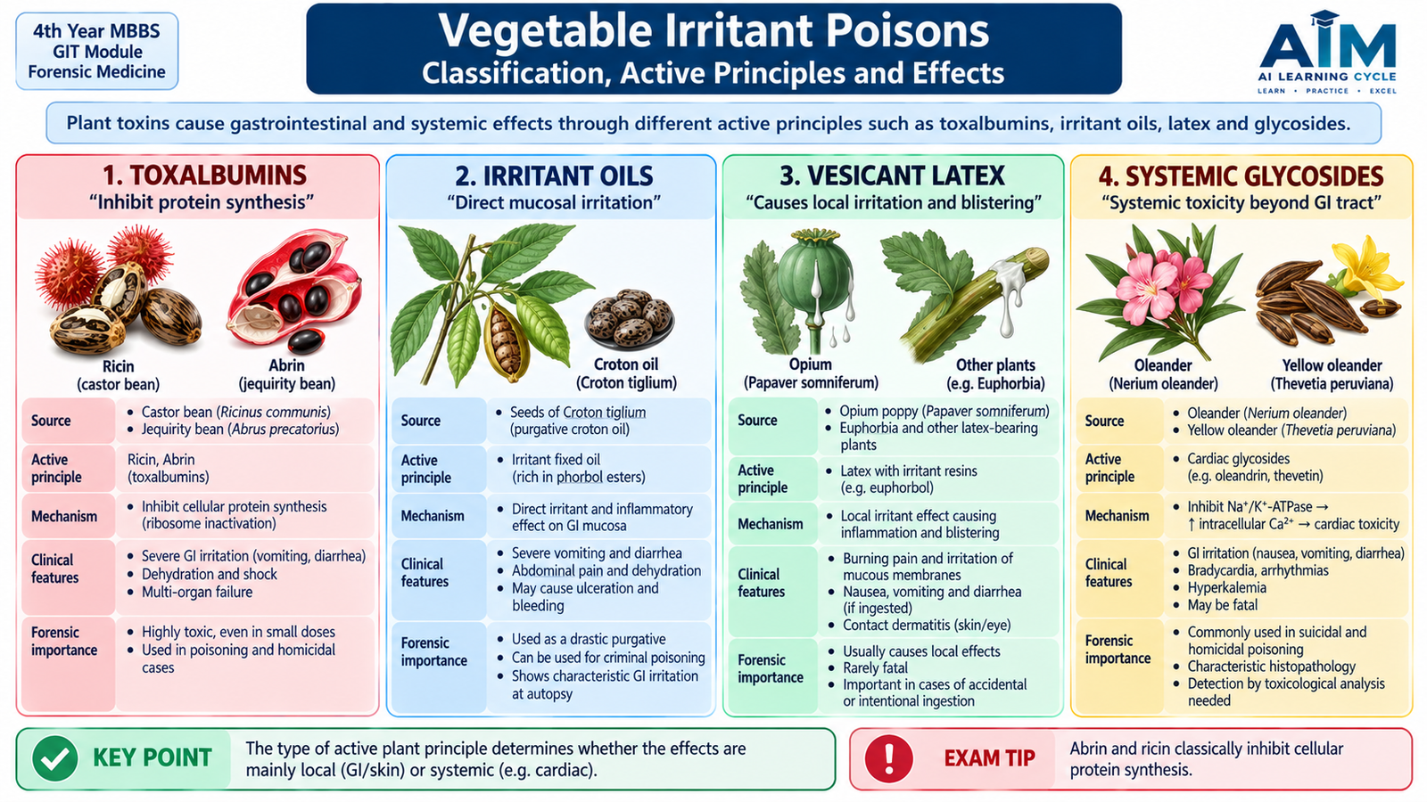
Vegetable irritants are poisonous plant products that damage tissues through substances such as toxalbumins, irritant oils, resins, latex or glycosides. The clinical picture commonly begins with gastrointestinal irritation, but particular plants may also produce cardiac, hepatic, renal or systemic cellular toxicity. The amount of toxin in a seed or plant part can vary widely, so traditional fatal-dose figures are approximate rather than absolute.
Abrus precatorius — Jequirity / Rosary Pea
The seeds are characteristically bright red with a black area. The important active principle is abrin, a highly toxic toxalbumin. Intact seeds may pass through the gastrointestinal tract, whereas crushed, chewed or otherwise disrupted seeds release the toxin.
- Mechanism: inhibition of cellular protein synthesis.
- Clinical features: severe vomiting, abdominal pain, diarrhea, dehydration and, in serious cases, hepatic, renal and circulatory failure.
- Traditional fatal dose/period: approximately one or two thoroughly crushed seeds have traditionally been regarded as potentially fatal; death, when it occurs, may follow over several days.
- Treatment: early supportive toxicology care, fluid and electrolyte replacement and management of organ dysfunction; no specific antidote is established.
- Postmortem: gastrointestinal inflammation and hemorrhage with nonspecific visceral congestion.
- Forensic importance: seeds have historical importance in deliberate poisoning and in poisoned needles or preparations used against animals.
Ricinus communis — Castor Plant
The seed contains ricin, another toxalbumin that inhibits protein synthesis. Properly processed castor oil does not contain the same clinically significant amount of ricin as the intact toxic seed.
- Clinical features: vomiting, severe diarrhea, abdominal pain and dehydration; severe poisoning can cause hepatic, renal and circulatory dysfunction.
- Fatal dose/period: the number of chewed seeds required to cause death varies substantially; traditional forensic texts describe several well-chewed seeds as potentially dangerous, with death developing over a period of days rather than at a fixed predictable time.
- Treatment: supportive care and correction of fluid and electrolyte loss; no specific antidote.
- Postmortem: congested and inflamed gastrointestinal mucosa with nonspecific visceral changes.
- Forensic importance: accidental ingestion is possible, particularly where the plant is readily available.
Croton tiglium
Croton seeds and croton oil contain powerful irritant constituents. Croton oil has historically been used as a drastic purgative but is unsafe for such use.
- Mechanism: intense local irritation of skin and gastrointestinal mucosa.
- Clinical features: burning pain, vomiting, profuse diarrhea, dehydration and shock.
- Fatal dose/period: traditional forensic teaching describes only a few seeds or a small quantity of croton oil as potentially dangerous; because potency varies, exact numbers are unreliable for individual case interpretation.
- Treatment: supportive treatment of gastrointestinal losses and shock; no specific antidote.
- Postmortem: marked gastrointestinal congestion and inflammation.
- Forensic importance: historical use as a purgative and irritant gives it accidental and intentional poisoning relevance.
Semecarpus anacardium — Marking Nut
The marking nut contains irritant phenolic substances, including bhilawanol-type compounds, that can produce intense skin inflammation and blistering.
- Clinical effects: contact dermatitis and vesication externally; ingestion may cause burning, vomiting, diarrhea and systemic illness.
- Uses: the dark juice has traditionally been used for marking fabrics and other materials.
- Fatal dose/period: no dependable single fatal quantity can be applied because plant preparations vary considerably.
- Treatment: removal of contamination, skin decontamination where appropriate and supportive care after ingestion.
- Postmortem: irritant lesions of contacted surfaces and gastrointestinal inflammation after ingestion.
- Forensic importance: it has traditionally been associated with fabricated skin lesions because its juice can produce dermatitis resembling injury.
Calotropis
Calotropis species produce a milky latex containing irritant substances and cardiac glycoside-like compounds such as calotropin. The latex is strongly irritating to skin and mucous membranes.
- Clinical features: irritation of the mouth and gastrointestinal tract, vomiting and diarrhea; serious systemic exposure may affect cardiac conduction and circulation.
- Uses: the plant has various traditional uses, which explains the possibility of accidental or deliberate exposure.
- Fatal dose/period: no reliable universally applicable fatal dose can be stated because plant potency and preparation vary.
- Treatment: supportive treatment with attention to gastrointestinal losses and cardiovascular abnormalities.
- Postmortem: gastrointestinal irritation and nonspecific visceral congestion may occur.
- Forensic importance: deliberate application or ingestion has historical medico-legal relevance, including alleged use in harmful traditional preparations.
F. Animal Poisons: Snakebite and Scorpion Envenomation
Animal poisons are delivered through structures such as fangs or stings rather than simply being swallowed. In snakebite, clinical assessment should focus on whether envenomation has occurred and which syndrome is developing. External appearance of the snake may help with identification, but it should never replace clinical assessment because traditional rules about head shape, pupil shape or markings are not completely reliable.
Poisonous versus Non-Poisonous Snakes
| Feature | Venomous Snake | Non-Venomous Snake |
|---|---|---|
| Venom apparatus | Specialized venom glands and fangs are present. | Specialized venom-delivery fangs are absent. |
| Bite marks | One or more fang punctures may be present. | Several small tooth marks may form an arc. |
| External morphology | Varies greatly by species. | Also varies greatly by species. |
| Forensic rule | Do not declare a snake venomous or non-venomous from a single superficial feature alone. | |
Classification of Snake Venom by Predominant Effect
Venoms contain complex mixtures of enzymes and toxins, so a species may produce more than one effect. For undergraduate understanding, the predominant clinical syndromes can be grouped as follows:
- Neurotoxic: interferes with neuromuscular transmission, producing ptosis, ophthalmoplegia, bulbar weakness and respiratory paralysis. Elapid bites commonly show this pattern.
- Hemotoxic / vasculotoxic: activates or consumes coagulation factors, damages endothelium and promotes bleeding, shock and renal injury. Viper bites commonly produce this pattern.
- Myotoxic: damages skeletal muscle, causing pain, weakness, rhabdomyolysis and myoglobinuria. This is particularly important in some sea-snake envenomations.
- Local cytotoxic effects: pain, swelling, blistering and tissue necrosis may accompany several venomous bites.
Clinical Assessment of Snakebite
Not every bite by a venomous snake results in significant venom injection. The patient must therefore be assessed for progressive local effects and systemic signs of envenomation.
- Fang marks, pain and swelling at the bite site.
- Progressive edema, blistering or necrosis.
- Ptosis, diplopia, difficulty swallowing, weak voice or respiratory weakness in neurotoxic envenomation.
- Spontaneous bleeding, prolonged clotting, hypotension or shock in hemotoxic envenomation.
- Dark urine due to hemoglobin or myoglobin in appropriate syndromes.
- Acute kidney injury may follow severe hemotoxic, myotoxic or shock-associated envenomation.
Snakebite Management
Early management aims to slow unnecessary movement of venom, identify systemic envenomation and provide organ support. The affected limb should be kept still, constricting items such as rings should be removed, and the patient should be transferred for medical assessment.
Hospital assessment includes airway and breathing, circulation, neurological examination, local swelling and appropriate coagulation, renal and hematological investigations. Antivenom is the specific treatment when clinically significant systemic envenomation or other accepted indications are present. It is not given merely because fang marks are visible. Respiratory failure requires ventilatory support; shock, bleeding, renal failure and wound complications are treated according to their clinical severity. Antivenom reactions must be recognized and treated promptly.
Postmortem and Medico-Legal Aspects of Snakebite
Examination may show fang punctures, swelling, bruising, blistering or local necrosis. Hemotoxic bites may cause widespread internal hemorrhage, while fatal neurotoxic bites may have relatively nonspecific internal findings such as congestion and pulmonary edema. Absence of dramatic local injury does not exclude fatal envenomation.
Most snakebites are accidental. Forensic assessment should document the bite site, clinical progression and treatment and preserve relevant material according to accepted local procedures. Identification of the species from the wound alone is unreliable. Cause of death should be based on the complete clinical, pathological and investigative evidence.
Scorpion Bite
Scorpion venom acts mainly on ion channels and may trigger intense autonomic nervous-system activity. Many stings cause only severe local pain, but dangerous species can produce widespread autonomic and cardiovascular toxicity.
- Immediate severe burning pain at the sting site.
- Sweating, salivation, nausea and vomiting.
- Tachycardia, hypertension or later hypotension.
- Agitation and autonomic overactivity.
- Severe cases may develop myocardial dysfunction and pulmonary edema.
Treatment includes pain control, monitoring and support of airway, respiration and circulation. Region-specific antivenom may be used where indicated and available. Management of severe autonomic or cardiovascular toxicity depends on the clinical syndrome and local toxicology protocols.

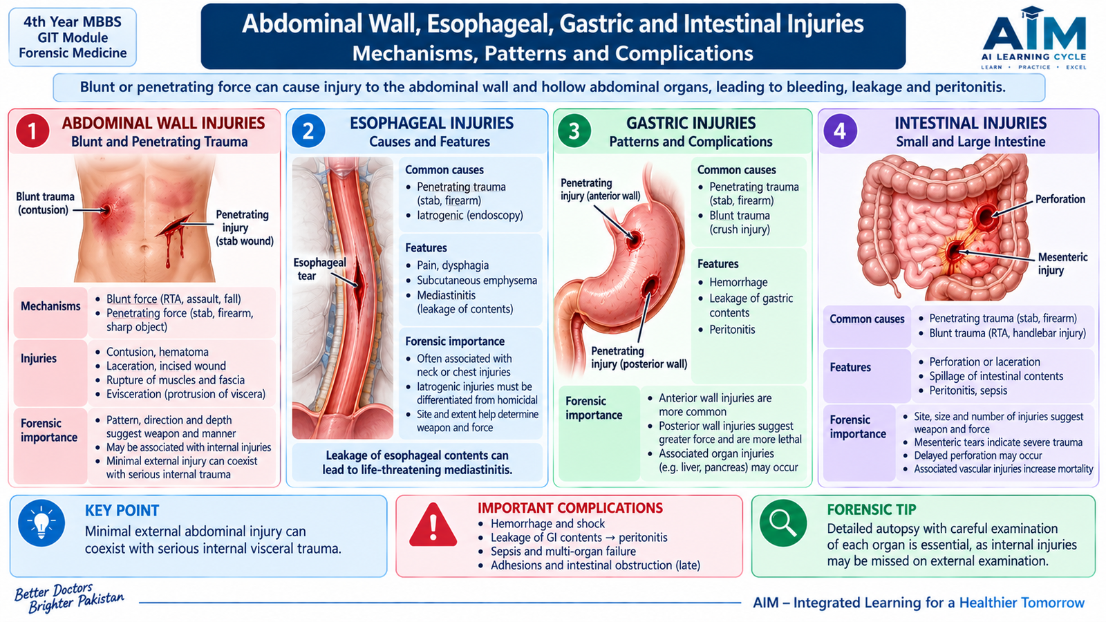
G. Abdominal Wall, Esophageal, Gastric and Intestinal Injuries
Abdominal injuries may be produced by blunt force or penetrating force. Blunt trauma compresses abdominal organs against the spine or posterior abdominal wall, stretches fixed attachments and produces rapid changes in pressure. Penetrating injuries produce a wound track that may involve several structures. A major forensic principle is that severe internal injury can exist even when external abdominal marks are slight or absent.
Injuries to the Abdominal Wall
The abdominal wall consists of skin, subcutaneous tissue, muscles and fascial layers. Injury depends on the nature, direction and magnitude of force.
- Abrasions may indicate friction against a rough surface.
- Contusions result from rupture of small blood vessels beneath intact skin.
- Patterned bruises may reproduce part of the shape of an impacting object and can provide useful reconstructive information.
- Muscle tears and hematomas may occur after substantial blunt force.
- Incised, stab or firearm wounds can penetrate the abdominal cavity and injure underlying organs.
The absence of external bruising does not exclude significant visceral trauma because force may be transmitted through the flexible abdominal wall to internal organs.
Esophageal Injuries
Traumatic rupture of the esophagus is less common than injury to many abdominal organs because much of the esophagus lies within the protected thorax. Injury can follow penetrating trauma, foreign bodies or instrumentation and may also occur after a sudden major rise in intraluminal pressure.
A perforation allows saliva, gastric contents and microorganisms to enter surrounding tissues. This explains the serious complications of mediastinal inflammation, infection, sepsis and shock when the thoracic esophagus is involved. Forensic examination should document the site and character of the tear and distinguish traumatic injury from postmortem artifact or disease-related perforation.
Gastric Injuries
The stomach may be bruised, torn or perforated by blunt or penetrating trauma. A distended stomach is more vulnerable to rupture because fluid or food contents cannot be compressed easily; a sudden external force therefore causes a rapid rise in intragastric pressure.
- Contusion of the gastric wall.
- Partial-thickness tears.
- Full-thickness rupture with leakage of gastric contents.
- Hemorrhage into the peritoneal cavity when associated vessels are damaged.
Leakage of gastric contents produces chemical irritation followed by bacterial peritonitis. The forensic examiner should record the size, location and margins of the injury and correlate it with the suspected mechanism.
Intestinal Injuries
The small and large intestines can be damaged directly by compression or indirectly by sudden stretching of bowel and mesentery. Fixed segments and attachment points are especially susceptible to shearing forces.
Patterns of Injury
- Serosal bruising or tears.
- Intramural hematoma.
- Mesenteric tears with bleeding.
- Partial or complete transection.
- Perforation with leakage of intestinal contents.
- Devascularization of a bowel segment after mesenteric vascular injury.
Why Delayed Perforation Can Occur
This sequence is important in forensic interpretation because a victim may initially remain alive and symptomatic for hours or longer before peritonitis or sepsis becomes clinically obvious.
Medico-Legal Interpretation
The examiner should correlate external injuries with internal findings, the direction of any penetrating track and the distribution of blood or intestinal contents. The presence or absence of an external mark cannot by itself determine the severity of the internal injury. Survival interval, treatment and possible complications must also be considered when explaining the cause and mechanism of death.
H. Liver and Spleen Injuries
The liver and spleen are highly vascular solid organs. Trauma to either organ can therefore cause major internal hemorrhage even when little blood is visible externally. The forensic significance lies not only in identifying the injured organ but also in determining the pattern of rupture, associated blood loss and whether the injury could have produced immediate or delayed deterioration.
Liver Injuries
The liver is large, relatively fixed by its ligamentous attachments and partly protected by the ribs. The right lobe is particularly exposed because of its size. Blunt force may compress the liver against the vertebral column or lower ribs, whereas penetrating trauma can directly cut or perforate the organ.
Patterns
- Subcapsular hemorrhage or hematoma.
- Capsular tears.
- Superficial or deep lacerations.
- Central parenchymal disruption.
- Major vascular injury with rapid hemoperitoneum.
Clinical and Forensic Consequences
Severe liver injury produces intra-abdominal hemorrhage, hypovolemic shock and death if bleeding is not controlled. A subcapsular hematoma may initially remain contained. If the capsule later ruptures, a delayed hemoperitoneum may occur. This provides a mechanism for a clinically important survival interval between trauma and sudden deterioration.
At postmortem examination, the extent and orientation of lacerations, blood in the peritoneal cavity and associated rib or abdominal injuries should be documented. The examiner should also distinguish true traumatic laceration from handling or postmortem artifacts.
Spleen Injuries
The spleen is highly vascular and lies beneath the left lower ribs. Blunt trauma to the left upper abdomen or lower chest can cause capsular tearing, parenchymal laceration or a subcapsular hematoma.
Patterns
- Capsular tears and superficial lacerations.
- Deep parenchymal rupture.
- Subcapsular hematoma.
- Fragmentation in severe trauma.
- Associated hemoperitoneum.
Delayed Rupture
A subcapsular hematoma can temporarily contain splenic bleeding. Subsequent rupture of the capsule may release a large quantity of blood into the peritoneal cavity. This classical concept of delayed splenic rupture is important when a victim initially appears relatively stable but later develops shock.
Cause, Mechanism and Manner of Death
These terms should not be confused. A traumatic liver or splenic laceration may be the injury responsible for death; massive internal hemorrhage and hypovolemic shock describe the physiological mechanism through which death occurred. The manner of death depends on the circumstances—such as accident, homicide or another category—and cannot be decided from organ morphology alone.

Integrated Mechanism Flow — Acute Irritant Poisoning
Poison contacts gastrointestinal, respiratory or other tissues.
Inflammation, corrosion or mechanical damage develops.
Pain, vomiting, diarrhea, bleeding or respiratory irritation appears.
Absorbed toxin produces its organ-specific biochemical effect.
Shock, organ failure, respiratory failure or death may follow.
⭐ AIM High-Yield Review
Gastrointestinal & Abdominal Forensic Medicine
Use these videos after completing the AIM learning material to reinforce irritant poisons, aluminium phosphide poisoning and abdominal injuries.
Video 1 — Classification of Poisons & Irritant Poisons
Useful for understanding the basic classification of corrosive, irritant and systemic poisons before studying individual toxic agents.
Video 2 — Aluminium Phosphide Poisoning
Focuses on sources, clinical features, treatment principles and postmortem findings of aluminium phosphide poisoning.
Video 3 — Abdominal Injuries in Forensic Medicine
Reinforces forensic concepts related to abdominal trauma and injuries involving abdominal and pelvic structures.