
This chapter follows the supplied KMU learning outcomes and builds upper GI bleeding from recognition to evaluation and management. First understand the clinical sequence and the portal-pressure mechanisms; then use the AIM High-Yield Review for rapid revision.
Upper GI and Variceal Bleeding: Evaluation and Pharmacological Management
1. Topic Introduction
Upper gastrointestinal bleeding means bleeding arising from the gastrointestinal tract proximal to the ligament of Treitz, most commonly from the esophagus, stomach or duodenum. It may present dramatically with hematemesis or circulatory shock, or more subtly with melena and anemia. The first clinical task is to recognize the severity of blood loss and stabilize the patient while the likely source is being identified. Variceal hemorrhage is a particularly important form of upper GI bleeding because increased portal pressure produces fragile dilated veins that may bleed heavily. This chapter explains the causes and clinical features of upper GI bleeding, the investigation and management pathway, the role of endoscopy, and the mechanisms of the major drugs used to lower portal pressure during or around variceal bleeding.
A. Upper GI Bleeding: Causes and Clinical Recognition
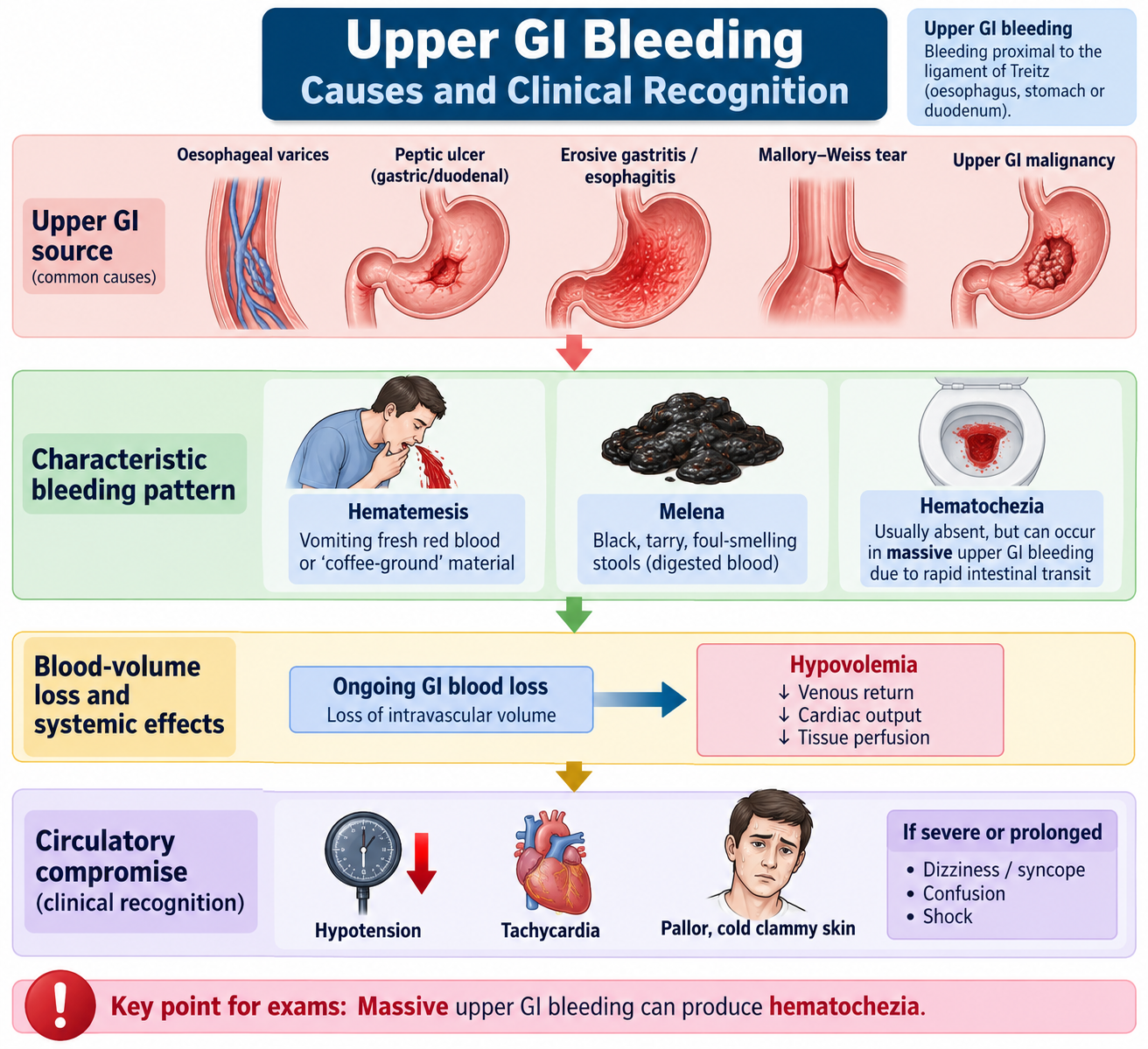
Upper GI bleeding is a clinical syndrome rather than a single disease. Blood enters the lumen of the esophagus, stomach or proximal duodenum because a vessel or vascular lesion has been disrupted. The appearance of the bleeding depends on its site, rate and duration. A slowly bleeding lesion may produce melena, whereas brisk bleeding may cause obvious hematemesis and rapid circulatory compromise.
Etiology
Several lesions can produce upper GI bleeding. Understanding the broad groups helps the student interpret the history and plan endoscopy rather than assuming that every episode of hematemesis has the same cause.
- Peptic ulcer disease: an ulcer may erode into an underlying artery and produce significant bleeding.
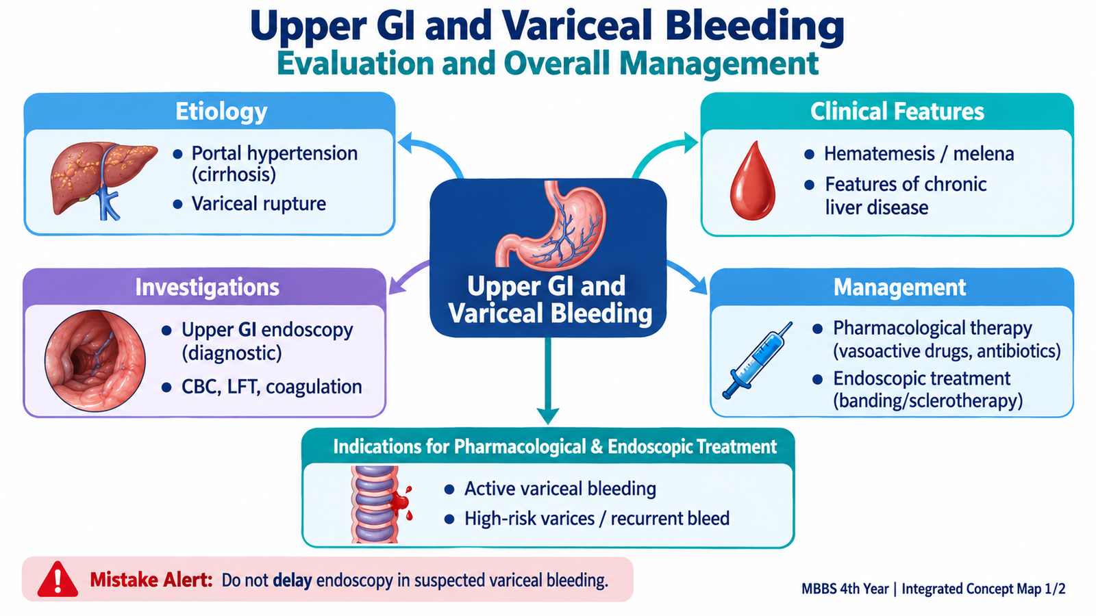
- Esophageal or gastric varices: portal hypertension produces dilated collateral veins that may rupture.
- Erosive gastritis, duodenitis or esophagitis: inflamed and damaged mucosa may ooze blood.
- Mallory–Weiss tear: forceful retching may tear the mucosa near the gastroesophageal junction.
- Upper GI malignancy: a friable ulcerated tumor may bleed.
Clinical Features
The characteristic manifestations result from blood entering the GI tract and from the reduction in circulating blood volume.
- Hematemesis: vomiting of fresh red blood usually suggests active or relatively brisk upper GI bleeding.
- Coffee-ground vomiting: blood has remained in the stomach long enough for gastric acid to alter hemoglobin, producing a dark granular appearance.
- Melena: black, tarry stool develops after digestion of blood as it passes through the intestine.
- Hematochezia: passage of fresh or maroon blood per rectum usually suggests lower GI bleeding, but it can occur in very rapid, severe upper GI hemorrhage because intestinal transit becomes fast.
- Dizziness, weakness or syncope: these occur when blood loss reduces effective circulating volume and cerebral perfusion.
- Tachycardia and hypotension: increasing blood loss activates compensatory cardiovascular responses; severe loss eventually causes inadequate tissue perfusion and shock.
- Pallor: may reflect significant blood loss or preceding anemia.
Clues to a Variceal Source
A history of chronic liver disease or portal hypertension increases the possibility of variceal bleeding. Findings such as jaundice, ascites, splenomegaly or other features of chronic liver disease support this possibility, but they do not prove that the varices are the bleeding source. Endoscopy is required to identify the lesion responsible.
B. Evaluation and Investigations in Upper GI Bleeding
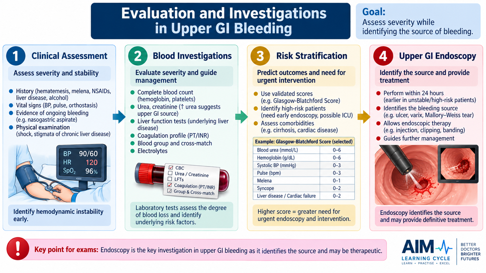
Evaluation has two simultaneous aims: determine how severely the patient has been affected by blood loss and identify the likely source of bleeding. The patient with active bleeding must therefore be assessed clinically before laboratory or endoscopic results are available.
History
History helps distinguish the likely cause and identifies factors that can increase the severity of bleeding. Important points include the form and amount of bleeding, previous episodes, symptoms suggesting ulcer disease, known liver disease or portal hypertension, and use of drugs that promote mucosal injury or impair hemostasis.
- Hematemesis, coffee-ground vomiting or melena.
- Approximate onset and progression.
- Previous ulcer or GI bleeding.
- Known chronic liver disease or portal hypertension.
- Use of NSAIDs, antiplatelet drugs or anticoagulants.
- Alcohol history where liver disease is relevant.
- Associated syncope, weakness, abdominal symptoms or reduced urine output.
Examination
The examination first looks for circulatory instability. Tachycardia, falling blood pressure, cool peripheries, altered mental state and reduced urine output suggest progressively impaired tissue perfusion. The abdomen and general examination are then used to look for clues to the underlying source, including features of chronic liver disease.
Laboratory Investigations
Laboratory tests assess blood loss, organ function and factors that may complicate treatment. Results must always be interpreted with the clinical condition because the initial hemoglobin concentration can underestimate the significance of very recent acute blood loss.
- Complete blood count: assesses hemoglobin, hematocrit and platelet count.
- Blood group and compatibility testing: prepares for transfusion when clinically required.
- Urea, creatinine and electrolytes: assess renal function and consequences of hypovolemia. Urea may rise because digested blood protein is absorbed and because reduced circulating volume decreases renal perfusion.
- Liver function tests: are especially relevant when chronic liver disease and portal hypertension are suspected.
- Coagulation profile: identifies impaired coagulation that may accompany liver disease or anticoagulant treatment.
Upper GI Endoscopy
Upper gastrointestinal endoscopy is central because it can directly identify the bleeding lesion and, in many cases, treat it during the same procedure. Endoscopy can demonstrate an ulcer, varix, mucosal erosion, tear or tumor and can show whether active bleeding or recent signs of bleeding are present.
Bleeding presentation → assess severity → laboratory evaluation → endoscopic localization → cause-specific treatment.
C. Initial Management of Upper GI Bleeding
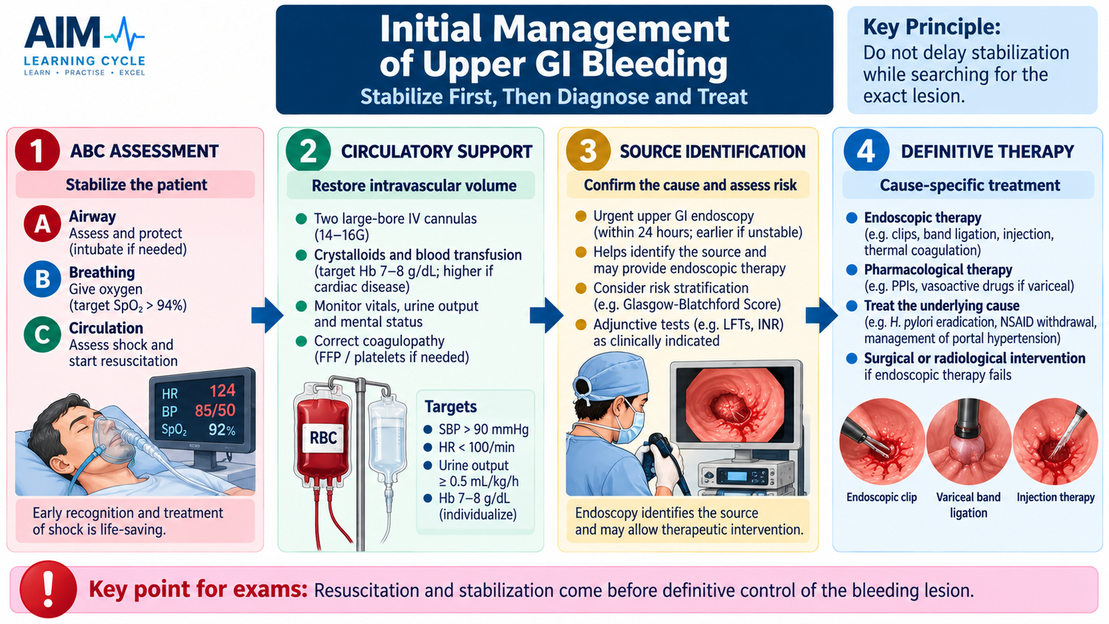
The immediate priority in significant upper GI bleeding is not to name the exact lesion but to preserve oxygen delivery and circulation. Diagnostic and definitive treatment follow stabilization. Management therefore proceeds from resuscitation to identification of the source and then to cause-specific pharmacological or endoscopic therapy.
Immediate Priorities
Airway, breathing and circulation are assessed first. A patient with massive hematemesis or impaired consciousness may be unable to protect the airway. Circulatory assessment determines whether intravenous fluid or blood replacement is required.
- Assess and protect the airway when necessary.
- Assess breathing and oxygenation.
- Obtain adequate intravenous access.
- Restore circulating volume according to clinical need.
- Arrange blood products when significant blood loss requires transfusion.
- Monitor pulse, blood pressure, mental status and urine output.
- Obtain the important blood investigations without delaying resuscitation.
Cause-Directed Treatment
Once resuscitation is underway, treatment is directed toward the suspected source. Acid-suppressive therapy is relevant when an ulcer-related non-variceal bleed is suspected. When variceal hemorrhage is suspected, a portal-pressure-reducing vasoactive drug is started promptly and definitive endoscopic assessment and treatment are arranged.
This distinction is important: general resuscitation treats the consequences of hemorrhage, while pharmacological and endoscopic therapy treat the bleeding mechanism or lesion itself.
D. Variceal Hemorrhage and Endoscopic Treatment
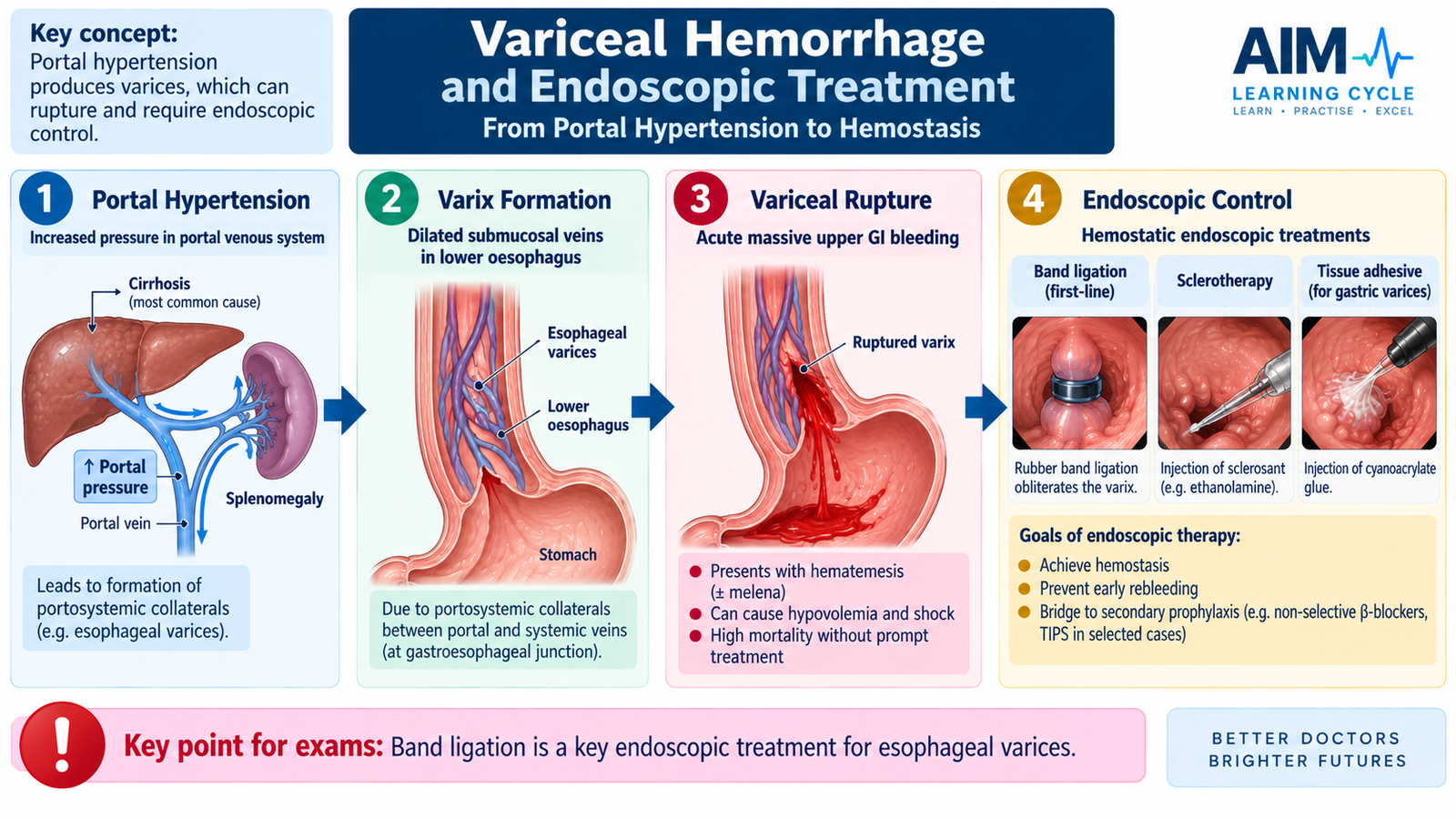
Varices are abnormally dilated veins that develop when portal venous pressure is increased. At the gastroesophageal region, portal venous blood is diverted through collateral channels. These veins become enlarged and thin-walled. When a varix ruptures, bleeding may be rapid because it is occurring from a high-flow collateral circulation.
Pharmacological Treatment: Why It Is Started
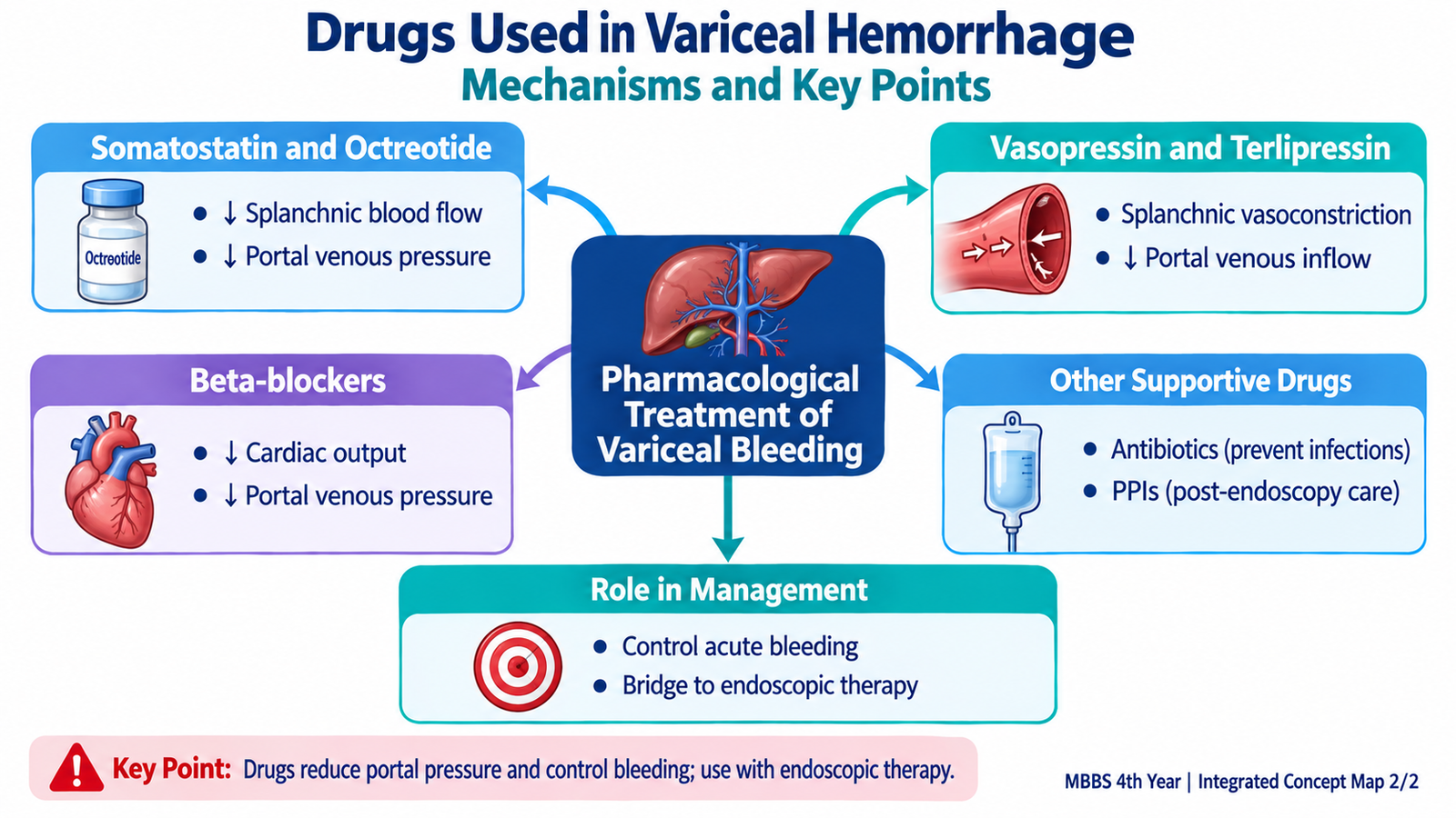
Vasoactive therapy is used when acute variceal bleeding is suspected because reducing blood flow into the portal circulation lowers variceal pressure. This can decrease ongoing hemorrhage and supports endoscopic control. Pharmacological therapy and endoscopic therapy therefore work together rather than serving as competing alternatives.
Endoscopic Treatment
Endoscopy confirms whether varices are responsible for bleeding and allows direct treatment. The procedure chosen depends on the location and type of varix.
- Endoscopic variceal ligation: bands are placed around bleeding esophageal varices. The band mechanically occludes the varix, leading to thrombosis and subsequent obliteration.
- Endoscopic sclerotherapy: a sclerosant is injected into or near the varix, producing thrombosis and tissue reaction. It is mainly an alternative when effective band ligation cannot be performed.
- Tissue adhesive injection: may be used for bleeding gastric varices because their anatomy often makes conventional band ligation less suitable.
When Endoscopic Treatment Is Indicated
A patient with suspected significant upper GI bleeding requires endoscopic assessment after initial stabilization. When the examination demonstrates active or recent variceal hemorrhage, endoscopic hemostatic treatment is performed. Continued or recurrent bleeding despite appropriate pharmacological and endoscopic treatment indicates severe disease and requires urgent specialist escalation.
E. Vasoactive Drugs in Acute Variceal Hemorrhage
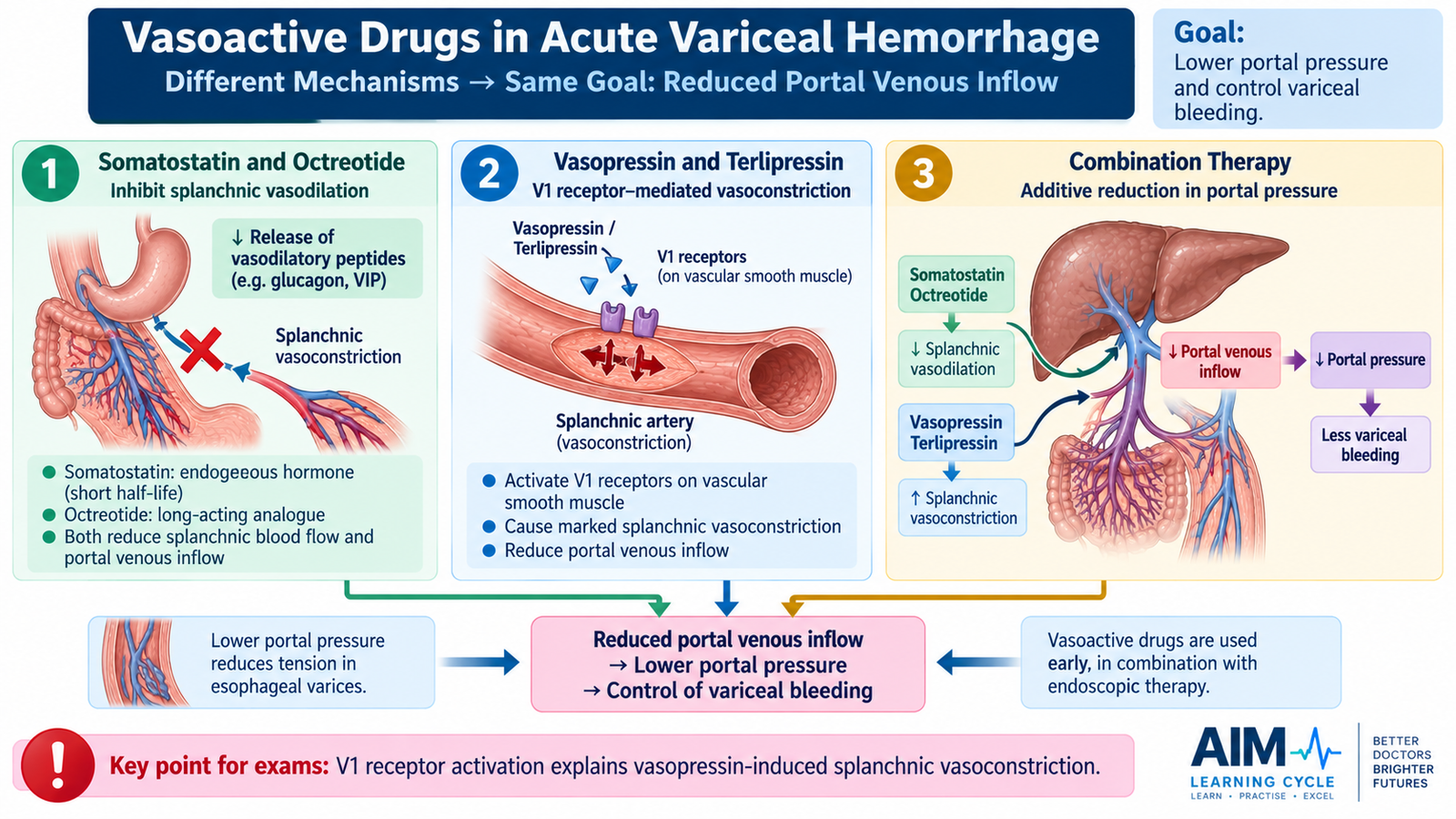
The principal pharmacological objective in acute variceal hemorrhage is to reduce portal venous inflow. Less blood entering the portal circulation means lower pressure within the varices and therefore less driving force for continued bleeding. Somatostatin, octreotide, vasopressin and terlipressin achieve this final effect through different receptor and vascular mechanisms.
Somatostatin
Somatostatin is an endogenous inhibitory peptide. In the splanchnic circulation it reduces the release and action of vasodilatory mediators, including glucagon. The resulting reduction in splanchnic blood flow decreases the amount of blood entering the portal venous system.
Octreotide
Octreotide is a synthetic somatostatin analogue with a longer duration of action than natural somatostatin. It produces the same clinically useful final effect: reduced splanchnic circulation and reduced portal inflow. It can therefore support hemostasis during acute variceal hemorrhage while definitive endoscopic therapy is arranged and performed.
Vasopressin
Vasopressin acts on V1 receptors on vascular smooth muscle. V1-receptor stimulation causes powerful vasoconstriction, including constriction of mesenteric arterioles. This decreases blood flow through the splanchnic circulation and therefore decreases portal venous inflow.
Because vasopressin can also constrict systemic vessels, its vasoconstrictor action may affect the coronary, peripheral and mesenteric circulations. This explains why ischemic cardiovascular and intestinal adverse effects are important limitations of the drug.
Terlipressin
Terlipressin is a longer-acting vasopressin analogue. It ultimately stimulates vascular V1 receptors, producing splanchnic vasoconstriction and reducing portal venous inflow. Its longer action makes the portal-pressure effect more sustained than that of vasopressin itself. However, because its therapeutic action still depends on vasoconstriction, ischemic complications remain an important caution.
F. Beta-Blockers in Variceal Bleeding
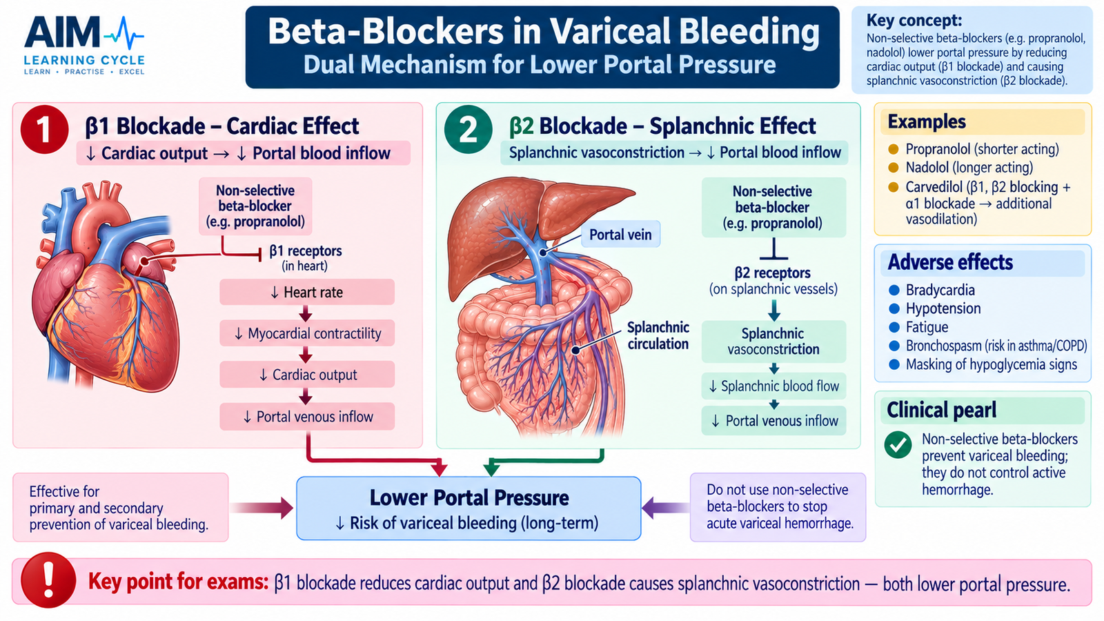
Non-selective beta-blockers reduce portal pressure through two complementary actions. They are important in the longer-term control of portal hypertension and prevention of variceal bleeding or rebleeding, but they are not drugs for rapidly stopping an actively bleeding varix in a hemodynamically unstable patient.
Mechanism of Action
Drugs such as propranolol and nadolol block both β1 and β2 receptors. Each component contributes to the reduction in portal venous inflow.
- β1 blockade in the heart reduces heart rate and cardiac output. Less blood is therefore delivered to the splanchnic circulation.
- β2 blockade in splanchnic vessels removes β2-mediated vasodilation. Alpha-mediated vascular tone becomes relatively unopposed, producing splanchnic vasoconstriction.
- The combined result is reduced splanchnic blood flow → reduced portal venous inflow → reduced portal pressure.
Carvedilol also blocks α1 receptors. This additional action can reduce intrahepatic vascular resistance, further lowering portal pressure. The central principle for the student, however, remains that non-selective beta-blockade reduces the pressure transmitted to gastroesophageal varices.
Integrated Mechanism Flow
Important Comparison: Drugs Affecting Portal Pressure
| Drug/Class | Main Mechanism | Portal Effect | Main Clinical Role |
|---|---|---|---|
| Somatostatin | Reduces splanchnic vasodilatory influences | ↓ Splanchnic flow and portal inflow | Acute variceal hemorrhage |
| Octreotide | Somatostatin-receptor mediated reduction of splanchnic flow | ↓ Portal venous inflow | Acute variceal hemorrhage |
| Vasopressin | V1-mediated vascular smooth-muscle constriction | Marked ↓ splanchnic inflow | Acute portal-pressure reduction; systemic vasoconstriction limits use |
| Terlipressin | Longer-acting vasopressin analogue acting mainly through V1 effects | ↓ Splanchnic and portal inflow | Acute variceal hemorrhage |
| Non-selective β-blockers | β1 blockade + β2 blockade | ↓ Cardiac output and splanchnic flow | Prevention of variceal bleeding/rebleeding rather than acute hemostasis |
⭐ AIM High-Yield Review
Upper Gastrointestinal Bleeding
Review the clinical presentation, evaluation and management principles of upper GI bleeding.