
15. Pancreatic Disorders: Pancreatitis, Pseudocyst and Pancreatic Carcinoma
A pathology-and-surgery learning chapter covering congenital pancreatic anomalies, acute and chronic pancreatitis, pancreatic pseudocyst and pancreatic carcinoma.
Topic Introduction
The pancreas is vulnerable to both inflammatory injury and malignant disease. In acute pancreatitis, pancreatic enzymes become activated inappropriately and begin damaging the gland itself. In chronic pancreatitis, repeated or persistent injury causes permanent fibrosis, loss of pancreatic tissue and progressive functional failure. Collections produced by pancreatitis may form a pancreatic pseudocyst. The pancreas also develops important congenital abnormalities and an aggressive malignant tumor, pancreatic ductal adenocarcinoma. Understanding these disorders becomes easier when the cause, mechanism, structural change, clinical presentation, investigation and complication are connected in sequence.
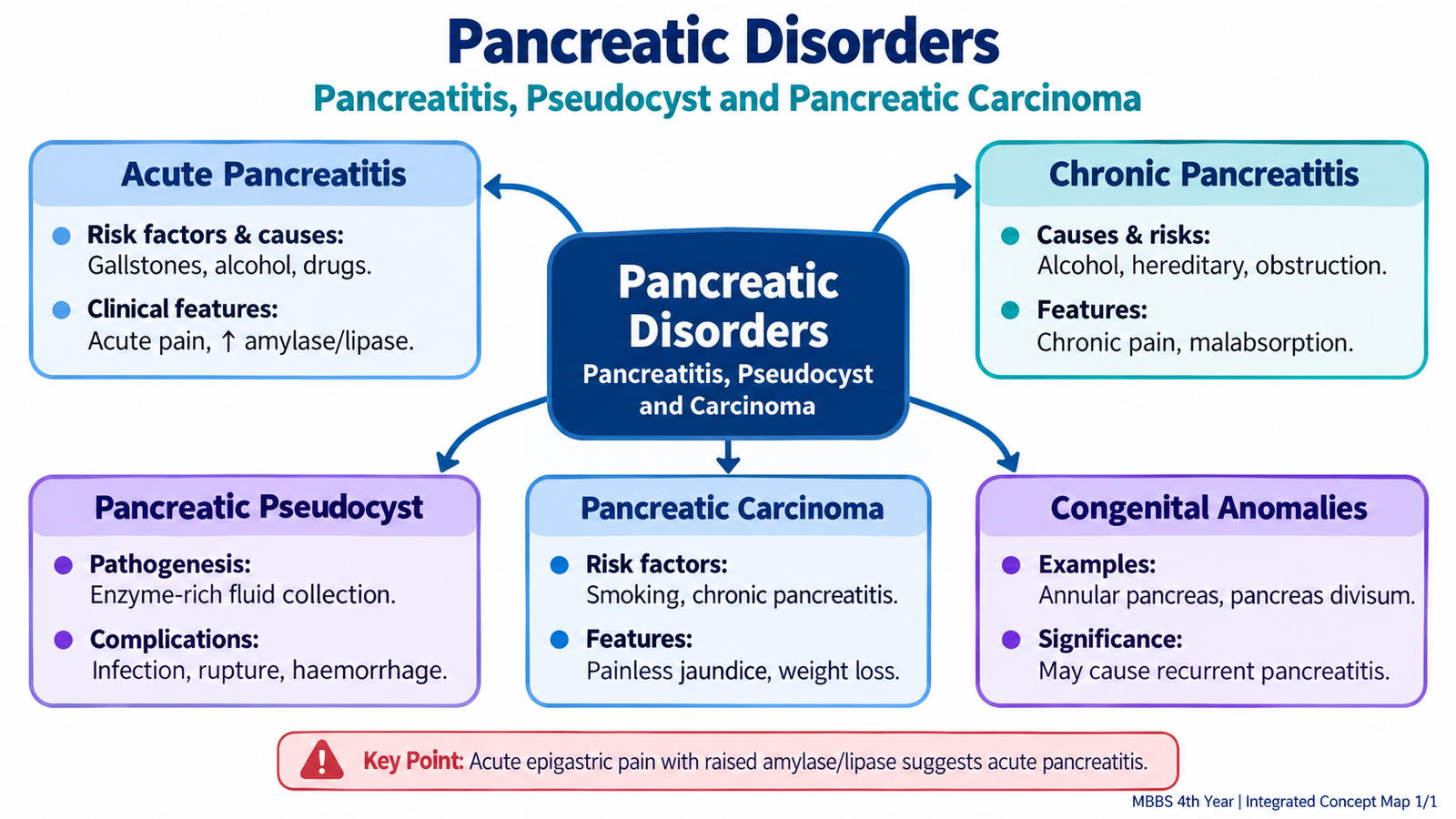
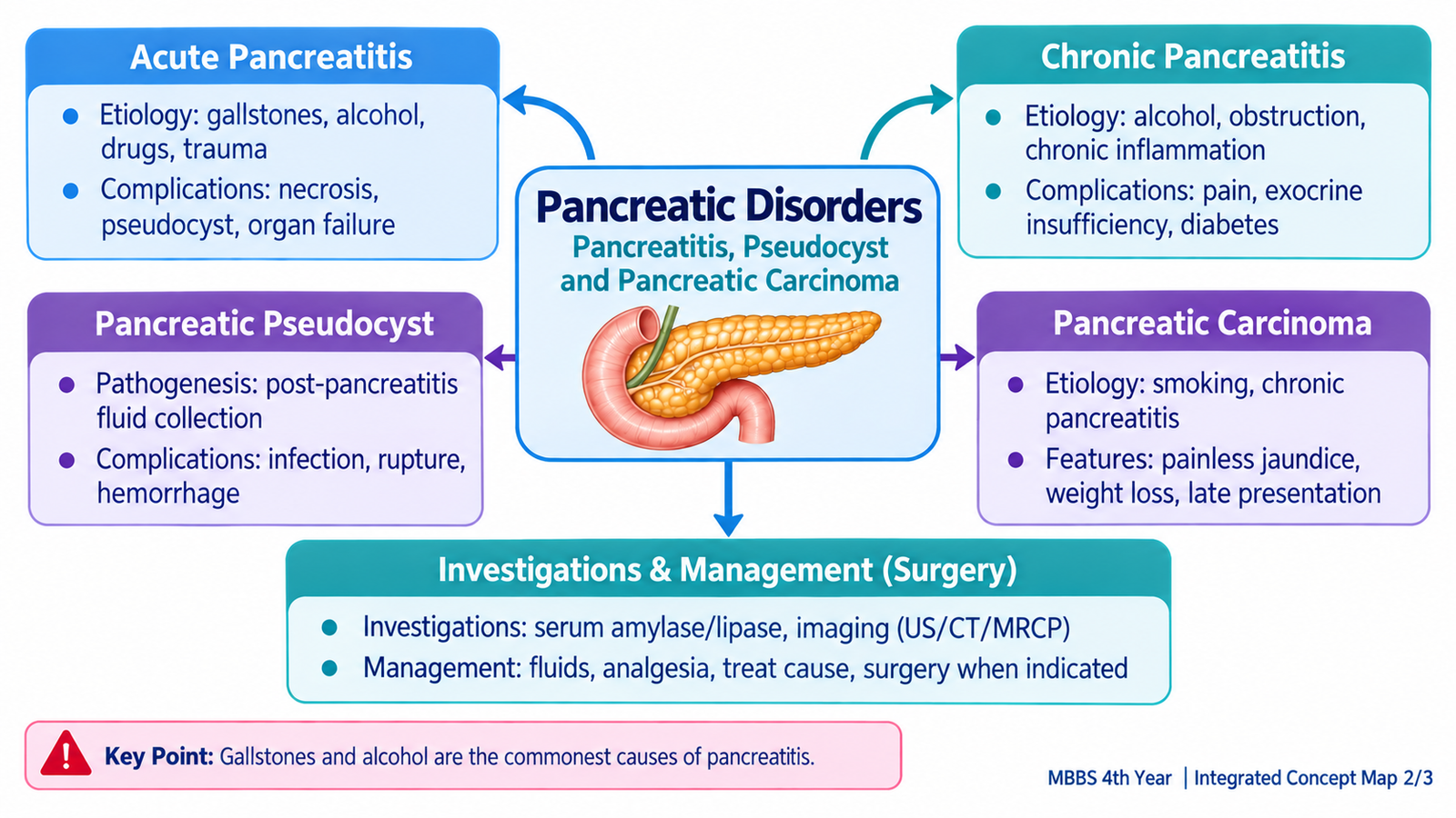
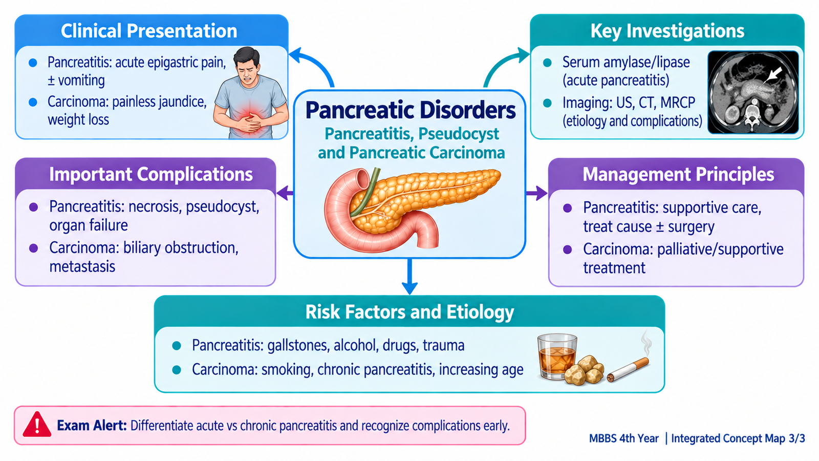
A. Congenital Anomalies of the Pancreas
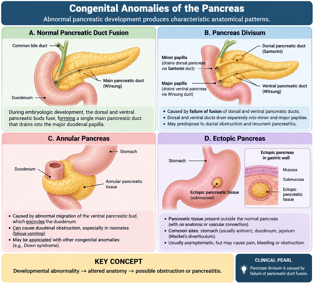
Most pancreatic congenital anomalies result from abnormal development, fusion or migration of the dorsal and ventral pancreatic buds. Many remain asymptomatic, but some alter pancreatic drainage or compress nearby structures and therefore become clinically important.
Pancreas divisum
Pancreas divisum occurs when the dorsal and ventral pancreatic duct systems fail to fuse normally. As a result, most pancreatic secretions drain through the smaller minor papilla rather than the major papilla. This drainage arrangement is usually harmless. In some individuals, however, relatively restricted drainage through the minor papilla may predispose to recurrent pancreatic inflammation.
Annular pancreas
In annular pancreas, pancreatic tissue forms a ring around the second part of the duodenum. This results from abnormal migration of the ventral pancreatic bud. If the ring significantly narrows the duodenum, it can produce duodenal obstruction, particularly in infancy, although milder cases may present later.
Ectopic pancreas
Ectopic or heterotopic pancreatic tissue is pancreatic tissue located outside the normal pancreas without a direct anatomical connection to it. Common sites include the stomach, duodenum and jejunum. It is usually incidental but can occasionally become inflamed or cause local symptoms.
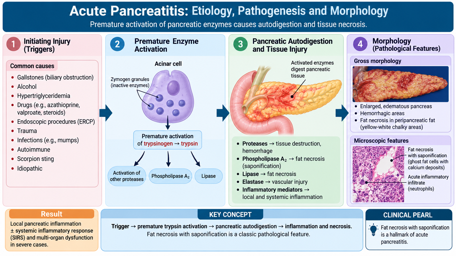
B. Acute Pancreatitis: Etiology, Pathogenesis and Morphology
Acute pancreatitis is an acute inflammatory injury of the pancreas caused by inappropriate activation of digestive enzymes within the gland. The central pathological problem is therefore autodigestion: enzymes that normally digest food begin damaging pancreatic tissue, surrounding fat and blood vessels.
Etiology and risk factors
The common causes act through different mechanisms, but each can ultimately promote premature enzyme activation or pancreatic duct obstruction.
- Gallstones: obstruction near the ampulla can interfere with pancreatic drainage.
- Alcohol: promotes pancreatic injury and abnormal secretory processes and may contribute to duct obstruction.
- Hypertriglyceridemia: marked triglyceride elevation can produce toxic free fatty acids and pancreatic injury.
- Drugs: certain medications can cause pancreatitis as an adverse reaction.
- Endoscopic or operative procedures: pancreatic injury may follow procedures involving the pancreaticobiliary tract, especially ERCP.
- Abdominal trauma: direct injury can damage pancreatic tissue and ducts.
- Metabolic and less common causes: hypercalcemia, infections and hereditary predisposition may contribute in selected patients.
Core pathogenesis
Digestive enzymes are normally produced as inactive precursors and activated only after reaching the intestine. In acute pancreatitis, this protective separation fails. Premature activation of trypsin within pancreatic acinar cells or ducts activates additional enzymes. Proteases damage pancreatic tissue, lipases digest fat, and elastase can damage vessel walls.
Morphology
The appearance of the pancreas varies with severity. Mild disease may be predominantly edematous, whereas severe disease may show extensive pancreatic and peripancreatic necrosis.
Gross morphology
- Pancreatic swelling and edema.
- Areas of chalky-white fat necrosis in and around the pancreas.
- Foci of pancreatic tissue necrosis.
- Hemorrhage in severe destructive pancreatitis.
The chalky deposits in fat necrosis result when released fatty acids bind calcium, producing calcium soaps. This process is called saponification.
Microscopic morphology
- Acinar cell necrosis.
- Acute inflammatory infiltrate, particularly neutrophils.
- Fat necrosis with calcium deposition.
- Interstitial edema.
- Vascular destruction and hemorrhage in severe cases.
C. Acute Pancreatitis: Clinical Features, Investigations, Treatment and Complications
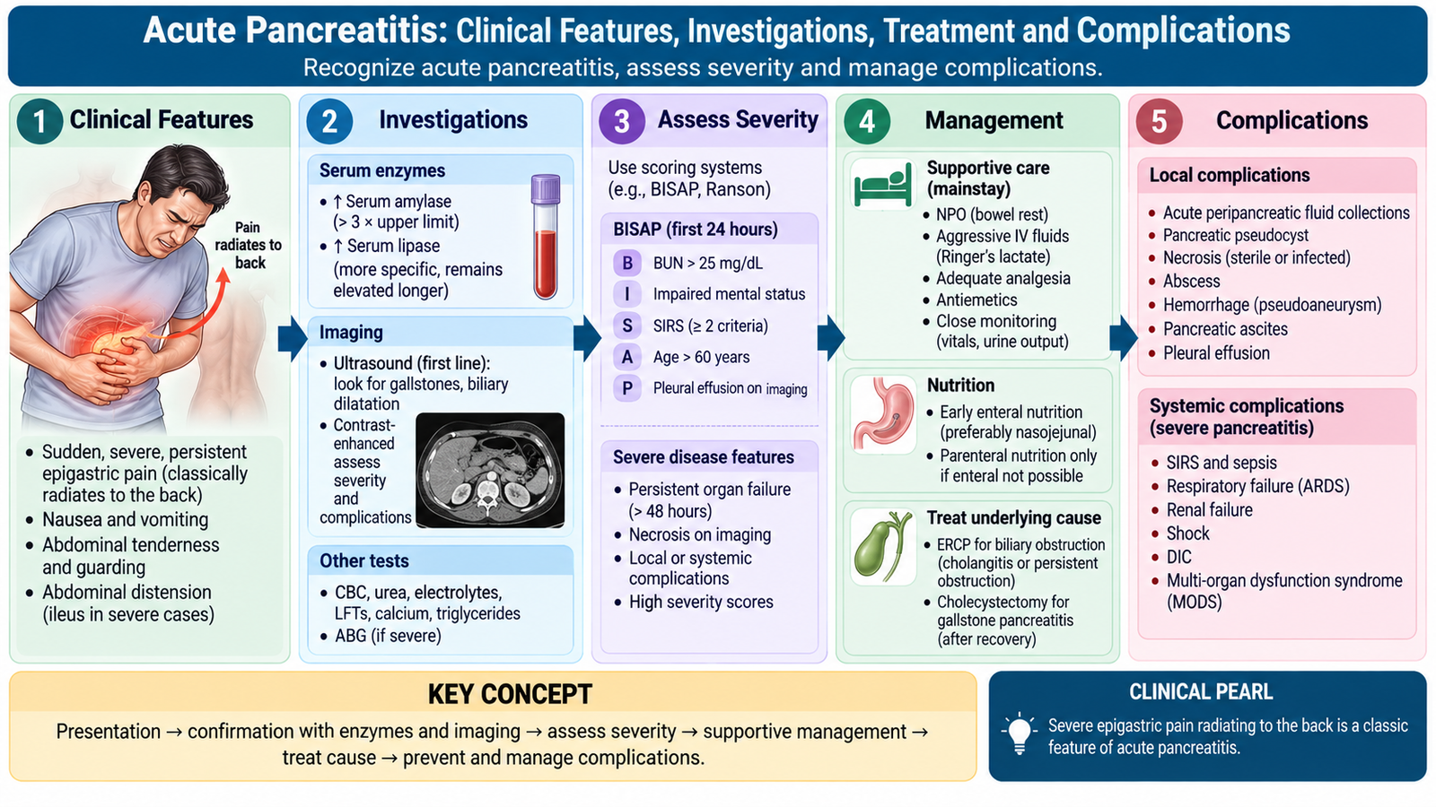
Clinically, acute pancreatitis ranges from a self-limited inflammatory illness to a severe systemic disease with organ failure. The combination of characteristic abdominal pain, pancreatic enzyme elevation and appropriate imaging usually establishes the diagnosis.
Clinical features
The typical symptom is sudden severe epigastric pain, often radiating to the back. The pain reflects inflammation of the retroperitoneal pancreas and surrounding tissues.
- Severe persistent epigastric or upper abdominal pain.
- Pain commonly radiating to the back.
- Nausea and vomiting.
- Upper abdominal tenderness and guarding.
- Abdominal distension or reduced bowel sounds due to ileus.
- Fever and tachycardia caused by the inflammatory response.
- Hypotension or shock in severe disease due to fluid loss, vasodilatation and systemic inflammation.
Extensive retroperitoneal hemorrhage may rarely produce discoloration around the umbilicus or flanks. These findings indicate severe hemorrhagic disease rather than uncomplicated pancreatitis.
Investigations
Investigations have three purposes: support the diagnosis, identify the cause and assess severity or complications.
- Serum lipase: rises in pancreatic acinar injury and is generally more pancreas-specific than amylase.
- Serum amylase: may also rise but is less specific because other conditions can increase it.
- Liver biochemical tests: help identify a biliary cause when abnormal in the appropriate clinical setting.
- Full blood count, renal function, electrolytes and glucose: help assess systemic effects and severity.
- Serum calcium and triglycerides: may reveal important metabolic causes.
Imaging
- Ultrasound: useful for detecting gallstones and biliary dilatation.
- Contrast-enhanced CT: useful when the diagnosis is uncertain, disease is severe or complications such as necrosis and collections must be assessed.
- MRCP or endoscopic ultrasound: may help evaluate suspected biliary or ductal disease in selected patients.
Treatment principles
Most patients are managed initially with supportive treatment while the underlying cause is identified. The aim is to maintain circulation and organ function while pancreatic inflammation settles.
- Fluid replacement: corrects intravascular volume loss caused by inflammation and fluid shift into tissues.
- Analgesia: controls the severe abdominal pain.
- Oxygen and physiological monitoring: are important in patients with significant systemic illness.
- Enteral nutrition: is preferred when nutritional support is required because maintaining gut function is beneficial.
- Treat the underlying cause: for example, address biliary obstruction or metabolic abnormalities.
In gallstone pancreatitis, urgent biliary endoscopic intervention is particularly relevant when there is associated cholangitis or persistent biliary obstruction. Definitive management of gallstones reduces the risk of recurrent attacks. Surgery or drainage for pancreatic necrosis is reserved for selected complicated cases, especially infected necrosis or persistent symptomatic collections. Routine prophylactic antibiotics are not used simply because pancreatic necrosis is present; antibiotics are appropriate when infection is suspected or demonstrated.
Complications
Local complications
- Pancreatic and peripancreatic necrosis.
- Infection of necrotic tissue.
- Pancreatic pseudocyst.
- Hemorrhage due to vascular erosion.
- Persistent fluid collections.
Systemic complications
- Hypovolemic or inflammatory shock.
- Acute respiratory distress syndrome.
- Acute kidney injury.
- Disseminated intravascular coagulation in severe systemic disease.
- Metabolic abnormalities, including hypocalcemia associated with extensive fat necrosis.
- Multiorgan failure in severe pancreatitis.
D. Chronic Pancreatitis: Etiology, Pathogenesis and Morphology
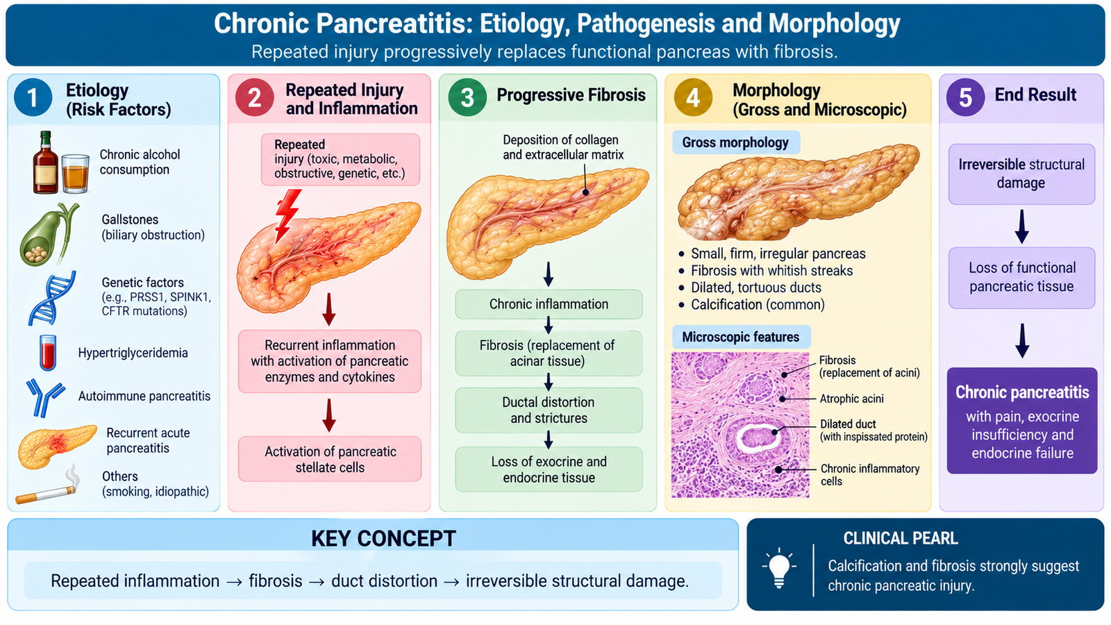
Chronic pancreatitis is a progressive inflammatory disorder in which repeated or persistent pancreatic injury leads to irreversible fibrosis, loss of acinar tissue and eventually impairment of both exocrine and endocrine pancreatic function. Unlike acute pancreatitis, structural damage in established chronic pancreatitis does not completely return to normal.
Etiology and risk factors
Several different insults can produce chronic pancreatic injury. Alcohol exposure and cigarette smoking are important acquired risk factors, while duct obstruction and genetic abnormalities are important in selected patients.
- Long-term heavy alcohol exposure.
- Cigarette smoking.
- Long-standing obstruction of the pancreatic duct.
- Hereditary or genetic predisposition.
- Recurrent episodes of acute pancreatitis.
- Certain metabolic and idiopathic forms.
Pathogenesis
Repeated pancreatic injury stimulates inflammation and activation of fibrogenic pathways. Collagen is deposited within and around pancreatic lobules, acinar cells are progressively lost, and ducts may become distorted or obstructed. Protein plugs and calcification can further impair ductal drainage.
Morphology
Gross morphology
- The pancreas becomes firm and fibrotic.
- Pancreatic ducts may be irregularly dilated.
- Intraductal protein plugs and calcifications may be present.
- Advanced disease may produce an irregular, shrunken gland.
Microscopic morphology
- Loss and atrophy of pancreatic acini.
- Dense fibrosis within and between lobules.
- Variable chronic inflammatory infiltrate.
- Ductal distortion and dilatation.
- Relative preservation of islets early in the disease, followed by endocrine loss in advanced disease.
The relationship between morphology and function is important: loss of acinar cells reduces digestive enzyme secretion, while later damage to pancreatic islets can impair insulin secretion and produce diabetes.
E. Chronic Pancreatitis: Clinical Features, Investigations, Treatment and Complications
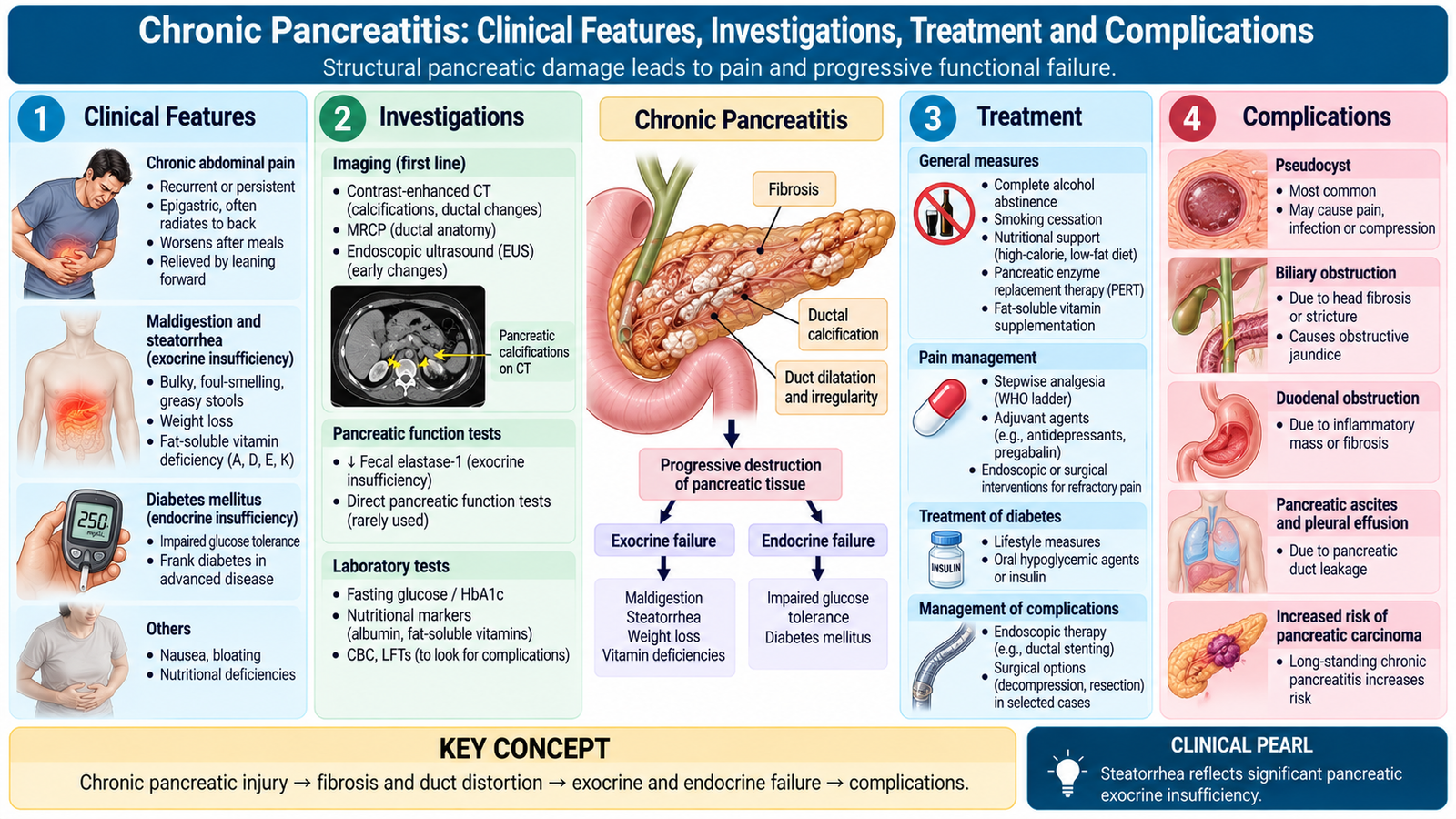
Patients with chronic pancreatitis may initially present with recurrent upper abdominal pain. As pancreatic tissue is progressively destroyed, the clinical picture shifts toward maldigestion, weight loss and diabetes. The investigation strategy therefore looks for structural pancreatic disease and evidence of functional failure.
Clinical features
- Recurrent or persistent epigastric pain, often radiating to the back.
- Weight loss due to reduced intake, maldigestion and malabsorption.
- Steatorrhea when loss of exocrine pancreatic function becomes significant.
- Diabetes mellitus in advanced disease due to endocrine pancreatic destruction.
- Features related to complications such as biliary obstruction or pseudocyst formation.
Steatorrhea occurs because insufficient pancreatic lipase reaches the intestine. Dietary fat is therefore poorly digested and absorbed, producing bulky, greasy stools and contributing to nutritional deficiency.
Investigations
Unlike acute pancreatitis, serum pancreatic enzymes may be normal or only mildly abnormal in chronic pancreatitis because the damaged gland may no longer release large quantities of enzymes.
- Plain imaging or CT: may show pancreatic calcification, ductal dilatation, gland atrophy or complications.
- Ultrasound: may demonstrate structural change, although the pancreas may be difficult to visualize in some patients.
- MRCP: can demonstrate pancreatic duct abnormalities non-invasively.
- Endoscopic ultrasound: can detect structural changes when conventional imaging is inconclusive.
- Tests of exocrine function: may demonstrate pancreatic insufficiency when clinically suspected.
- Blood glucose assessment: detects endocrine dysfunction.
Treatment principles
Treatment aims to reduce further pancreatic injury, relieve pain, replace lost digestive function, manage diabetes and correct structural complications when present.
- Stop alcohol exposure when relevant.
- Stop smoking because it promotes progression and recurrent pancreatic injury.
- Provide appropriate nutritional support.
- Use analgesic therapy for pain control.
- Replace pancreatic enzymes when exocrine insufficiency causes maldigestion.
- Treat diabetes when endocrine failure develops.
- Use endoscopic or surgical intervention for selected patients with duct obstruction, persistent debilitating pain or important local complications.
Complications
- Exocrine pancreatic insufficiency and malnutrition.
- Diabetes mellitus.
- Pancreatic pseudocyst.
- Biliary or duodenal obstruction due to inflammation and fibrosis.
- Vascular complications around the pancreas.
- Increased risk of pancreatic carcinoma in long-standing chronic pancreatitis.
F. Pancreatic Pseudocyst
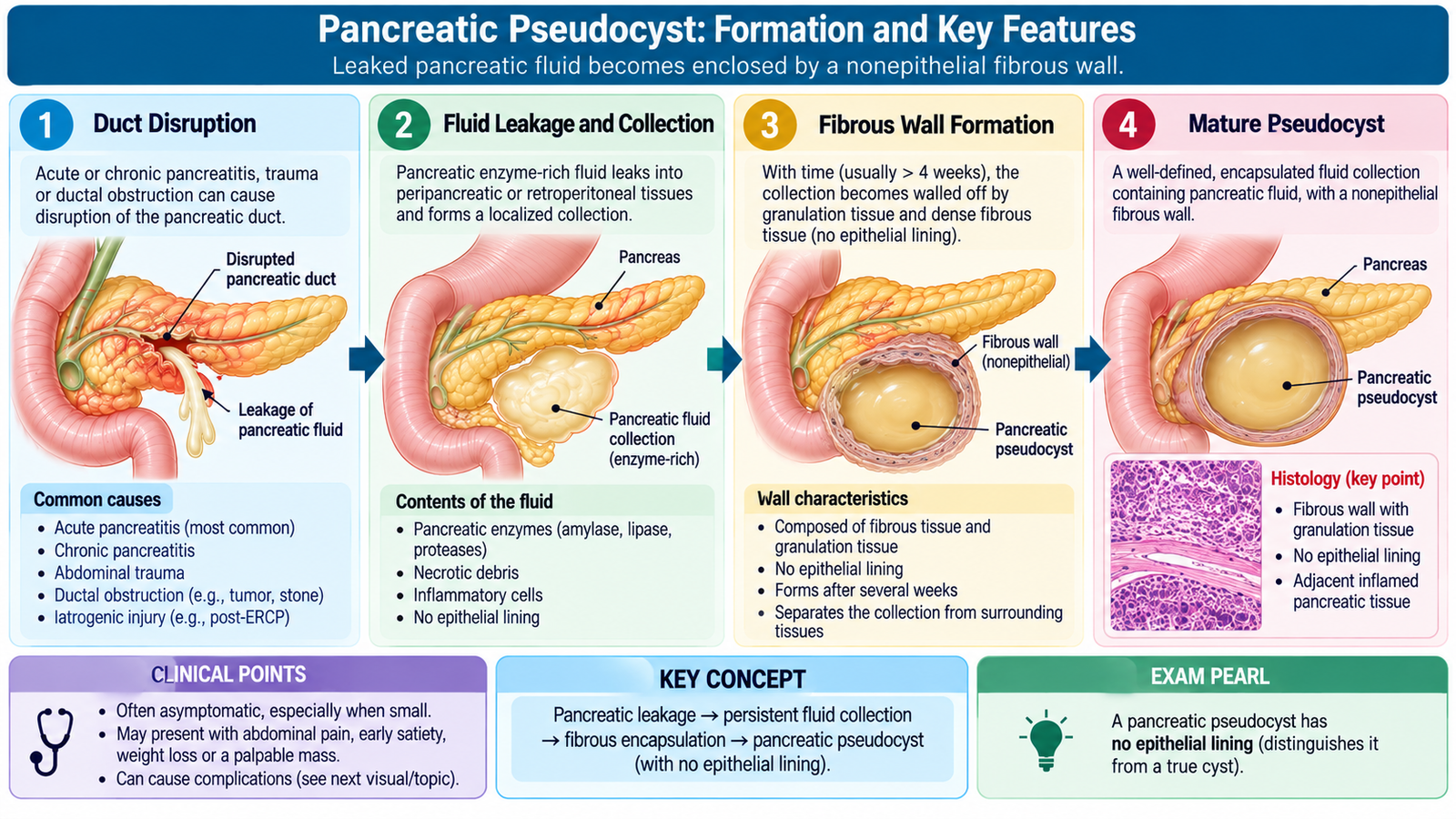
A pancreatic pseudocyst is a localized collection of pancreatic fluid surrounded by a wall of fibrous and granulation tissue. It usually develops after pancreatitis or pancreatic duct injury. The word pseudo is important because, unlike a true cyst, its wall has no epithelial lining.
Pathogenesis
Pancreatic inflammation or duct disruption allows enzyme-rich pancreatic fluid and tissue breakdown products to escape into surrounding tissues. If the collection persists, the body gradually walls it off with granulation tissue and fibrosis.
Pseudocysts may occur after acute pancreatitis and are also associated with chronic pancreatitis. Their contents may include pancreatic enzymes, inflammatory fluid and necrotic material.
Complications
Complications occur because a pseudocyst can enlarge, become infected, erode nearby vessels or communicate with surrounding organs.
- Infection of the collection.
- Hemorrhage if pancreatic enzymes or inflammation erode a nearby blood vessel.
- Rupture into the peritoneal cavity or an adjacent hollow organ.
- Obstruction of the stomach, duodenum or biliary tract due to mass effect.
- Persistent abdominal pain or an enlarging abdominal mass.
G. Pancreatic Carcinoma
The most important malignant epithelial tumor of the pancreas is pancreatic ductal adenocarcinoma. It is an aggressive malignancy that usually arises from pancreatic ductal epithelial precursor lesions and often becomes clinically evident only after local invasion or spread has occurred.
Risk factors and etiology
Pancreatic carcinoma develops through accumulation of genetic alterations in pancreatic ductal epithelial cells. Environmental and inherited factors can increase this risk.
- Increasing age.
- Cigarette smoking.
- Long-standing chronic pancreatitis.
- Obesity and related metabolic factors.
- Family history and selected inherited cancer-predisposition syndromes.
- Some patients have no identifiable major risk factor.
Pathogenesis
Many pancreatic ductal adenocarcinomas develop through stepwise progression of microscopic precursor lesions called pancreatic intraepithelial neoplasia. Genetic abnormalities accumulate as these lesions progress toward invasive carcinoma. Important molecular abnormalities include activation of KRAS and loss of tumor-suppressor pathways involving genes such as CDKN2A, TP53 and SMAD4. At undergraduate level, the important principle is that increasing genetic damage allows abnormal ductal epithelial cells to escape normal growth control and become invasive.
Morphology
Gross morphology
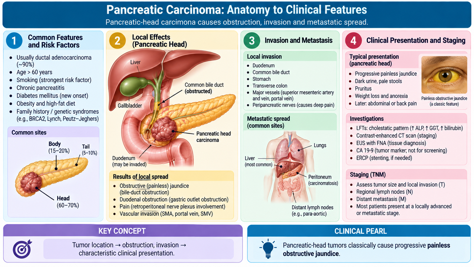
Most pancreatic ductal adenocarcinomas arise in the head of the pancreas. They are typically firm, poorly defined infiltrative masses because the tumor stimulates a dense fibrous stromal response known as desmoplasia.
- Hard, irregular and infiltrative mass.
- Frequently located in the pancreatic head.
- May obstruct the distal common bile duct or pancreatic duct.
- Often extends into surrounding soft tissue and adjacent structures.
Microscopic morphology
- Malignant gland-forming epithelial cells.
- Irregular infiltrating glands within dense desmoplastic stroma.
- Cellular atypia and abnormal glandular architecture.
- Perineural invasion is common and contributes to pain and local spread.
- Lymphatic and vascular invasion may be present.
Clinical features
The presentation depends strongly on tumor location. A tumor in the pancreatic head can obstruct the common bile duct relatively early, whereas tumors in the body or tail often become large before causing obvious obstructive symptoms.
- Progressive painless jaundice: particularly characteristic of tumors in the head obstructing the distal common bile duct.
- Dark urine, pale stools and pruritus due to obstructive cholestasis.
- Weight loss and reduced appetite.
- Persistent upper abdominal or back pain, especially with locally advanced disease.
- Palpable, distended gallbladder may occur with malignant distal biliary obstruction.
- New metabolic disturbance, including diabetes, may accompany pancreatic carcinoma.
- Migratory thrombophlebitis may occur because some adenocarcinomas create a hypercoagulable state.
Staging
Pancreatic ductal adenocarcinoma is staged using the TNM system. The system evaluates the primary tumor, regional lymph nodes and distant metastasis. The exact anatomical stage determines the extent of disease and is closely related to whether the tumor can be completely removed.
| Component | Undergraduate interpretation |
|---|---|
| T | Describes the size and local extent of the primary tumor; major arterial involvement represents advanced local disease. |
| N | Describes spread to regional lymph nodes. |
| M | M1 indicates distant metastasis and therefore stage IV disease. |
In practical terms, early stages are localized to the pancreas without distant spread; intermediate stages include increasing tumor extent or regional nodal involvement; advanced local disease involves critical nearby vessels or extensive regional disease; and stage IV indicates distant metastasis.
Complications and spread
- Biliary obstruction and obstructive jaundice.
- Pancreatic duct obstruction.
- Local invasion into surrounding tissues and nerves.
- Regional lymph-node metastasis.
- Distant metastasis, particularly to the liver and peritoneal surfaces.
- Thromboembolic complications associated with malignancy.
- Progressive weight loss and systemic effects of advanced cancer.
Integrated Mechanism Flow
Important Comparison: Acute versus Chronic Pancreatitis
| Feature | Acute Pancreatitis | Chronic Pancreatitis |
|---|---|---|
| Basic process | Acute enzyme-mediated pancreatic injury | Progressive irreversible fibrosis and tissue loss |
| Major morphology | Edema, acute inflammation, fat necrosis, hemorrhage | Fibrosis, acinar loss, duct distortion, calcification |
| Pancreatic function | Usually preserved after recovery from mild disease | Progressive exocrine and later endocrine failure |
| Pancreatic enzymes | Typically elevated during an acute attack | May be normal in advanced disease |
| Characteristic imaging clue | Inflammation, edema, collections or necrosis | Calcification, duct abnormalities and gland atrophy |
| Important long-term consequence | Pseudocyst or complications of severe acute injury | Steatorrhea, malnutrition and diabetes |
⭐ AIM High-Yield Review
- Pancreas divisum results from failure of fusion of the dorsal and ventral duct systems.
- Annular pancreas is pancreatic tissue encircling the duodenum and may cause duodenal obstruction.
- The central event in acute pancreatitis is premature activation of pancreatic digestive enzymes, especially trypsin.
- Acute pancreatitis can produce fat necrosis with saponification, pancreatic necrosis and hemorrhage.
- Severe epigastric pain radiating to the back with elevated pancreatic enzymes is a classic diagnostic pattern.
- Ultrasound is particularly useful for looking for gallstones, while CT helps assess severe disease and complications.
- Initial treatment of acute pancreatitis is primarily supportive, with specific intervention directed toward the cause or complications.
- Chronic pancreatitis causes irreversible fibrosis, acinar loss, duct distortion and often pancreatic calcification.
- Exocrine failure in chronic pancreatitis causes maldigestion and steatorrhea; endocrine failure can cause diabetes.
- A pancreatic pseudocyst has no epithelial lining; its wall consists of granulation and fibrous tissue.
- Pancreatic pseudocysts may become infected, bleed, rupture or compress adjacent structures.
- Most pancreatic carcinomas are ductal adenocarcinomas with a dense desmoplastic stroma.
- A carcinoma in the head of the pancreas can cause progressive painless obstructive jaundice by compressing or invading the distal common bile duct.
- Pancreatic carcinoma commonly spreads locally, to regional lymph nodes, and distantly to sites such as the liver and peritoneum.
- Distant metastasis defines stage IV disease in the TNM staging framework.
Pancreatic Disorders
Use these videos after reading the AIM Learning Material to reinforce pancreatitis, pancreatic complications and pancreatic carcinoma.
1. Acute Pancreatitis
Etiology • Pathophysiology • Clinical Features • Diagnosis • Treatment
2. Chronic Pancreatitis
Fibrosis • Calcification • Exocrine Failure • Complications • Treatment
3. Pancreatic Carcinoma
Risk Factors • Pathophysiology • Clinical Features • Diagnosis • Complications