
24. Hemorrhoids, Anal Fissure, Fistula and Anorectal Infections
A clear approach to the pathology, clinical presentation and management of common anorectal disorders, including infectious and sexually transmitted conditions.
Topic Introduction
Hemorrhoids, anal fissure, perianal fistula and anorectal infections are common disorders of the anal canal and surrounding tissues. Although they may share symptoms such as pain, bleeding or discharge, their underlying mechanisms are different. Hemorrhoids arise from enlargement and downward displacement of the normal anal vascular cushions. An anal fissure is a painful linear tear of the anoderm, while a perianal fistula is an abnormal tract that commonly develops after an anorectal abscess. Anorectal infections range from local pyogenic abscesses to sexually transmitted infections affecting the rectum and perianal region. Understanding the relationship between mechanism, morphology and symptoms makes these conditions much easier to distinguish and manage.
A. Hemorrhoids: Definition, Classification, Morphology and Pathogenesis
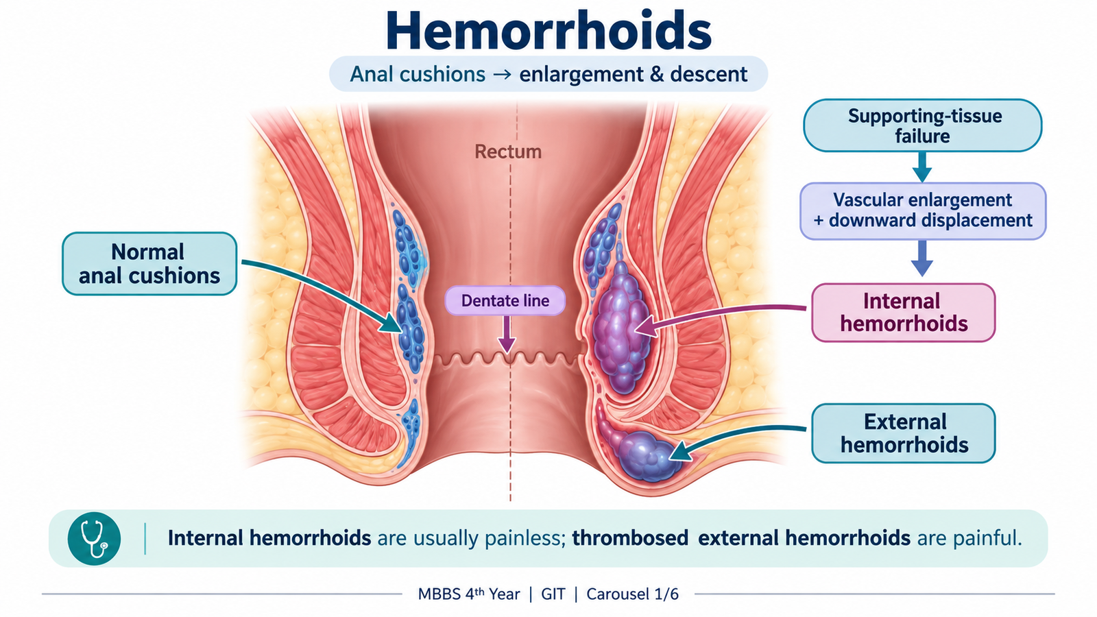
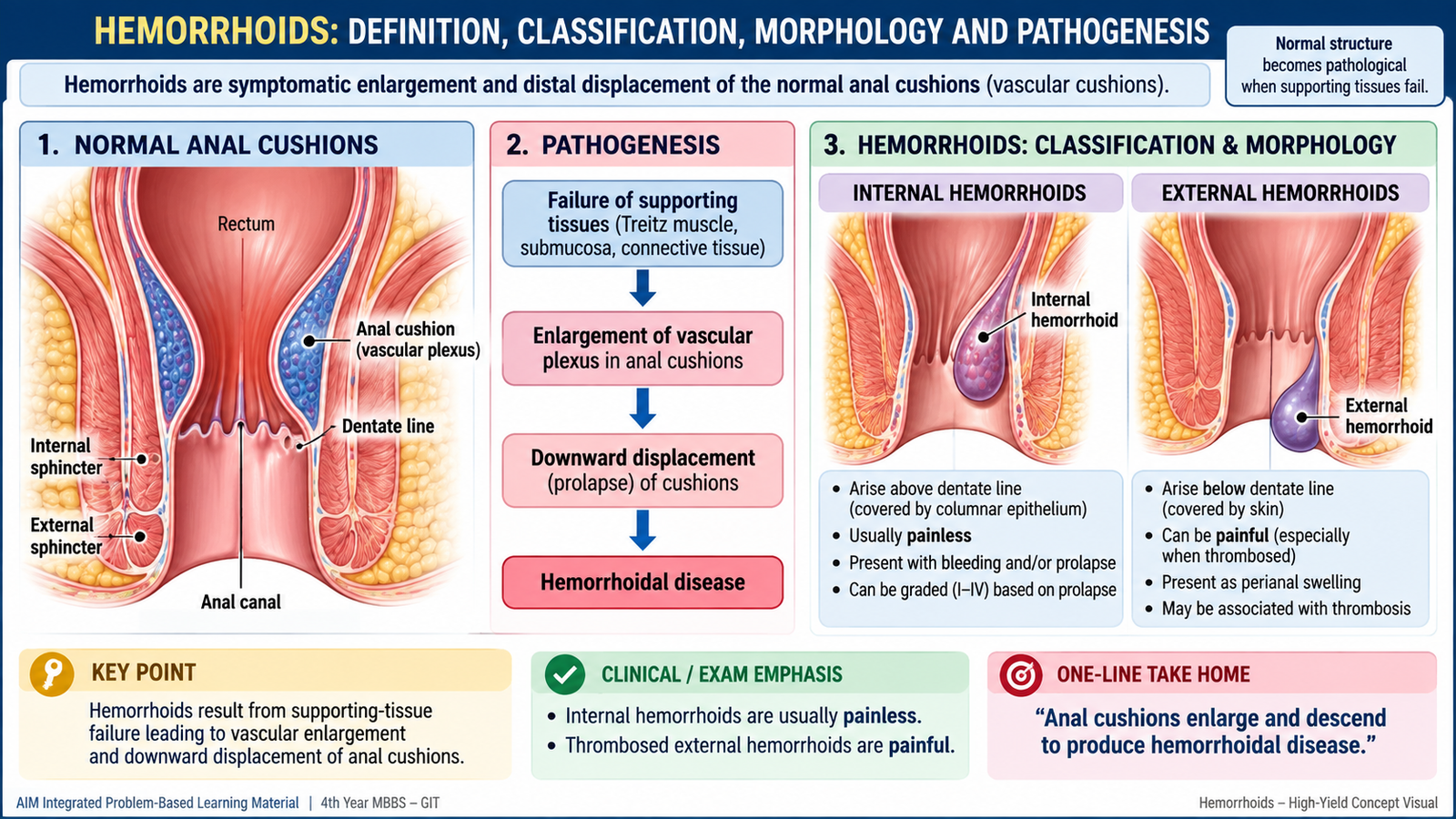
Hemorrhoids are symptomatic enlargement and displacement of the normal anal cushions. These cushions are vascular and connective-tissue structures that normally contribute to fine control of continence. Therefore, hemorrhoids are not simply “varicose veins of the anus.” The disease develops when the supporting tissues weaken and the vascular cushions become enlarged, congested and displaced downward.
Definition and Classification
Hemorrhoids are classified mainly according to their relationship to the dentate line, because this determines their epithelial covering, sensory innervation and typical symptoms.
- Internal hemorrhoids: arise above the dentate line and are covered by rectal or transitional mucosa. Because this region has predominantly visceral sensory innervation, uncomplicated internal hemorrhoids are usually painless.
- External hemorrhoids: occur below the dentate line and are covered by anoderm or skin. This area has somatic pain sensation, so thrombosis of an external hemorrhoid can cause intense pain.
Morphology
The pathological lesion consists of enlarged vascular channels together with supporting connective tissue. Repeated congestion and mechanical stress may produce prolapse, thrombosis, inflammation or surface injury.
- Bulging vascular cushions within or around the anal canal.
- Internal lesions may progressively protrude through the anal opening.
- Thrombosed external hemorrhoids appear as a tense, tender, bluish perianal swelling.
- Dilated, thin-walled vascular spaces.
- Variable thrombosis within vascular channels.
- Supporting connective tissue may show fibrosis and inflammation.
- Surface erosion or ulceration can contribute to bleeding.
Pathogenesis
The normal anal cushions are supported in position by connective tissue and smooth-muscle elements. With repeated straining, raised pressure and progressive weakening of this support, the cushions become congested and slide downward. The vascular channels enlarge, and the protruding mucosa becomes vulnerable to trauma during defecation.
B. Hemorrhoids: Clinical Features, Risk Factors and Management
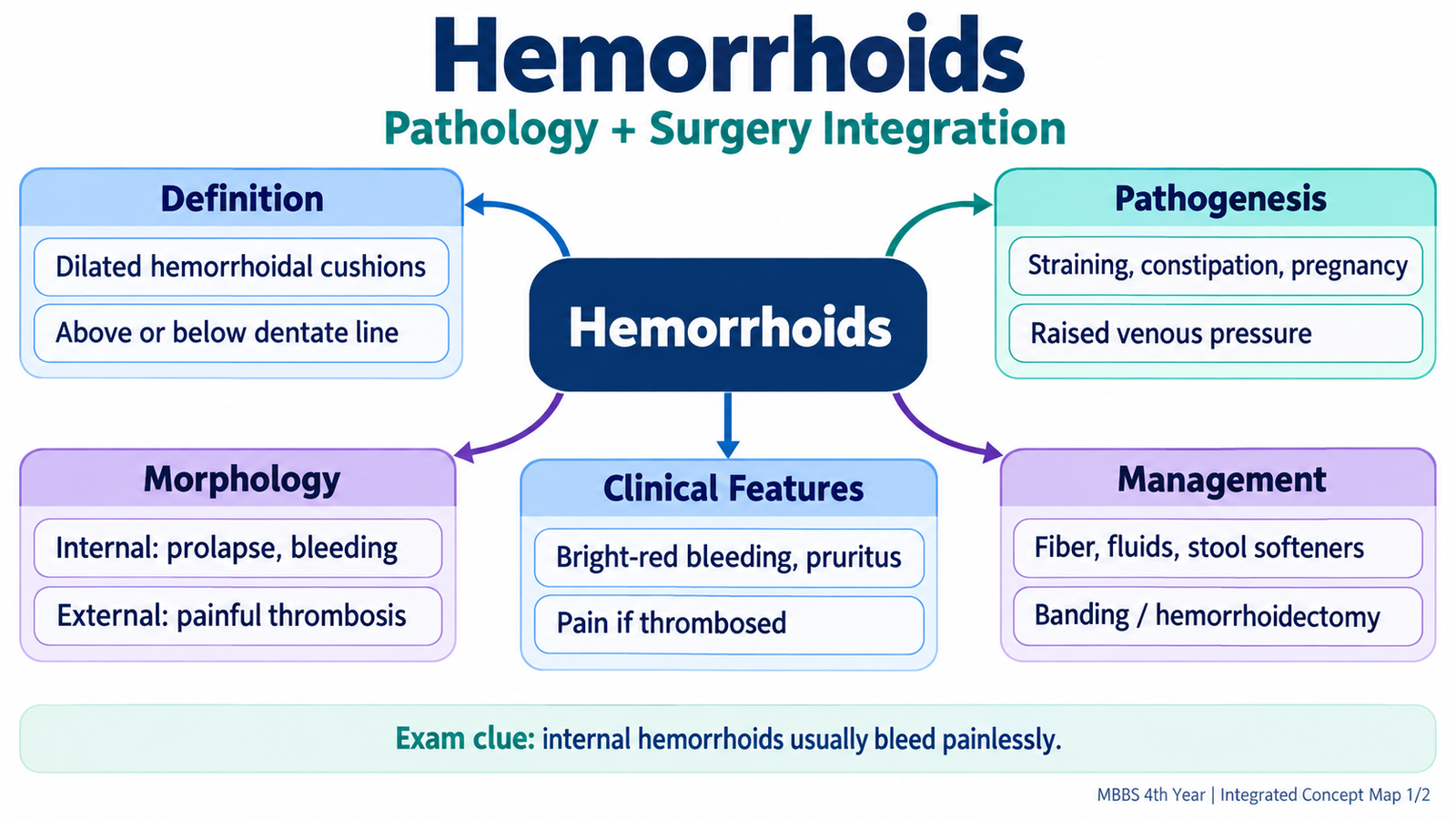
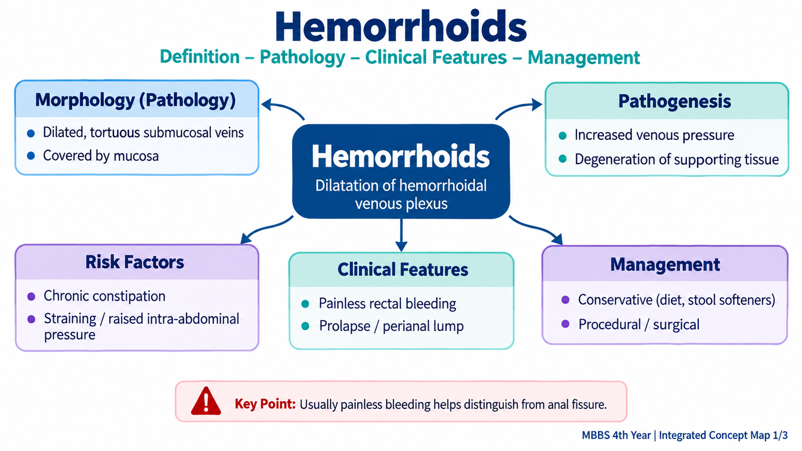
The symptoms of hemorrhoids depend mainly on whether the affected cushions are internal or external and whether complications such as prolapse or thrombosis have occurred. Internal hemorrhoids classically produce bleeding and prolapse, while thrombosed external hemorrhoids are characteristically painful.
Risk Factors
Conditions that repeatedly increase pressure within the hemorrhoidal cushions or increase straining can promote symptomatic hemorrhoidal disease.
- Chronic constipation and repeated straining during defecation.
- Passage of hard stool.
- Prolonged time spent during defecation.
- Pregnancy, due to increased pelvic pressure and venous congestion.
- Age-related weakening of the tissues supporting the anal cushions.
Clinical Features
Internal hemorrhoids commonly cause painless, bright-red rectal bleeding. Blood may be noticed on toilet paper, on the surface of the stool or after defecation. Increasing downward displacement produces prolapse, mucus discharge, difficulty maintaining local cleanliness and pruritus. External hemorrhoids may remain asymptomatic until thrombosis occurs. Acute thrombosis stretches the pain-sensitive anoderm and produces a sudden, very tender perianal swelling.
Grades of Internal Hemorrhoids
| Grade | Characteristic feature |
|---|---|
| I | Bleeding may occur, but there is no prolapse outside the anal canal. |
| II | Prolapse occurs during straining but reduces spontaneously. |
| III | Prolapse occurs and requires manual reduction. |
| IV | Prolapsed hemorrhoids remain irreducible. |
Management
Management depends on the severity of symptoms, degree of prolapse and presence of complications. Treatment should begin by reducing trauma and excessive straining during defecation.
- Conservative treatment: adequate dietary fibre, appropriate fluid intake, maintaining soft stool and avoiding prolonged straining.
- Office procedures: persistent internal hemorrhoids may be treated by procedures such as rubber-band ligation, injection sclerotherapy or infrared coagulation. These treatments reduce blood flow or produce fixation and fibrosis of the hemorrhoidal tissue.
- Surgical treatment: considered for large or persistent prolapsing hemorrhoids, especially advanced disease, recurrent symptoms or failure of less invasive treatment. Hemorrhoidectomy removes symptomatic hemorrhoidal tissue while preserving normal anal function.
- Thrombosed external hemorrhoid: pain control and local supportive treatment are important; selected patients with severe recent thrombosis may benefit from surgical treatment.
C. Anal Fissure: Core Concept, Risk Factors and Management
An anal fissure is a linear tear or ulcer in the anoderm of the anal canal, usually extending from the anal verge toward the dentate line. Most primary fissures occur in the midline, especially posteriorly. The severe pain of a fissure is explained by its location in the pain-sensitive anoderm below the dentate line.
How a Fissure Develops
A common initiating event is trauma from passage of a hard stool. The tear causes severe pain, which triggers spasm of the internal anal sphincter. Persistent sphincter spasm reduces local blood flow, especially in the posterior midline, and poor perfusion makes healing difficult. This creates a self-perpetuating pain–spasm–ischemia cycle.
Risk Factors
- Constipation and passage of hard stool.
- Repeated straining.
- Episodes of frequent diarrhea causing repeated local irritation.
- Trauma during childbirth.
- Underlying inflammatory or infectious disease in secondary fissures.
Clinical Recognition
The characteristic symptom is sharp pain during defecation that may continue afterward. A small amount of bright-red blood may be seen on the stool or toilet paper. Chronic fissures develop persistent structural changes such as a visible ulcer, a sentinel skin tag at the external end and hypertrophy of the anal papilla internally.
Management
The main objectives are to prevent further trauma, keep the stool soft and break the cycle of internal sphincter spasm and ischemia.
- Conservative management: increased fibre, appropriate fluid intake, treatment of constipation, stool-softening measures and warm sitz baths.
- Medical sphincter relaxation: topical nitrates or topical calcium-channel blockers may reduce internal sphincter tone, improve blood flow and allow healing.
- Botulinum toxin: may be used in selected persistent fissures to produce temporary sphincter relaxation.
- Surgery: lateral internal sphincterotomy is an effective treatment for selected chronic fissures that fail conservative treatment. The principle is controlled reduction of excessive internal sphincter tone while minimizing the risk of impaired continence.
- Secondary fissures: management must also address the underlying disease.
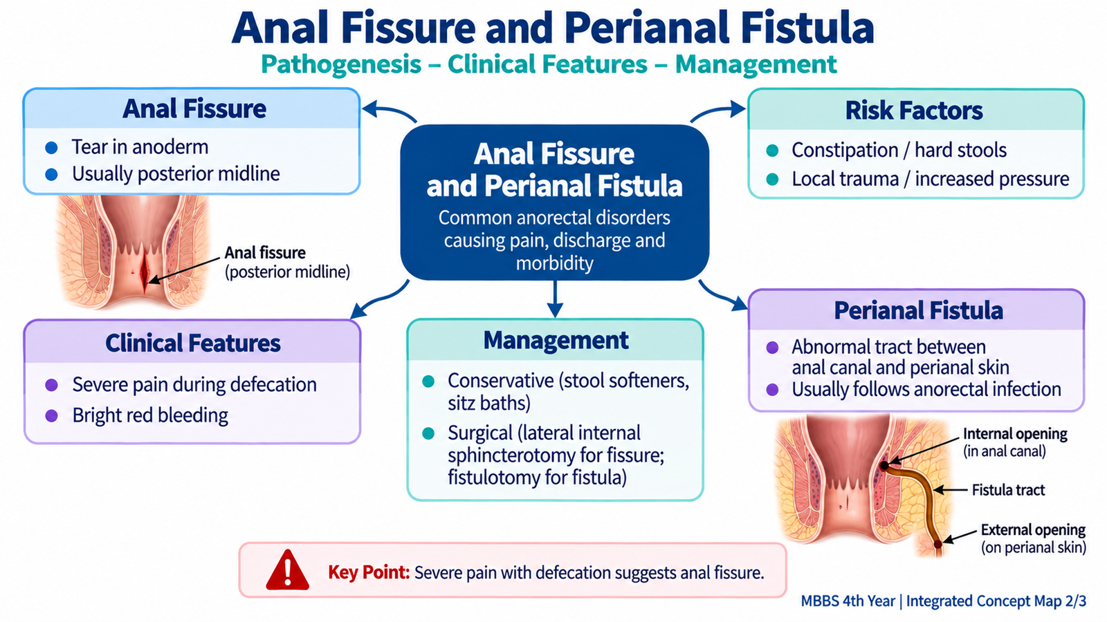
D. Perianal Fistula: Core Concept, Risk Factors and Management
A perianal fistula, commonly called fistula-in-ano, is an abnormal tract connecting the anal canal or rectum with the perianal skin. Most cases develop after infection of an anal gland produces an anorectal abscess. Even when the abscess drains, either spontaneously or surgically, a persistent tract may remain between the original internal opening and the skin.
Risk Factors and Causes
Cryptoglandular infection is the usual mechanism, but persistent or recurrent fistula may also be associated with underlying disease.
- Previous or recurrent anorectal abscess.
- Inflammatory bowel disease, particularly Crohn disease.
- Tuberculosis and other chronic infections in appropriate clinical settings.
- Previous anorectal surgery or local trauma.
- Less commonly, malignancy or other chronic inflammatory processes.
Clinical Pattern
A fistula often causes intermittent or persistent purulent discharge from an opening near the anus. Patients may describe repeated cycles of swelling, discomfort and discharge. Pain becomes more prominent if the tract becomes obstructed and another abscess forms. Examination may reveal an external opening, surrounding granulation tissue or induration.
Assessment
Many straightforward fistulas can be recognized clinically. Imaging such as MRI is particularly useful when the tract is recurrent, complex, high, associated with Crohn disease or difficult to define clinically because the relationship between the fistula and the anal sphincters determines safe treatment.
Management
The objective is not simply to close the external opening. The entire fistulous pathway must be controlled while preserving anal sphincter function and continence.
- An associated acute abscess requires adequate drainage.
- Simple low fistulas may be treated by fistulotomy, in which the tract is opened so that it heals from its base.
- High or complex fistulas require techniques that protect the anal sphincter; a seton may be used in selected cases.
- Recurrent or complex disease should be assessed carefully before intervention.
- Any underlying condition such as Crohn disease or chronic infection must also be managed.
E. Anorectal Infections: Classification and Clinical Patterns
Anorectal infections include infections of the anal glands, perianal tissues, anal canal and rectum. Their presentation depends on the site and causative organism. A deep pyogenic infection usually produces an abscess and severe localized pain, whereas infection of the rectal mucosa may produce proctitis with pain, tenesmus and discharge. Sexually transmitted organisms are important causes of proctitis and perianal lesions.
Classification
Usually results from infection of anal glands and may form perianal, ischioanal, intersphincteric or deeper abscesses.
Includes infections causing proctitis, ulceration or perianal lesions, such as gonorrhea, chlamydial infection, herpes and syphilis.
Superficial bacterial, fungal or opportunistic infections are more likely when local skin integrity or host immunity is impaired.
Anorectal Abscess
Many anorectal abscesses begin when bacteria enter and obstruct an anal gland. Pus then collects in the surrounding tissue. Because this collection is under pressure in an inflamed, pain-sensitive region, the patient typically develops constant or throbbing pain rather than pain occurring only during defecation.
- Severe localized anal or perianal pain.
- Tender swelling when the abscess is superficial.
- Redness and local warmth.
- Fever or systemic symptoms may occur.
- Deep abscesses may cause severe pain with little visible external swelling.
Sexually Transmitted Clinical Patterns
Sexually transmitted infection may affect the perianal skin or rectal mucosa. Proctitis typically produces rectal discomfort, tenesmus, discharge or bleeding. The appearance of lesions can give additional diagnostic clues.
| Infection | Typical anorectal pattern |
|---|---|
| Gonorrhea | Proctitis with pain, tenesmus and mucopurulent discharge. |
| Chlamydial infection | May cause proctitis; lymphogranuloma venereum may produce a more inflammatory proctocolitis with pain, discharge, bleeding or ulceration. |
| Herpes simplex | Painful vesicles or ulcers with marked anorectal pain. |
| Syphilis | Primary anorectal lesions may be painless; later manifestations vary with disease stage. |
| Human papillomavirus | May produce anogenital warts rather than acute purulent proctitis. |
F. Anorectal Infections: Risk Factors and Management, Including STIs
Management of anorectal infection depends first on recognizing whether the main problem is a collection of pus requiring drainage, a mucosal infection requiring antimicrobial treatment, or a rapidly spreading infection requiring emergency care. Identifying relevant risk factors helps determine the likely cause and the possibility of complications.
Risk Factors
- Previous anorectal abscess or fistula.
- Diabetes mellitus or impaired host defense.
- Immunosuppression.
- Inflammatory bowel disease.
- Local trauma or disruption of the perianal skin.
- Sexual exposure that allows transmission of anorectal sexually transmitted infection.
- Previous sexually transmitted infection or exposure to a partner with an STI.
Cryptoglandular abscesses can also occur in otherwise healthy individuals, so absence of a systemic risk factor does not exclude an anorectal abscess.
Management of Anorectal Abscess
An abscess is a closed collection of pus. Antibiotics penetrate such collections poorly and cannot replace drainage when a drainable abscess is present. The main treatment is therefore prompt surgical drainage.
- Adequate incision and drainage of the collection.
- Pain control and local care.
- Antibiotics when there is an additional indication such as surrounding cellulitis, systemic infection or significant impairment of host defenses.
- Follow-up when persistent drainage or recurrent swelling suggests development of a fistula.
Management of Sexually Transmitted Anorectal Infection
The diagnosis should be guided by a respectful sexual history, examination and appropriate microbiological or serological testing. Treatment should be directed toward the identified or strongly suspected organism rather than using the same treatment for all forms of proctitis.
- Obtain appropriate samples for organism-specific testing where indicated.
- Use pathogen-directed antimicrobial or antiviral treatment according to the identified infection.
- Assess for coexisting sexually transmitted infections when clinically appropriate.
- Provide appropriate partner evaluation and management because untreated partners can lead to continuing transmission or reinfection.
- Provide nonjudgmental counselling regarding prevention of further transmission and barrier protection.
- Refer or seek specialist input when disease is severe, recurrent, diagnostically uncertain or complicated.
Red Flags
Severe systemic illness or rapidly spreading perineal infection requires urgent assessment. Increasing pain out of proportion to visible findings, rapidly advancing swelling or skin discoloration, crepitus, tissue necrosis or systemic toxicity should raise concern for a severe necrotizing infection such as Fournier gangrene.
Integrated Mechanism Flow: Abscess to Fistula
The important relationship is that an abscess is an acute collection of pus, whereas a fistula is a persistent abnormal tract that may remain after the acute infection has drained.
Important Comparison: Common Anorectal Presentations
| Feature | Internal Hemorrhoids | Anal Fissure | Anorectal Abscess | Perianal Fistula |
|---|---|---|---|---|
| Main symptom | Painless bleeding ± prolapse | Severe pain with defecation | Constant throbbing pain | Recurrent discharge |
| Typical finding | Enlarged/prolapsing anal cushions | Linear midline tear | Tender swelling ± fever | External opening or draining tract |
| Bleeding | Common, bright red | Small amount, bright red | Not the dominant feature | Not usually dominant |
| Management focus | Reduce straining; procedures if persistent | Break pain–spasm cycle | Drain the pus | Treat tract while preserving sphincter |