🩺 Station 7 — Histology of Jejunum and Ileum
AIM OSPE/OSCE Lab — Practical Station | KMU Style | MBBS Practical + Viva
📌 Station Overview
Module: Cardiovascular System
Year: 1st Year MBBS
Focus: Identification • Procedure • Interpretation • Viva
Total Marks: 5
📋 Complete OSPE Station Content
Learning Target
By the end of this station, the student should be able to:
- Identify jejunum and ileum histology slides using key microscopic features.
- Differentiate jejunum from ileum by recognizing plicae circulares, villi, and Peyer’s patches.
Required Material
- Prepared histology slide of jejunum
- Prepared histology slide of ileum
- Light microscope
- Pointer / marker
- Labeled reference diagram for examiner
- OSPE response sheet
Student Task / Procedure
- Focus the given slide under low power.
- Identify whether the slide is jejunum or ileum.
- Observe the mucosal surface and look for villi.
- Identify plicae circulares if present.
- Look for lymphoid aggregates in the wall.
- Identify Peyer’s patches if present.
- Write one distinguishing feature of jejunum and one distinguishing feature of ileum.
- Mention one functional significance of villi or Peyer’s patches.
Observation / Identification Points
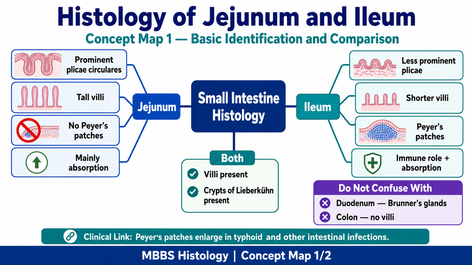
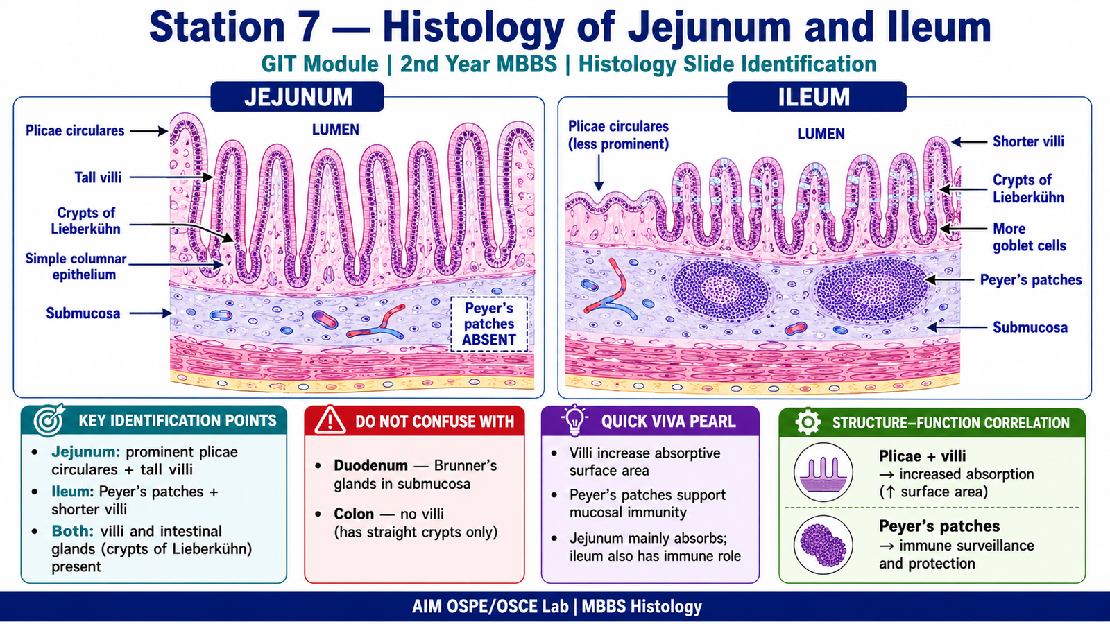
Jejunum
- Tall finger-like villi
- Prominent plicae circulares
- Simple columnar epithelium with goblet cells
- Intestinal glands / crypts of Lieberkühn
- No Peyer’s patches
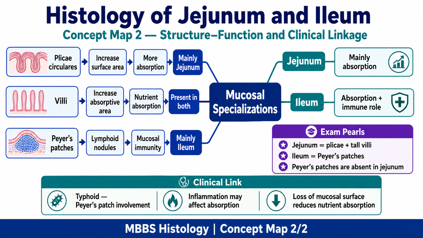
- Mainly specialized for absorption
Ileum
- Shorter villi compared with jejunum
- Less prominent plicae circulares
- More goblet cells
- Crypts of Lieberkühn present
- Peyer’s patches in lamina propria / submucosa
- Important for immune surveillance
Key Differentiating Point
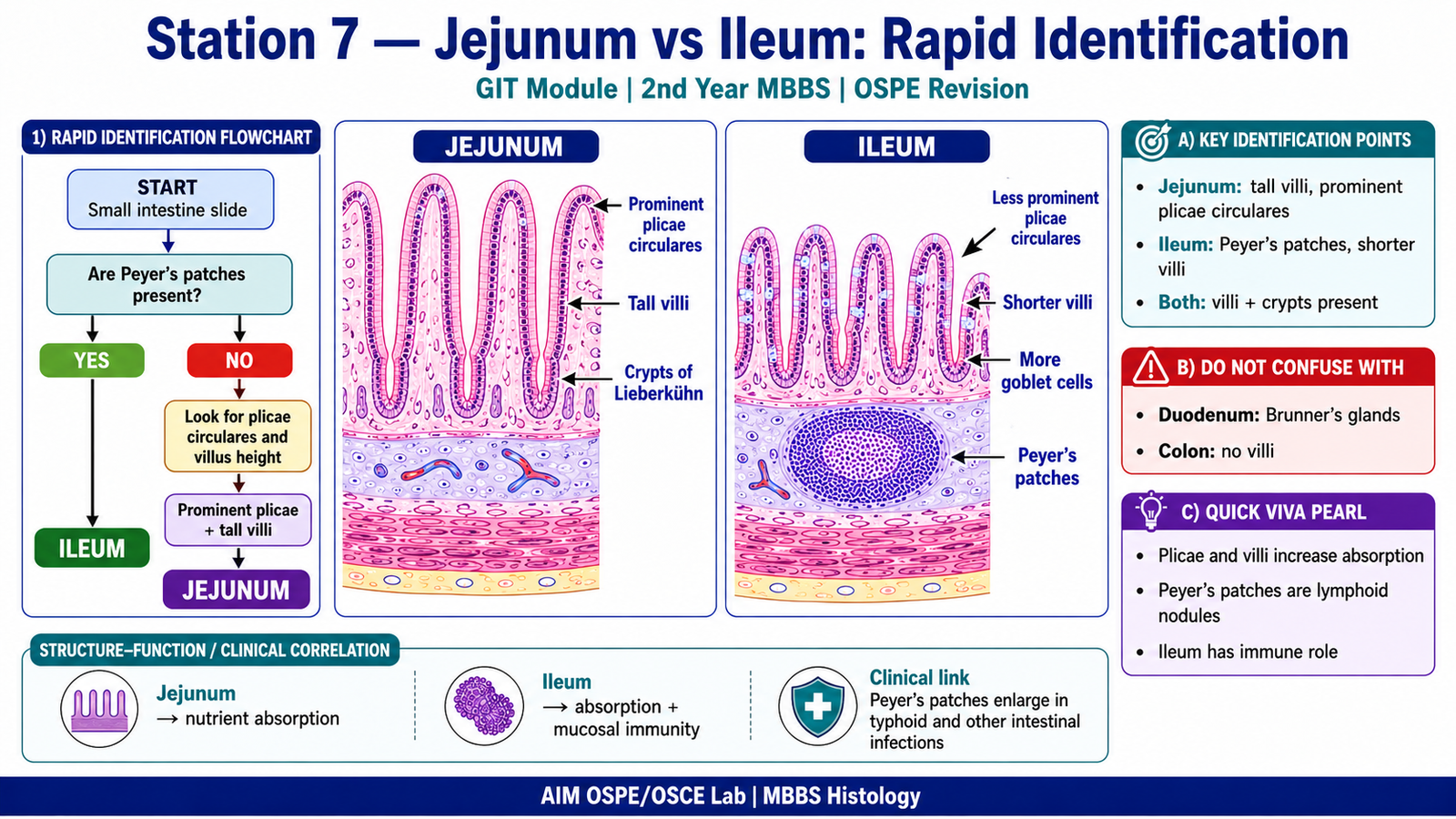
- Jejunum = prominent plicae circulares + tall villi
- Ileum = Peyer’s patches + shorter villi
Result / Interpretation
The slide is identified as jejunum if it shows tall villi and prominent plicae circulares without Peyer’s patches.
The slide is identified as ileum if it shows shorter villi and large lymphoid aggregates called Peyer’s patches.
Clinical significance:
Jejunum is mainly involved in nutrient absorption, while ileum is important for absorption and immune defense. Peyer’s patches are important in mucosal immunity and are commonly involved in intestinal infections such as typhoid fever.
Viva Questions
| Question | Short Ideal Answer |
|---|---|
| What is the most important identifying feature of jejunum? | Prominent plicae circulares with tall villi. |
| What is the most important identifying feature of ileum? | Peyer’s patches. |
| What are plicae circulares? | Permanent folds of mucosa and submucosa that increase absorptive surface area. |
| What is the function of villi? | They increase surface area for absorption. |
| Where are Peyer’s patches found? | In the ileum, mainly in lamina propria and submucosa. |
Marking Scheme
Total Marks: 5
| Component | Marks |
|---|---|
| Correct identification / performance | 2 |
| Key observation / procedure steps | 1 |
| Interpretation / principle | 1 |
| Viva answer | 1 |
Common Student Mistakes
- Confusing jejunum with ileum by looking only at villi.
- Forgetting that Peyer’s patches are the key feature of ileum.
- Calling plicae circulares “villi”; plicae are larger folds, while villi are mucosal projections.
AIM Feedback
To improve slide identification, first look at the overall mucosal pattern. If you see prominent plicae circulares and tall villi, think of jejunum. If you see lymphoid aggregates/Peyer’s patches, think of ileum. In OSPE, always identify the slide using one strong distinguishing feature rather than memorizing many minor points.
🖼️ Visual / Image Support.
🧩 Concept Map / Interpretation Support