🩺 Station 6 — Histology of Gall Bladder
AIM OSPE/OSCE Lab — Practical Station | KMU Style | MBBS Practical + Viva
📌 Station Overview
Module: Cardiovascular System
Year: 1st Year MBBS
Focus: Identification • Procedure • Interpretation • Viva
Total Marks: 5
📋 Complete OSPE Station Content
Learning Target
By the end of this station, the student should be able to:
- Identify the gall bladder histology slide using key microscopic features.
- Recognize folded mucosa, simple columnar epithelium, and absence of muscularis mucosa/submucosa.
Required Material
- Prepared histology slide of gall bladder
- Light microscope / digital histology image
- Pointer or labeled image marker
- Answer sheet / OSPE response sheet
Student Task / Procedure
- Observe the given histology slide under low power.
- Identify the organ/tissue.
- Point out the folded mucosa.
- Identify the epithelial lining.
- Mention two characteristic microscopic features of gall bladder.
- State one functional or clinical significance.
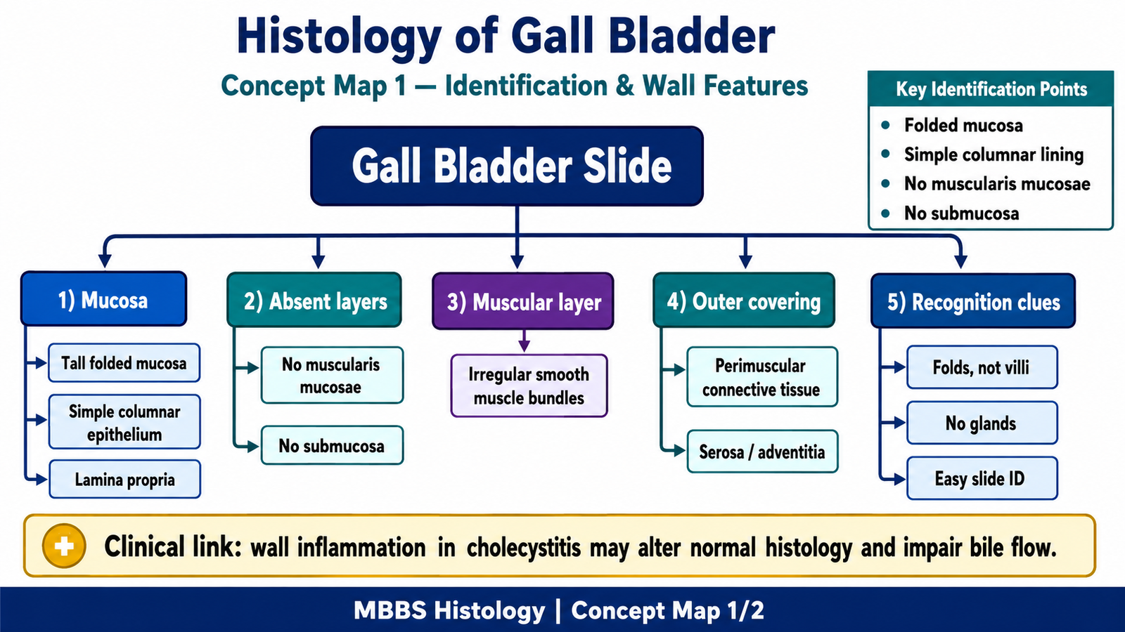
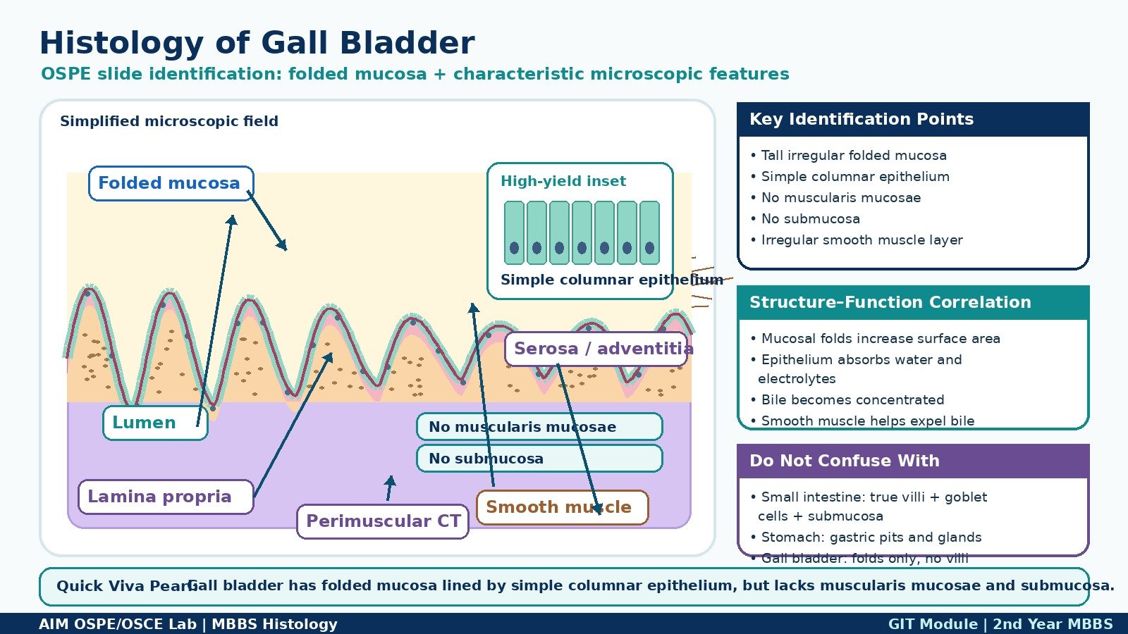
Observation / Identification Points
Students should observe:
- Tall, irregular folded mucosa projecting into the lumen
- Lining by simple columnar epithelium
- Lamina propria present beneath epithelium
- No muscularis mucosae
- No submucosa
- Irregular layer of smooth muscle fibers
- Outer perimuscular connective tissue / serosa or adventitia
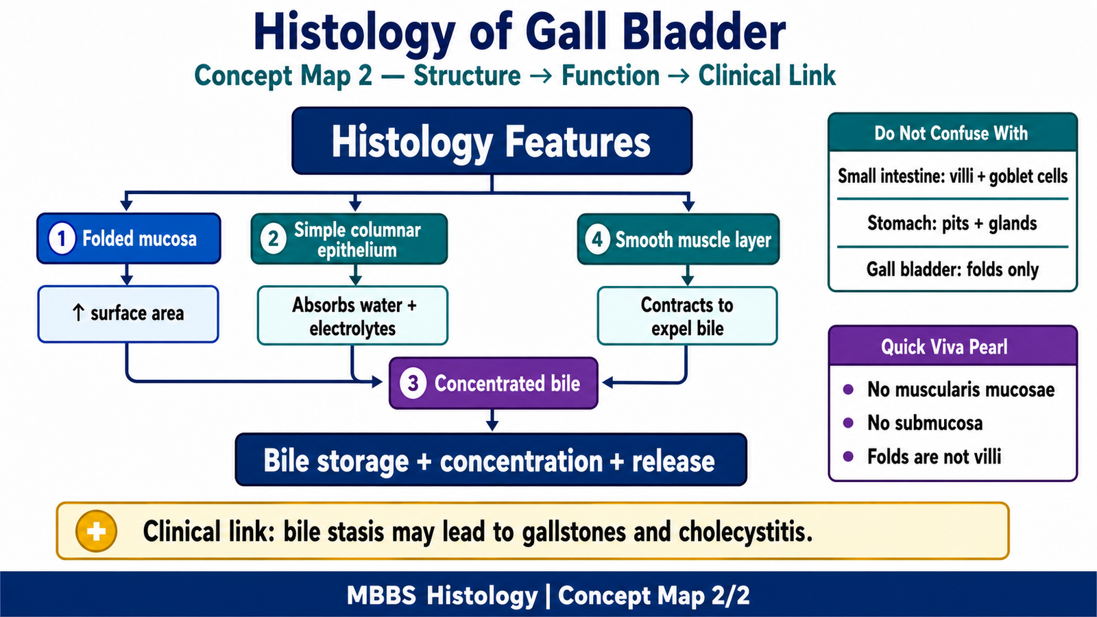
Result / Interpretation
The slide is identified as gall bladder.
The folded mucosa and simple columnar epithelium help the gall bladder absorb water and electrolytes, thereby concentrating bile.
Absence of muscularis mucosae and submucosa is an important identifying feature. Clinically, gall bladder inflammation may be associated with gallstones and cholecystitis.
Viva Questions
1. Which epithelium lines the gall bladder?
Simple columnar epithelium.
2. What is the most characteristic microscopic feature of gall bladder mucosa?
Tall, irregular mucosal folds.
3. Which two layers are absent in gall bladder wall?
Muscularis mucosae and submucosa.
4. What is the main function of gall bladder epithelium?
Absorption of water and electrolytes to concentrate bile.
5. How will you differentiate gall bladder from small intestine histologically?
Gall bladder has folded mucosa but no villi, no goblet cells, no muscularis mucosae, and no submucosa.
Marking Scheme
Total Marks: 5
| Component | Marks |
|---|---|
| Correct identification / performance | 2 |
| Key observation / procedure steps | 1 |
| Interpretation / principle | 1 |
| Viva answer | 1 |
Common Student Mistakes
- Confusing mucosal folds with intestinal villi
- Forgetting that gall bladder has no muscularis mucosae and no submucosa
- Identifying the slide only by epithelium without mentioning folded mucosa
AIM Feedback
To improve your identification, first look for the irregular folded mucosa under low power. Then confirm the slide by checking for simple columnar epithelium and absence of muscularis mucosae and submucosa. Remember: gall bladder mucosa is folded, but these folds are not intestinal villi.
🖼️ Visual / Image Support
🧩 Concept Map / Interpretation Support