🩺 Station 8 — Histology of Appendix
AIM OSPE/OSCE Lab — Practical Station | KMU Style | MBBS Practical + Viva
📌 Station Overview
Module: Cardiovascular System
Year: 1st Year MBBS
Focus: Identification • Procedure • Interpretation • Viva
Total Marks: 5
📋 Complete OSPE Station Content
Learning Target
By the end of this station, the student should be able to:
- Identify the appendix histology slide using lymphoid follicles, mucosa, and lumen features.
- Recognize the key microscopic features that differentiate appendix from colon and small intestine.
Required Material
- Prepared histology slide of appendix
- Light microscope / digital histology image
- Pointer or labeled slide image
- OSPE answer sheet
Student Task / Procedure
- Observe the given slide under low power.
- Identify the organ/tissue.
- Point out the lumen and mucosa.
- Identify the lymphoid follicles.
- Mention two characteristic microscopic features of appendix.
- State one functional or clinical significance.
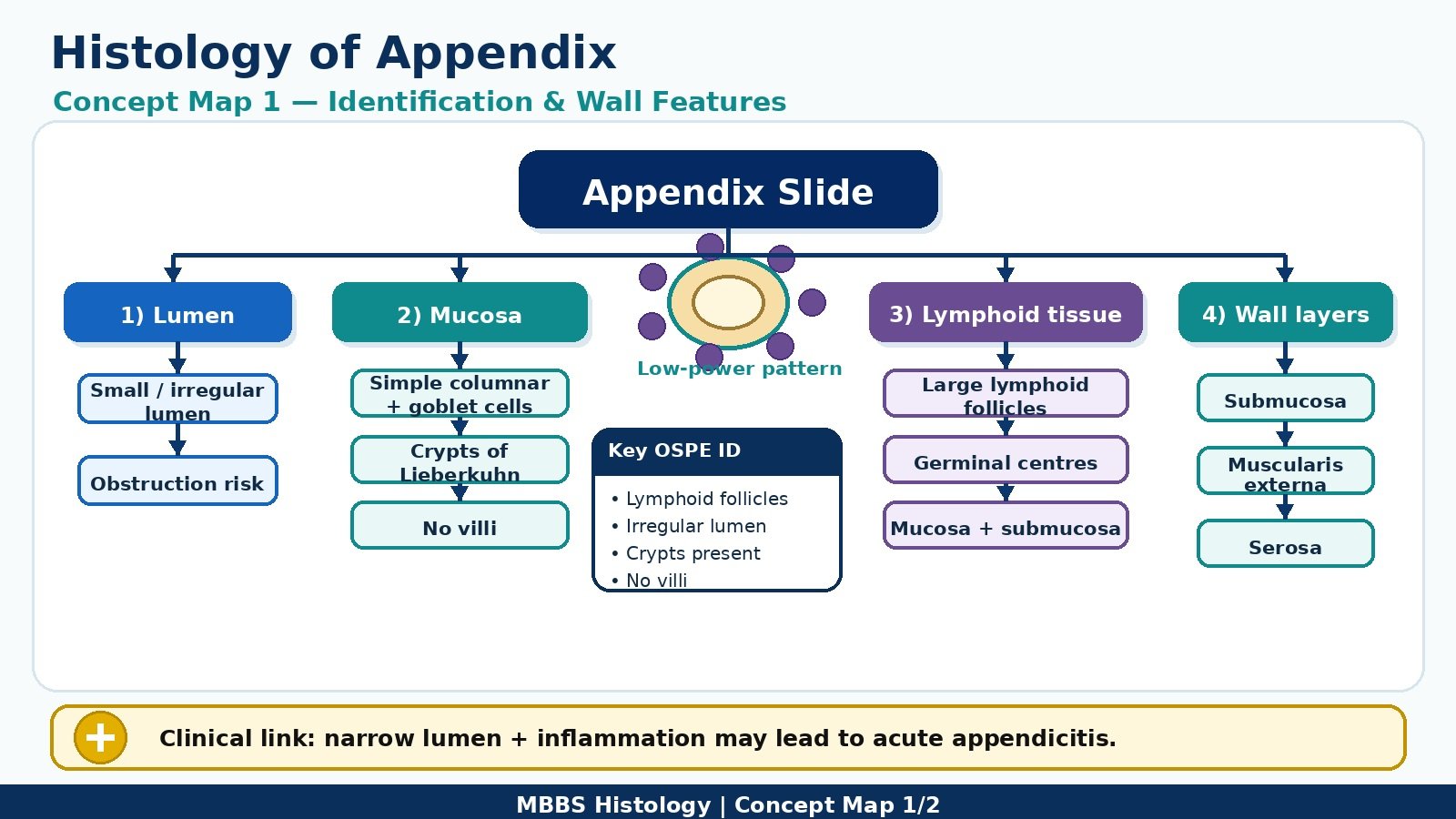
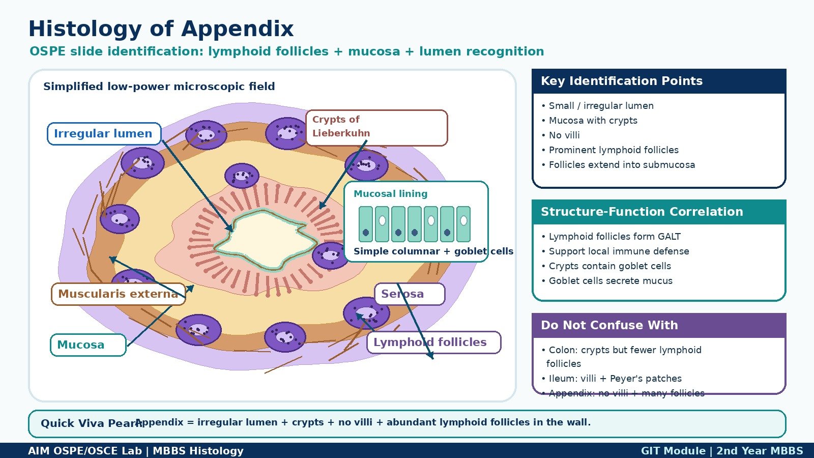
Observation / Identification Points
Students should observe:
- Narrow or irregular lumen
- Mucosa lined by simple columnar epithelium
- Crypts of Lieberkühn present
- No villi
- Numerous lymphoid follicles in mucosa and submucosa
- Muscularis externa present
- Outer serosa
Result / Interpretation
The slide is identified as appendix.
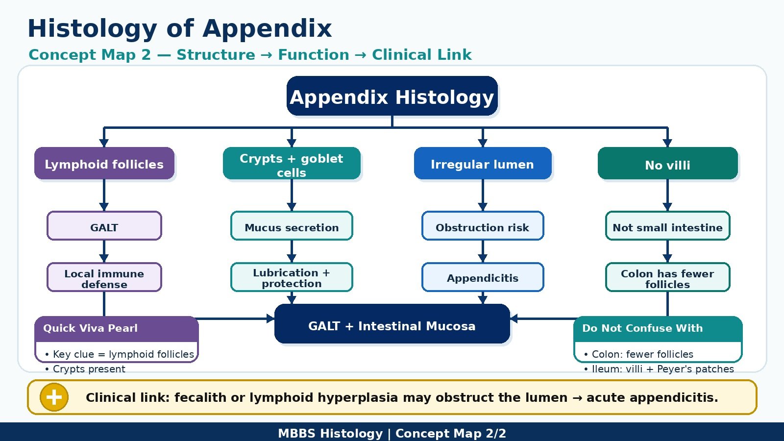
The most important identifying feature is the presence of prominent lymphoid follicles around the wall. The appendix has intestinal-type mucosa with crypts but no villi. It contains lymphoid tissue as part of gut-associated lymphoid tissue, helping in local immune defense.
Clinically, obstruction of the lumen with inflammation may lead to acute appendicitis.
Viva Questions
1. What is the key identifying feature of appendix histology?
Prominent lymphoid follicles in the mucosa and submucosa.
2. Which epithelium lines the appendix?
Simple columnar epithelium with goblet cells.
3. Are villi present in the appendix?
No, villi are absent.
4. What are crypts of Lieberkühn?
Tubular intestinal glands present in the mucosa.
5. What is the clinical importance of appendix obstruction?
It may cause acute appendicitis.
Marking Scheme
Total Marks: 5
| Component | Marks |
|---|---|
| Correct identification / performance | 2 |
| Key observation / procedure steps | 1 |
| Interpretation / principle | 1 |
| Viva answer | 1 |
Common Student Mistakes
- Confusing appendix with colon because both have crypts and goblet cells
- Forgetting that appendix has prominent lymphoid follicles
- Mistaking lymphoid follicles for glands or abnormal pathology
- Looking only at the lumen and missing the wall lymphoid tissue
AIM Feedback
To improve your slide identification, first scan the slide under low power and look for a small irregular lumen surrounded by abundant lymphoid follicles. Then confirm the mucosa by identifying simple columnar epithelium, crypts of Lieberkühn, and absence of villi. Remember: in appendix, lymphoid follicles are the main OSPE clue.
🖼️ Visual / Image Support
🧩 Concept Map / Interpretation Support