🩺 Station 3 — Histology of Stomach
AIM OSPE/OSCE Lab — Practical Station | KMU Style | MBBS Practical + Viva
📌 Station Overview
Module: Cardiovascular System
Year: 1st Year MBBS
Focus: Identification • Procedure • Interpretation • Viva
Total Marks: 5
📋 Complete OSPE Station Content
Learning Target
By the end of this station, the student should be able to:
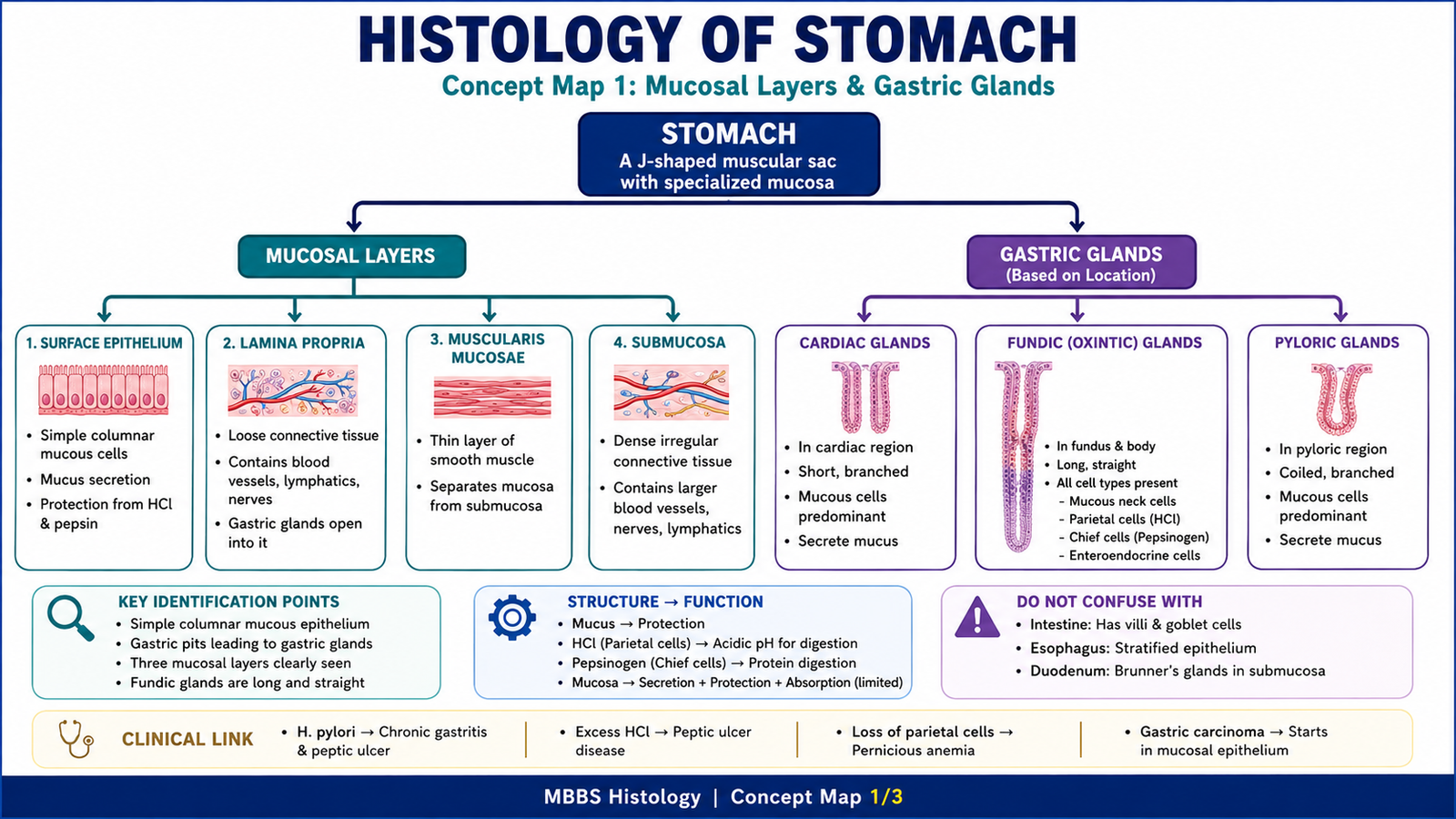
- Identify the stomach slide under the microscope using gastric pits, gastric glands, and mucosal layers.
- Explain the functional importance of gastric glands and mucosal protection at MBBS 2nd year level.
Required Material
- Prepared histology slide of stomach
- Light microscope
- Pointer / labeled photomicrograph
- Answer sheet
- Pencil
Student Task / Procedure
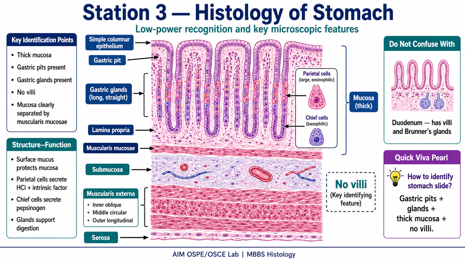
- Focus the slide under low power.
- Identify the tissue as stomach.
- Observe the mucosa, gastric pits, and gastric glands.
- Mention two key microscopic features supporting your identification.
- State one functional significance of gastric glands.
Observation / Identification Points
Students should observe and identify:
Low-Power Recognition Pattern
- Thick mucosa
- Gastric pits opening on mucosal surface
- Gastric glands extending deep into mucosa
- Absence of villi
Mucosal Layers
- Simple columnar surface epithelium
- Gastric pits
- Gastric glands
- Lamina propria
- Muscularis mucosae
Gastric Gland Features
- Mucous cells: secrete protective mucus
- Parietal cells: secrete HCl and intrinsic factor
- Chief cells: secrete pepsinogen
- Enteroendocrine cells: secrete hormones such as gastrin
Deeper Layers
- Submucosa
- Muscularis externa
- Serosa
Result / Interpretation
The slide is identified as stomach because it shows gastric pits, gastric glands, thick mucosa, and absence of villi.
Principle:
The stomach mucosa is specialized for secretion and protection. Gastric glands produce acid, enzymes, mucus, and hormones.
Clinical significance:
Damage to the protective mucus barrier may allow acid injury, leading to gastritis or peptic ulceration.
Viva Questions
Q1. What is the lining epithelium of the stomach?
Answer: Simple columnar mucous-secreting epithelium.
Q2. What are gastric pits?
Answer: Depressions in the gastric mucosa into which gastric glands open.
Q3. Which cells secrete hydrochloric acid?
Answer: Parietal cells.
Q4. Which cells secrete pepsinogen?
Answer: Chief cells.
Q5. Why does the stomach not digest itself normally?
Answer: Because the mucous barrier, bicarbonate, tight junctions, and epithelial renewal protect the mucosa.
Marking Scheme
Total Marks: 5
| Component | Marks |
|---|---|
| Correct identification / performance | 2 |
| Key observation / procedure steps | 1 |
| Interpretation / principle | 1 |
| Viva answer | 1 |
Common Student Mistakes
- Confusing stomach with duodenum; duodenum has villi and Brunner’s glands.
- Forgetting that stomach has gastric pits and glands but no villi.
- Mixing up parietal cells and chief cells.
AIM Feedback
To improve slide identification, first look at the surface pattern.
If you see gastric pits with deep glands and no villi, think of stomach.
Then confirm using gland cells: parietal cells for HCl and intrinsic factor, and chief cells for pepsinogen. Always connect microscopic structure with function.
🖼️ Visual / Image Support
🧩 Concept Map / Interpretation Support