
🩺 Station 11 — Estimation of Plasma Proteins
AIM OSPE/OSCE Lab — Practical Station | KMU Style | MBBS Practical + Viva
📌 Station Overview
Module: Cardiovascular System
Year: 1st Year MBBS
Focus: Identification • Procedure • Interpretation • Viva
Total Marks: 5
📋 Complete OSPE Station Content
Learning Target
By the end of this station, the student should be able to:
- Demonstrate correct handling of a blood/plasma sample for estimation of plasma proteins.
- Interpret total plasma protein results and relate them to liver function, nutrition, and clinical conditions.
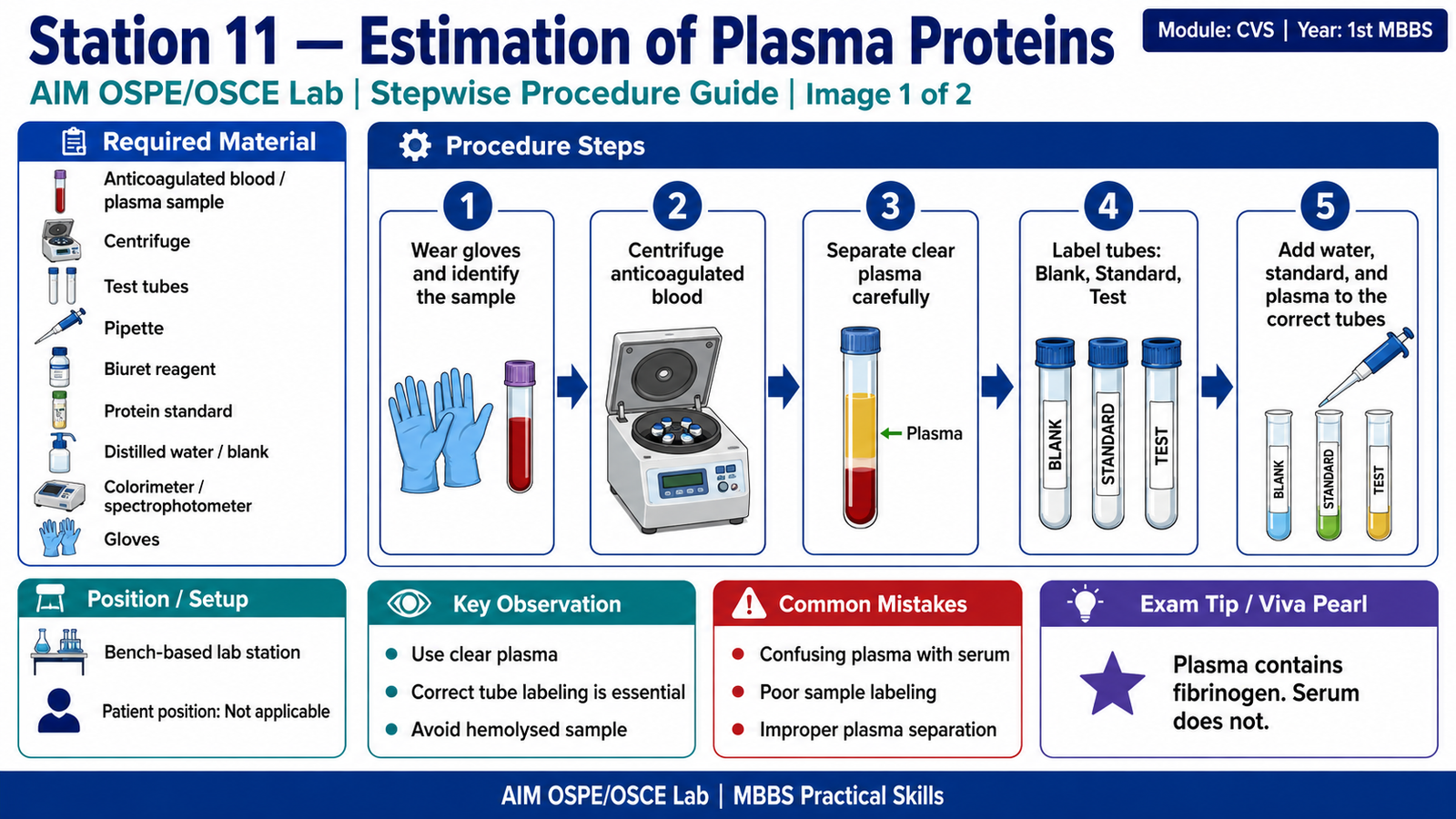
Required Material
- Anticoagulated blood sample / separated plasma sample
- Centrifuge
- Test tubes / cuvettes
- Pipettes or micropipettes
- Biuret reagent
- Protein standard solution
- Distilled water / blank
- Colorimeter / spectrophotometer
- Gloves and lab coat
- Waste container
- Marker / labels
Student Task / Procedure
- Wear gloves and identify the labeled anticoagulated blood sample.
- Centrifuge the sample to separate plasma from cells.
- Carefully transfer clear plasma into a labeled tube.
- Prepare three tubes: Blank, Standard, and Test.
- Add distilled water to blank, standard protein solution to standard tube, and plasma sample to test tube.
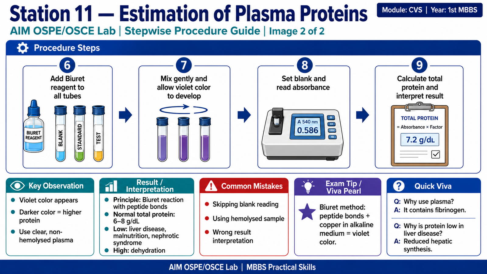
- Add Biuret reagent to all tubes.
- Mix gently and allow color development.
- Read absorbance using colorimeter against blank.
- Calculate total plasma protein using the standard.
- Interpret the result as normal, increased, or decreased.
Observation / Identification Points
The student should demonstrate or identify:
- Correct use of anticoagulated blood for plasma.
- Clear separation of plasma after centrifugation.
- Avoidance of hemolysed or lipemic sample.
- Correct labeling of blank, standard, and test tubes.
- Development of violet color with Biuret reagent.
- Correct use of blank before reading absorbance.
- Relationship between color intensity and protein concentration.
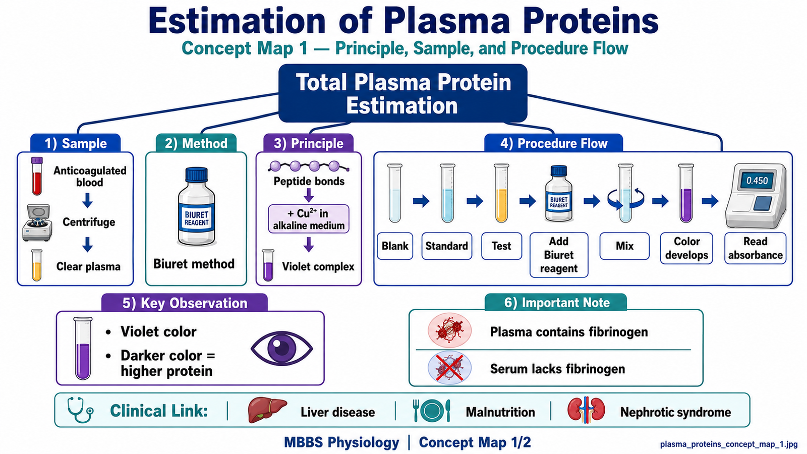
Result / Interpretation
Principle:
Plasma proteins react with copper ions in alkaline Biuret reagent to form a violet-colored complex. The intensity of color is proportional to protein concentration.
Formula:
Total plasma protein =
Absorbance of Test / Absorbance of Standard × Concentration of Standard
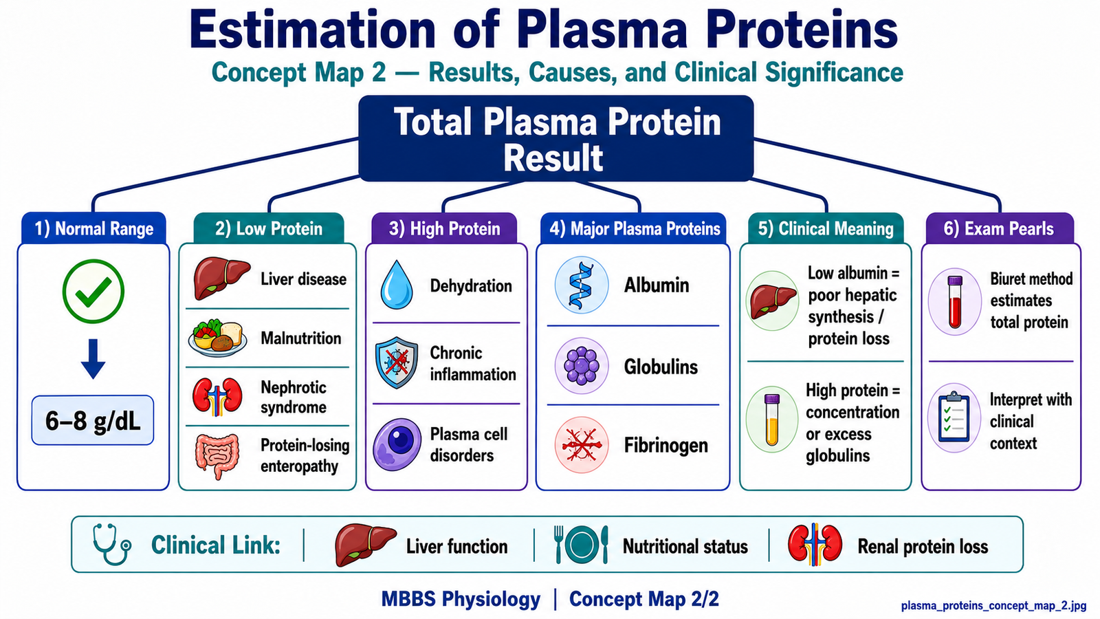
Normal Total Plasma Protein:
Approximately 6–8 g/dL
Clinical Significance:
- Decreased plasma proteins: liver disease, malnutrition, nephrotic syndrome, protein-losing enteropathy.
- Increased plasma proteins: dehydration, chronic inflammation, plasma cell disorders.
- GIT/Liver link: Albumin and many plasma proteins are synthesized in the liver, so low plasma protein may reflect impaired hepatic synthetic function.
Viva Questions
1. Which method is commonly used for estimation of total plasma proteins?
Answer: Biuret method.
2. What is the principle of the Biuret method?
Answer: Peptide bonds react with copper ions in alkaline medium to produce violet color.
3. What is the normal total plasma protein level?
Answer: Approximately 6–8 g/dL.
4. Why is plasma different from serum?
Answer: Plasma contains fibrinogen because anticoagulant prevents clotting; serum lacks fibrinogen.
5. Why may plasma proteins decrease in chronic liver disease?
Answer: The liver synthesizes albumin and many plasma proteins, so liver dysfunction reduces protein synthesis.
Marking Scheme
Total Marks: 5
| Component | Marks |
|---|---|
| Correct identification / performance | 2 |
| Key observation / procedure steps | 1 |
| Interpretation / principle | 1 |
| Viva answer | 1 |
Common Student Mistakes
- Using serum and plasma interchangeably without understanding the difference.
- Forgetting to run the blank before reading absorbance.
- Not relating low plasma proteins to liver disease, malnutrition, or renal protein loss.
AIM Feedback
Focus on three exam points: sample handling, Biuret principle, and clinical interpretation. Remember that plasma contains fibrinogen, while serum does not. In GIT integration, always link decreased plasma proteins with impaired liver synthesis, malnutrition, and protein loss.
🖼️ Visual / Image Support
🧩 Concept Map / Interpretation Support
🎥 Video Demonstration / Procedure Support
Watch this video to revise the Biuret method for total plasma/serum protein estimation, including reagent addition, color change, absorbance reading, and interpretation.