
🩺 Station 15 — Estimation of Serum Cholesterol
AIM OSPE/OSCE Lab — Practical Station | KMU Style | MBBS Practical + Viva
📌 Station Overview
Module: Cardiovascular System
Year: 1st Year MBBS
Focus: Identification • Procedure • Interpretation • Viva
Total Marks: 5
📋 Complete OSPE Station Content
Learning Target
By the end of this station, the student should be able to:
- Handle serum sample safely and perform basic serum cholesterol estimation using a colorimetric method.
- Calculate and interpret serum cholesterol level at MBBS practical examination level.
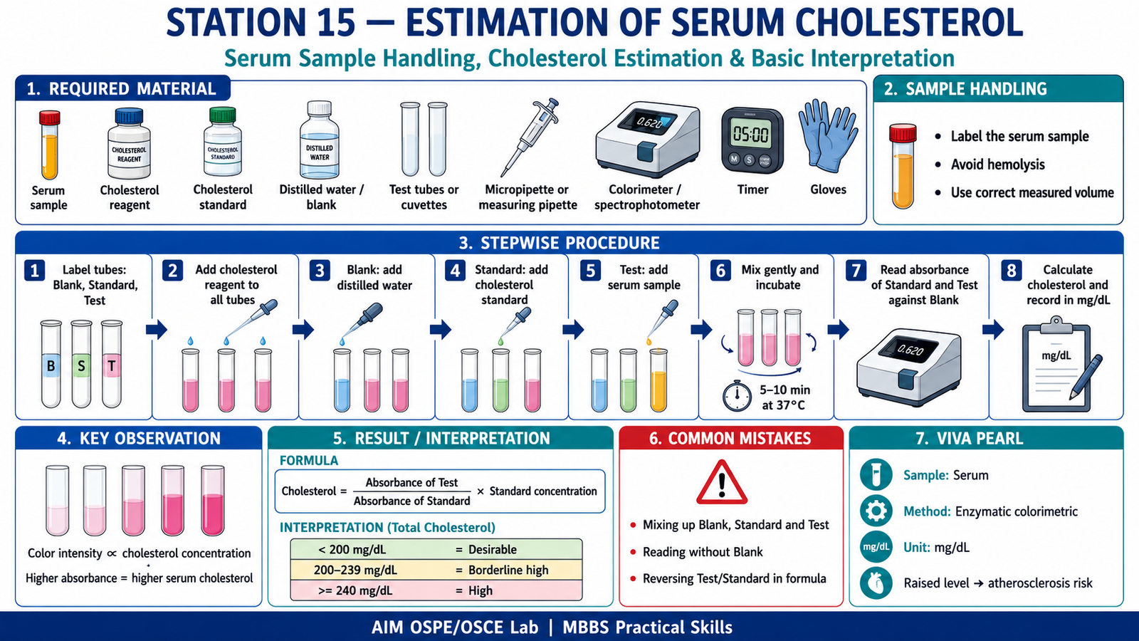
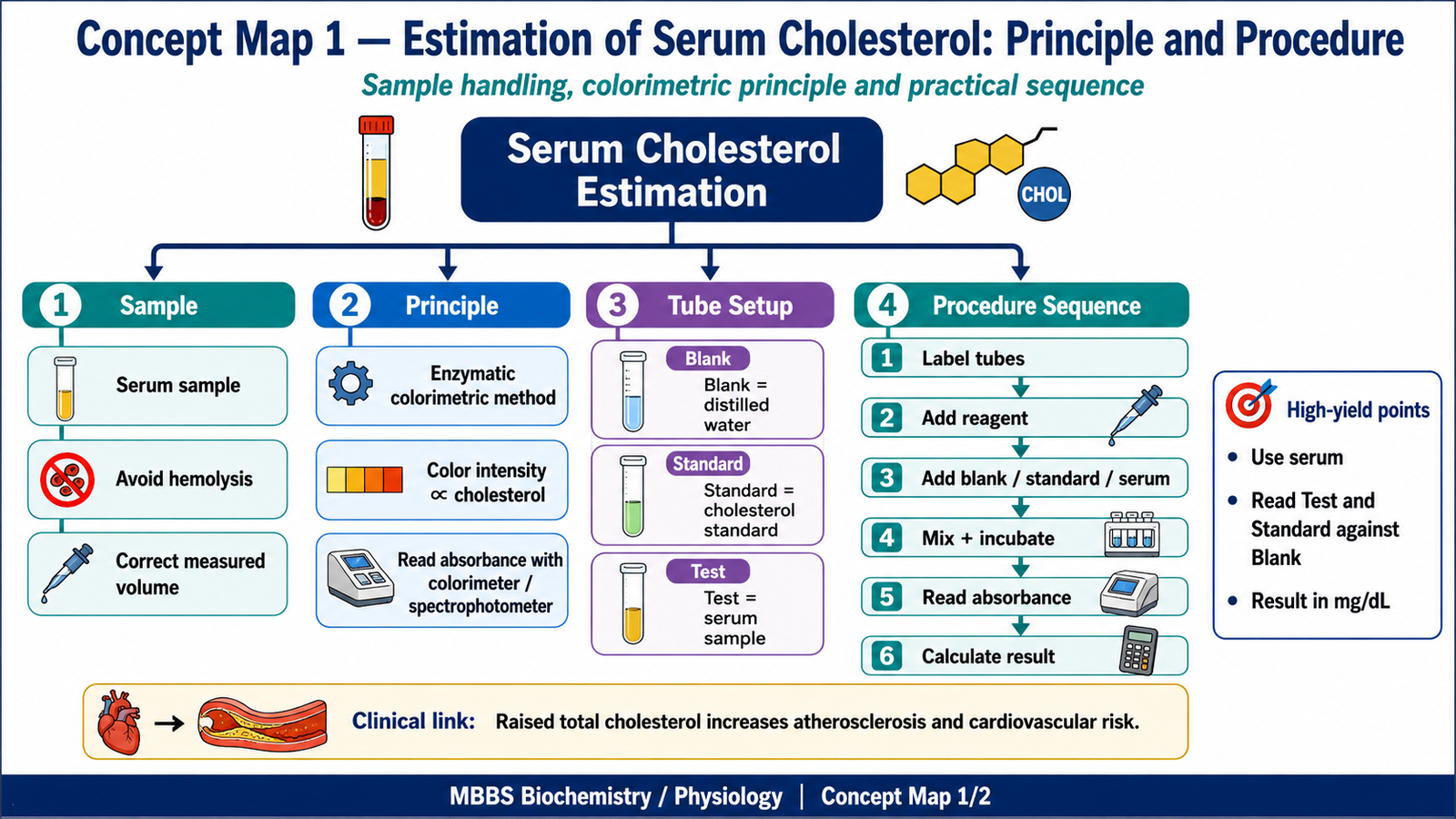
Required Material
- Serum sample
- Cholesterol reagent kit
- Cholesterol standard
- Distilled water / reagent blank
- Test tubes or cuvettes
- Micropipette / measuring pipette
- Colorimeter or spectrophotometer
- Water bath or incubator, if required
- Timer
- Gloves and waste container
Student Task / Procedure
- Wear gloves and check the labeled serum sample.
- Label three tubes: Blank, Standard, Test.
- Add cholesterol reagent to all three tubes.
- Add distilled water to the blank tube.
- Add cholesterol standard to the standard tube.
- Add serum sample to the test tube.
- Mix gently and incubate as instructed by the kit.
- Observe the development of color.
- Read absorbance of Standard and Test against Blank using colorimeter/spectrophotometer.
- Calculate serum cholesterol using the formula.
- Record the result in mg/dL.
- Interpret the result as normal, borderline high, or high.
Observation / Identification Points
The student should demonstrate:
- Safe handling of serum sample.
- Correct labeling of Blank, Standard, and Test tubes.
- Correct use of reagent, standard, and serum sample.
- Proper mixing and incubation.
- Correct absorbance reading against blank.
- Correct calculation of serum cholesterol.
- Basic interpretation of the cholesterol result.
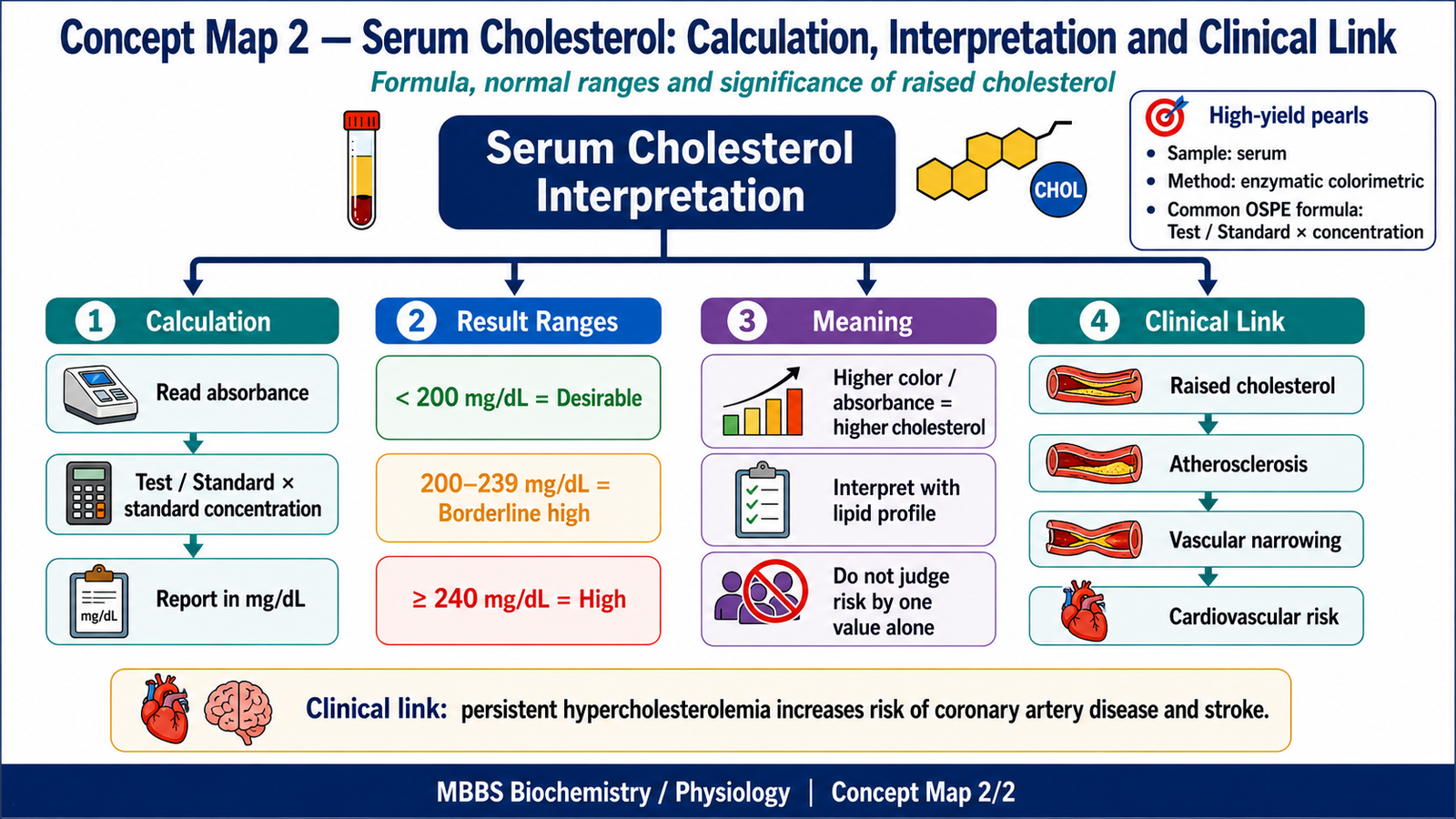
Result / Interpretation
Principle:
Serum cholesterol is estimated by an enzymatic colorimetric method. The intensity of the final colored compound is proportional to the cholesterol concentration in serum.
Formula:
Serum Cholesterol = Absorbance of Test / Absorbance of Standard × Concentration of Standard
Unit:
mg/dL
Basic Interpretation of Total Cholesterol:
| Serum Total Cholesterol | Interpretation |
|---|---|
| Less than 200 mg/dL | Desirable / Normal |
| 200–239 mg/dL | Borderline high |
| 240 mg/dL or above | High |
Clinical Significance:
Raised serum cholesterol is an important risk factor for atherosclerosis and cardiovascular disease. Interpretation should be done along with full lipid profile and clinical context.
Viva Questions
1. Which sample is used for cholesterol estimation?
Ideal Answer:
Serum sample is used.
2. What is the principle of serum cholesterol estimation?
Ideal Answer:
It is based on enzymatic colorimetric reaction where color intensity is proportional to cholesterol concentration.
3. Why is a blank tube used?
Ideal Answer:
Blank is used to set zero absorbance and eliminate reagent background color.
4. What is the formula for serum cholesterol calculation?
Ideal Answer:
Serum cholesterol = Absorbance of Test / Absorbance of Standard × Concentration of Standard.
5. What is the clinical importance of raised serum cholesterol?
Ideal Answer:
It increases the risk of atherosclerosis and cardiovascular disease.
Marking Scheme
Total Marks: 5
| Component | Marks |
| Correct identification / performance | 2 |
| Key observation / procedure steps | 1 |
| Interpretation / principle | 1 |
| Viva answer | 1 |
Checklist Breakdown
| Checklist Item | Marks |
| Handles serum sample safely and labels tubes correctly | 0.5 |
| Adds reagent, standard, and serum sample correctly | 0.75 |
| Performs mixing, incubation, and absorbance reading correctly | 0.75 |
| Uses blank correctly and records absorbance | 1 |
| Explains principle or calculation formula | 1 |
| Answers viva question correctly | 1 |
Common Student Mistakes
- Mixing up Blank, Standard, and Test tubes.
- Forgetting to read absorbance against blank.
- Writing the formula incorrectly by reversing Test and Standard absorbance.
AIM Feedback
In this station, remember the sequence: Blank → Standard → Test → Color development → Absorbance → Calculation → Interpretation. The most common exam mistake is formula reversal, so always place Test absorbance above Standard absorbance in the calculation.
🖼️ Visual / Image Support
🧩 Concept Map / Interpretation Support
🎥 Video Demonstration / Procedure Support
Watch this video to revise serum cholesterol estimation using the enzymatic colorimetric CHOD-POD method, including Blank–Standard–Test setup, absorbance reading, calculation, and interpretation.