🩺 Station 5 — Histology of Liver
AIM OSPE/OSCE Lab — Practical Station | KMU Style | MBBS Practical + Viva
📌 Station Overview
Module: Cardiovascular System
Year: 1st Year MBBS
Focus: Identification • Procedure • Interpretation • Viva
Total Marks: 5
📋 Complete OSPE Station Content
Learning Target
By the end of this station, the student should be able to:
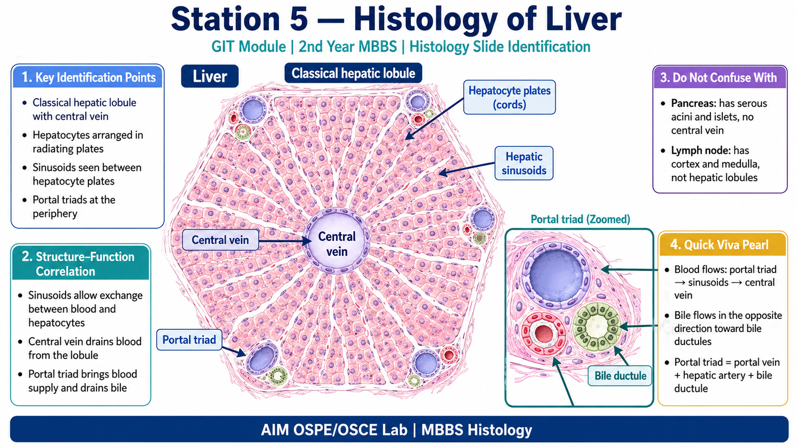
- Identify a liver histology slide under the microscope.
- Recognize the hepatic lobule, central vein, hepatic sinusoids, hepatocyte plates, and portal triad.
Required Material
- Prepared histology slide of liver
- Light microscope
- Pointer / marker
- Labeled diagram of liver lobule for examiner reference
- Answer sheet / OSPE response sheet
Student Task / Procedure
- Focus the given slide under low power.
- Identify the organ as liver.
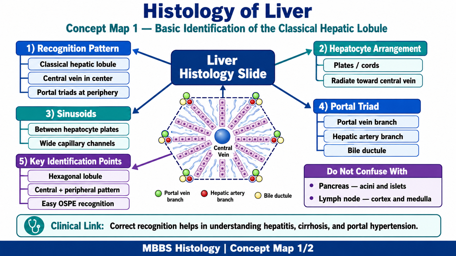
- Locate a classical hepatic lobule.
- Identify the central vein.
- Trace hepatocyte plates radiating toward the central vein.
- Identify hepatic sinusoids between hepatocyte plates.
- Locate the portal triad at the periphery of the lobule.
- Write one functional significance of the portal triad or hepatic lobule.
Observation / Identification Points
Students should identify:
- Liver slide
- Polygonal hepatic lobules
- Hepatocytes arranged in plates/cords
- Sinusoids between hepatocyte plates
- Classical hepatic lobule
- Hexagonal arrangement
- Central vein in the center
- Hepatocyte plates radiating toward central vein
- Portal triad
- Located at the periphery of lobule
- Contains:
- Branch of portal vein
- Branch of hepatic artery
- Bile ductule
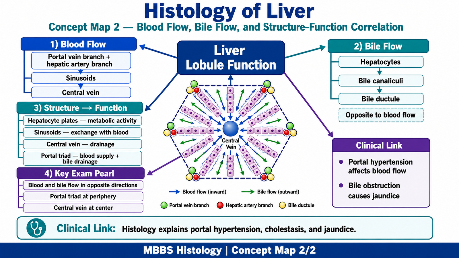
- Direction of flow
- Blood flows from portal triad → sinusoids → central vein
- Bile flows from hepatocytes → bile canaliculi → bile ductule in portal triad
Result / Interpretation
The slide is identified as liver because it shows hepatic lobules with a central vein, radiating hepatocyte plates, sinusoids, and portal triads at the periphery.
Clinical significance:
Liver histology helps in understanding liver function, bile formation, portal circulation, and pathological changes such as hepatitis, cirrhosis, fatty liver, and portal hypertension.
Viva Questions
| Question | Short Ideal Answer |
|---|---|
| What is the structural unit of the liver seen in routine histology? | Classical hepatic lobule. |
| What is present at the center of a classical hepatic lobule? | Central vein. |
| Name the three components of the portal triad. | Branch of portal vein, branch of hepatic artery, and bile ductule. |
| In which direction does blood flow in the hepatic lobule? | From portal triad through sinusoids toward the central vein. |
| In which direction does bile flow? | Opposite to blood flow, from hepatocytes toward bile ductules in portal triads. |
Marking Scheme
Total Marks: 5
| Component | Marks |
|---|---|
| Correct identification / performance | 2 |
| Key observation / procedure steps | 1 |
| Interpretation / principle | 1 |
| Viva answer | 1 |
Common Student Mistakes
- Confusing central vein with a vessel of the portal triad.
- Forgetting that the portal triad is at the periphery of the hepatic lobule.
- Mixing up direction of blood flow and bile flow.
AIM Feedback
To improve, first identify the central vein, then look outward for radiating hepatocyte plates and sinusoids. After that, search the periphery for the portal triad. Always remember: blood flows toward the central vein, while bile flows away from the central vein toward the portal triad.
🖼️ Visual / Image Support
🧩 Concept Map / Interpretation Support