
🩺 Station 1 — Histology of Lips and Tongue
AIM OSPE/OSCE Lab — Practical Station | KMU Style | MBBS Practical + Viva
📌 Station Overview
Module: Cardiovascular System
Year: 1st Year MBBS
Focus: Identification • Procedure • Interpretation • Viva
Total Marks: 5
📋 Complete OSPE Station Content
Learning Target
By the end of this station, the student should be able to:
- Identify lip and tongue slides under the microscope using key microscopic features.
- Differentiate lip from tongue histologically by observing epithelium, glands, papillae, taste buds, and skeletal muscle.
Required Material
- Prepared histology slide of lip
- Prepared histology slide of tongue
- Light microscope
- Pointer / labeled photomicrograph
- Pencil and answer sheet
- Optional LMS support: labeled histology image guide
Student Task / Procedure
- Focus the given slide under low power first.
- Identify whether the slide is lip or tongue.
- Observe the epithelial covering and deeper tissue.
- Write two key microscopic features supporting your identification.
- Mention one functional or clinical significance of the structure.
Observation / Identification Points
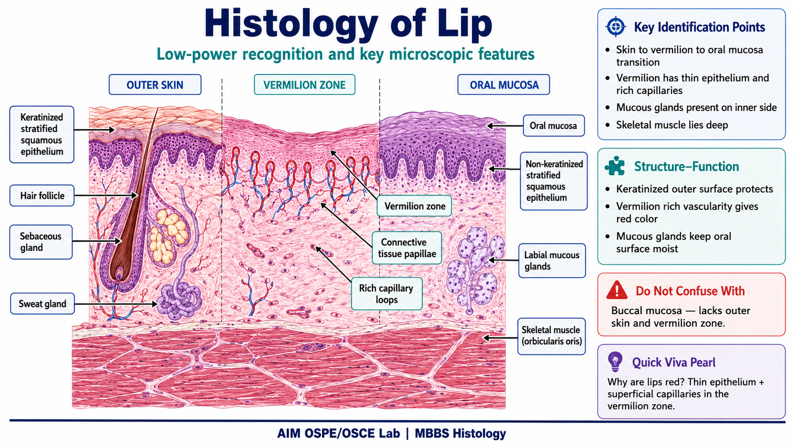
Slide 1 — Lip
Students should identify:
- Outer skin surface
- Keratinized stratified squamous epithelium
- Hair follicles, sebaceous glands, sweat glands may be seen
- Vermilion zone
- Thin epithelium
- Numerous connective tissue papillae
- Rich capillary network giving red color to lips
- Inner oral mucosal surface
- Non-keratinized stratified squamous epithelium
- Labial mucous salivary glands
- Skeletal muscle fibers of orbicularis oris
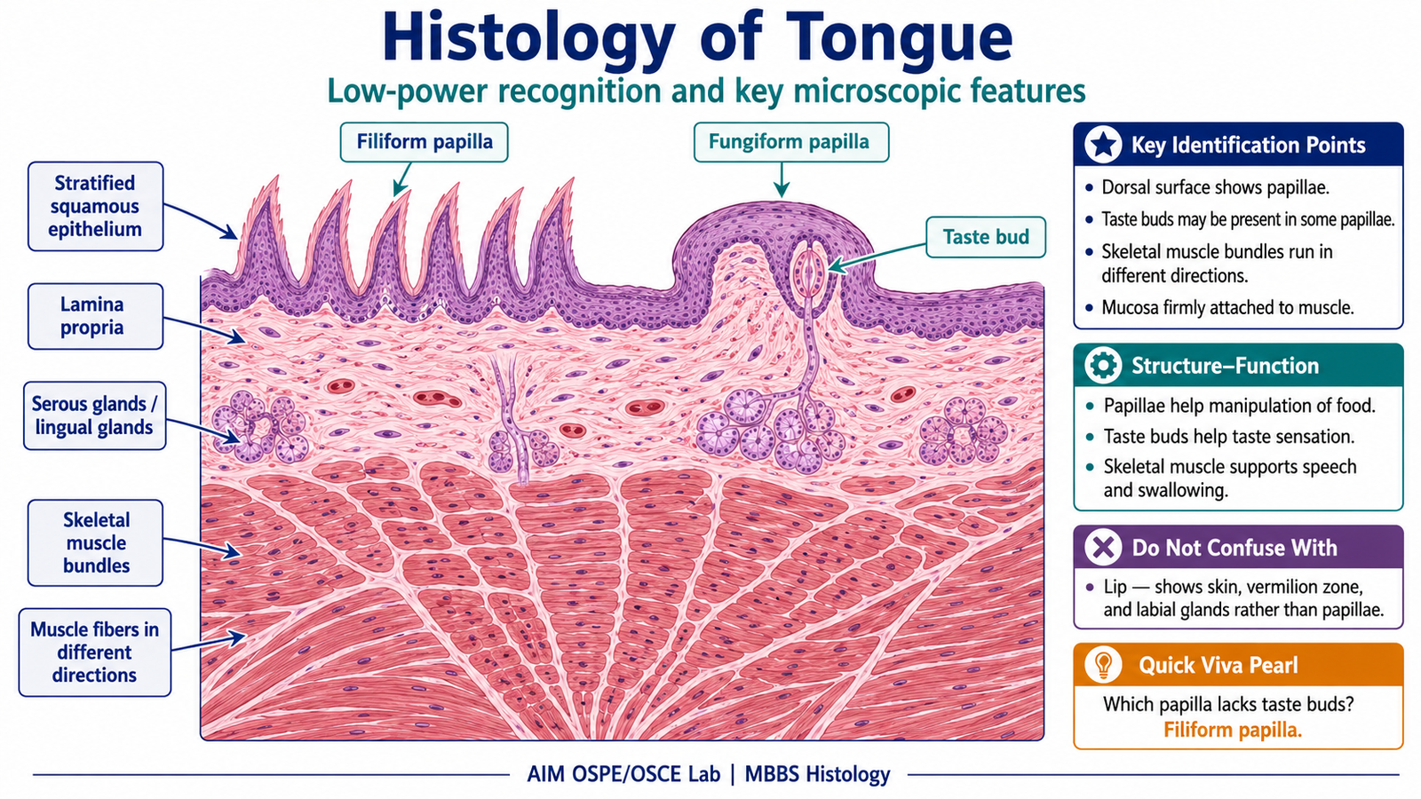
Slide 2 — Tongue
Students should identify:
- Stratified squamous epithelium
- Lingual papillae on dorsal surface
- Taste buds, especially near circumvallate or fungiform papillae if present
- Skeletal muscle bundles arranged in different directions
- Lingual salivary glands may be present
Result / Interpretation
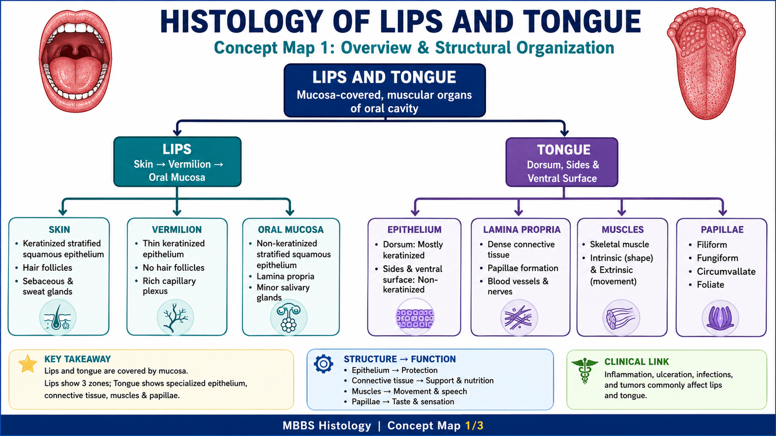
The lip is identified by the presence of a transition from skin to oral mucosa, vermilion zone, labial glands, and skeletal muscle.
The tongue is identified by lingual papillae, possible taste buds, and skeletal muscle bundles arranged in multiple directions.
Principle:
Histological identification is based on recognizing the relationship between epithelium, connective tissue, glands, muscle, and specialized surface structures.
Clinical significance:
Keratinization protects against friction, taste buds help in taste sensation, and labial and lingual glands keep the oral mucosa moist.
Viva Questions
Q1. What type of epithelium lines the oral surface of the lip?
Answer: Non-keratinized stratified squamous epithelium.
Q2. Why does the vermilion border of the lip appear red?
Answer: Because the epithelium is thin and connective tissue papillae contain many superficial capillaries.
Q3. What is the main identifying feature of tongue histology?
Answer: Presence of lingual papillae and skeletal muscle bundles arranged in different directions.
Q4. Which papillae commonly contain taste buds?
Answer: Fungiform, foliate, and circumvallate papillae.
Q5. Why is skeletal muscle prominent in tongue tissue?
Answer: It allows movements needed for speech, chewing, swallowing, and manipulation of food.
Marking Scheme
Total Marks: 5
| Component | Marks |
|---|---|
| Correct identification / performance | 2 |
| Key observation / procedure steps | 1 |
| Interpretation / principle | 1 |
| Viva answer | 1 |
Common Student Mistakes
- Confusing lip oral mucosa with general oral cavity mucosa and missing the skin/vermilion transition.
- Identifying tongue only by epithelium and ignoring papillae and skeletal muscle arrangement.
- Calling all papillae taste-bearing, although filiform papillae usually do not contain taste buds.
AIM Feedback
To improve, first scan the slide under low power and identify the overall pattern.
For lip, look for the skin–vermilion–oral mucosa transition.
For tongue, look for papillae, taste buds, and skeletal muscle bundles in different directions.
Do not memorize only one feature; use at least two microscopic clues before final identification.
🖼️ Visual / Image Support
🧩 Concept Map / Interpretation Support
🎥 Video Demonstration / Procedure Support