🩺 Station 2 — Histology of Esophagus
AIM OSPE/OSCE Lab — Practical Station | KMU Style | MBBS Practical + Viva
📌 Station Overview
Module: Cardiovascular System
Year: 1st Year MBBS
Focus: Identification • Procedure • Interpretation • Viva
Total Marks: 5
📋 Complete OSPE Station Content
Learning Target
By the end of this station, the student should be able to:
- Identify the esophageal slide under microscope using key histological features.
- Recognize the epithelium, glands, and muscular layers of the esophagus and relate them to function.
Required Material
- Prepared histology slide of esophagus
- Light microscope
- Pointer / labeled slide image if available
- Answer sheet / checklist
- Pencil or pen
Student Task / Procedure
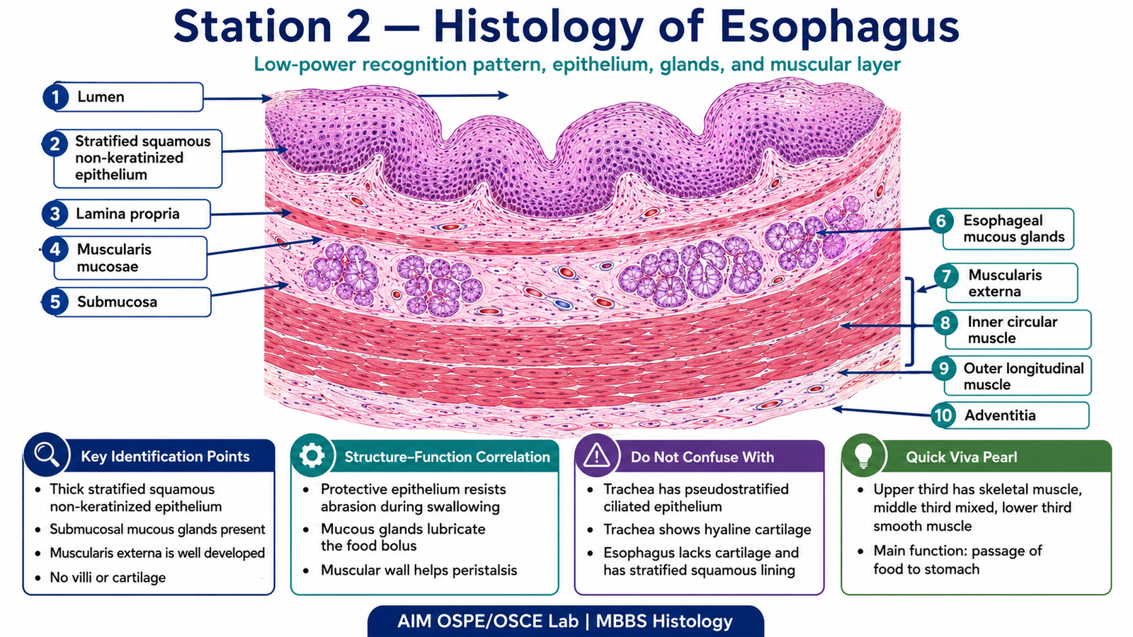
- Focus the slide first under low power.
- Identify the general arrangement of the esophageal wall.
- Observe the type of epithelium lining the lumen.
- Identify the mucosa, submucosa, and muscular layer.
- Look for mucous glands in the submucosa.
- State one functional importance of these histological features.
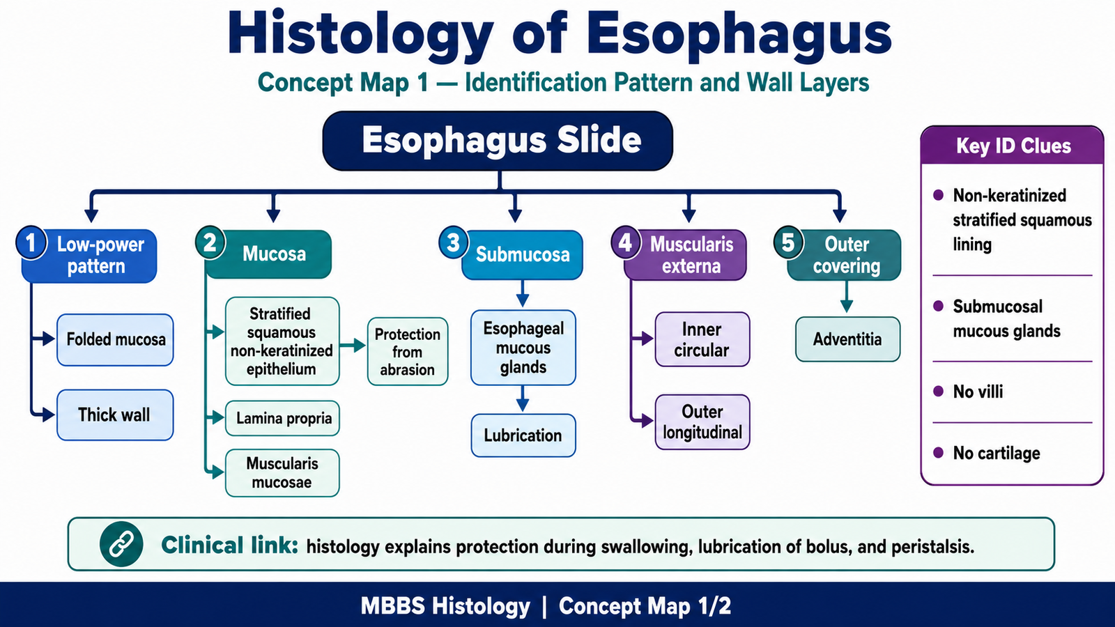
Observation / Identification Points
Students should identify:
- Lumen lined by mucosa
- Stratified squamous non-keratinized epithelium
- Lamina propria beneath the epithelium
- Muscularis mucosae
- Submucosa containing esophageal mucous glands
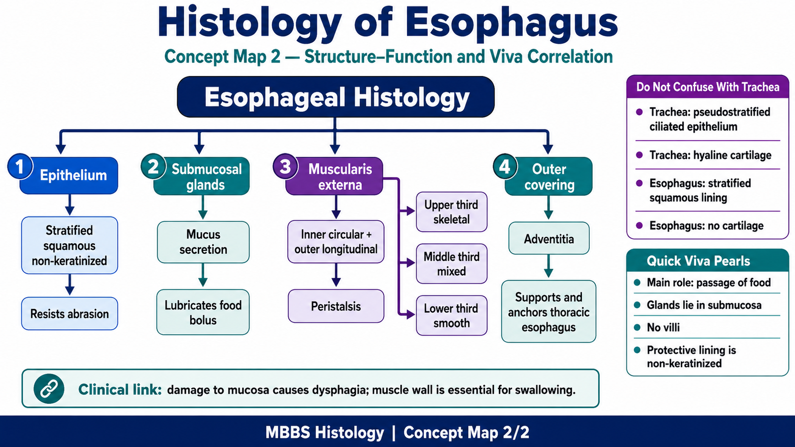
- Muscularis externa arranged mainly as inner circular and outer longitudinal layers
- Muscle type may vary according to level:
- Upper part: skeletal muscle
- Middle part: mixed skeletal and smooth muscle
- Lower part: smooth muscle
Result / Interpretation
The slide is identified as esophagus.
The esophagus is lined by stratified squamous non-keratinized epithelium, which protects against friction during swallowing. The submucosal mucous glands help in lubrication of food, while the muscular layer helps in peristaltic movement of the bolus toward the stomach.
Viva Questions
1. What type of epithelium lines the esophagus?
Stratified squamous non-keratinized epithelium.
2. Why is stratified squamous epithelium present in the esophagus?
It protects the esophageal wall from abrasion during swallowing.
3. Where are the esophageal glands commonly found?
Mainly in the submucosa.
4. What is the function of esophageal mucous glands?
They secrete mucus for lubrication and protection.
5. How does the muscle type change in the esophagus?
Upper third has skeletal muscle, middle third has mixed muscle, and lower third has smooth muscle.
Marking Scheme
Total Marks: 5
| Component | Marks |
|---|---|
| Correct identification / performance | 2 |
| Key observation / procedure steps | 1 |
| Interpretation / principle | 1 |
| Viva answer | 1 |
Common Student Mistakes
- Confusing esophagus with trachea due to presence of glands.
- Forgetting that esophageal epithelium is non-keratinized.
- Missing the submucosal glands, which are important for identification.
- Not recognizing the inner circular and outer longitudinal muscle arrangement.
AIM Feedback
To identify the esophagus correctly, first look at the lining epithelium. Stratified squamous non-keratinized epithelium suggests protection against friction. Then confirm the slide by finding submucosal mucous glands and a well-developed muscular layer for peristalsis. Always connect structure with function: protection, lubrication, and movement of food.
🖼️ Visual / Image Support
🧩 Concept Map / Interpretation Support