🩺 Station 9 — Histology of Colon and Rectum
AIM OSPE/OSCE Lab — Practical Station | KMU Style | MBBS Practical + Viva
📌 Station Overview
Module: Cardiovascular System
Year: 1st Year MBBS
Focus: Identification • Procedure • Interpretation • Viva
Total Marks: 5
📋 Complete OSPE Station Content
Learning Target
By the end of this station, the student should be able to:
- Identify colon and rectum histology slides using key microscopic features.
- Recognize goblet cells and intestinal glands/crypts and explain their functional significance.
Required Material
- Prepared histology slide of colon
- Prepared histology slide of rectum
- Light microscope
- Pointer / marker
- Labeled reference diagram for examiner
- OSPE response sheet
Student Task / Procedure
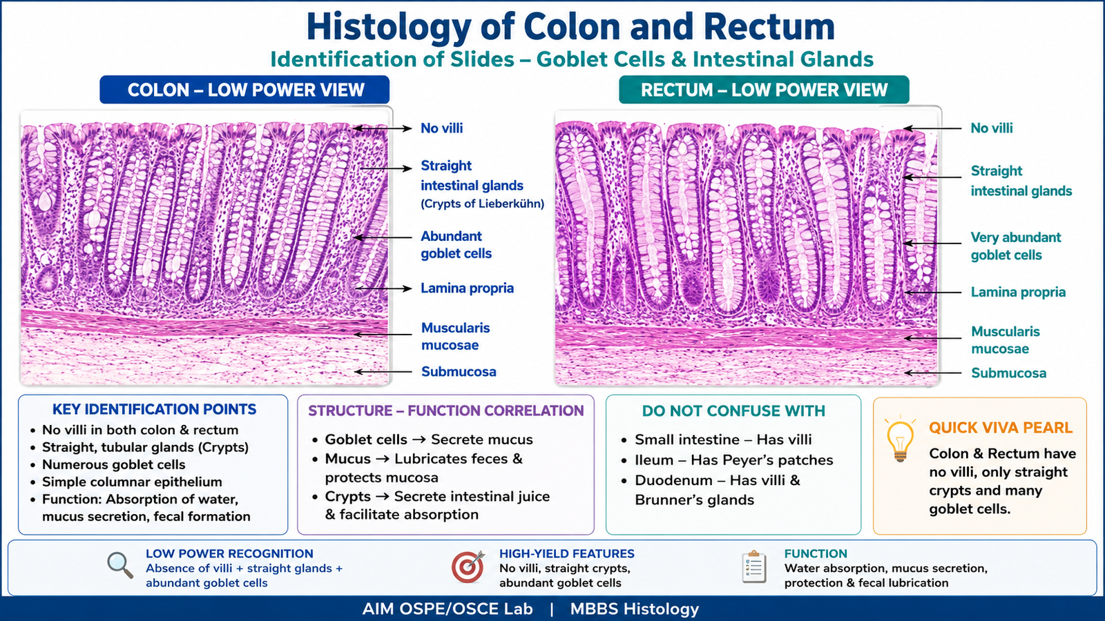
- Focus the given slide under low power.
- Identify whether the slide is colon or rectum.
- Observe the mucosa and check whether villi are absent.
- Identify straight tubular intestinal glands / crypts of Lieberkühn.
- Look for abundant goblet cells in the epithelium.
- Observe the mucosa, submucosa, and muscular layer.
- Write one key feature used to identify the slide.
- Mention one functional significance of goblet cells.
Observation / Identification Points
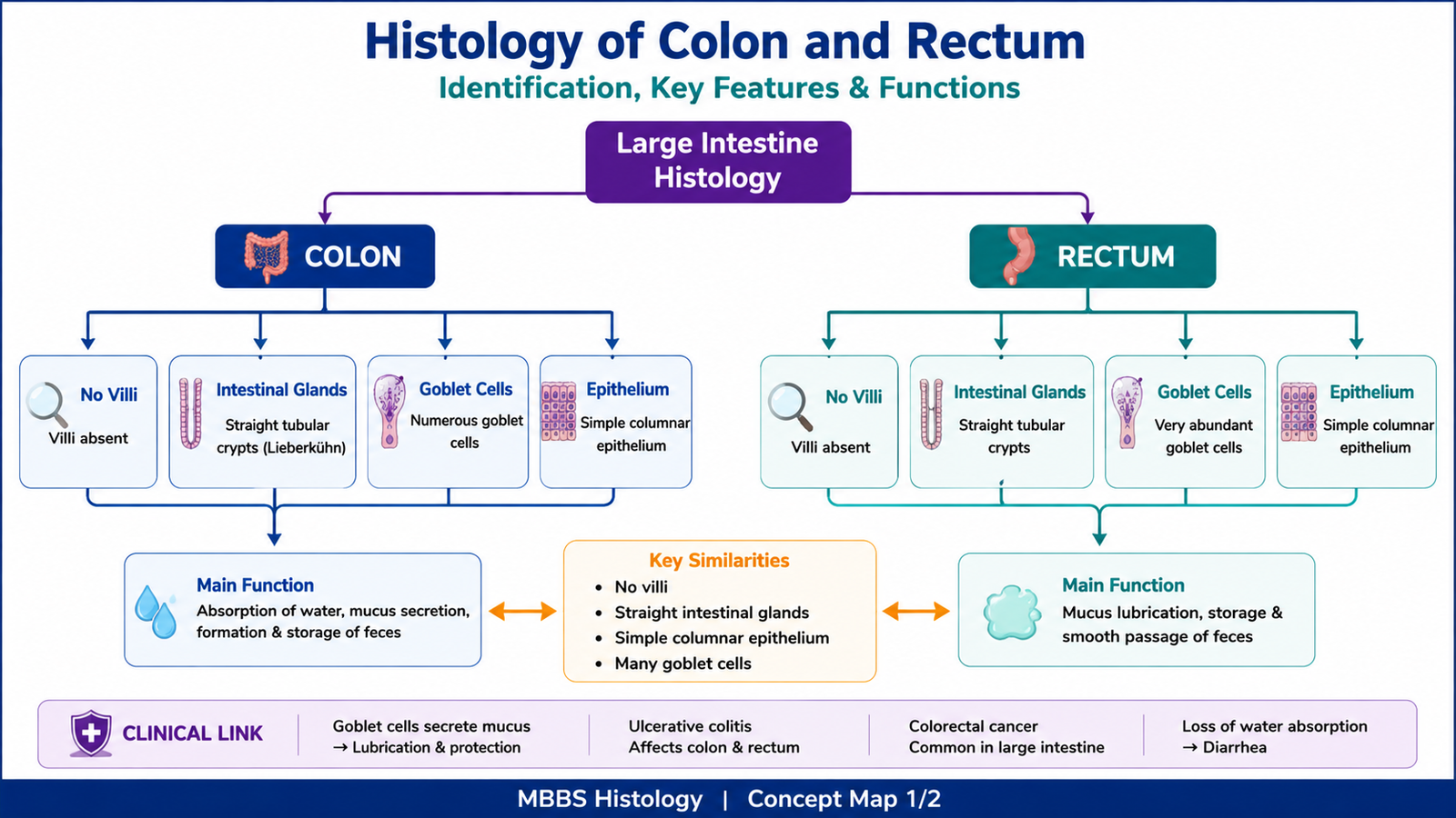
Colon
- No villi
- Straight tubular intestinal glands / crypts of Lieberkühn
- Numerous goblet cells
- Simple columnar epithelium
- Lamina propria between crypts
- Muscularis mucosae and submucosa present
- Function mainly related to water absorption and mucus lubrication
Rectum
- No villi
- Straight intestinal glands similar to colon
- Very abundant goblet cells
- Mucosa resembles colon but has more mucus-secreting cells
- Lower rectum shows transition toward anal canal epithelium
- Function related to fecal storage, mucus secretion, and lubrication
Key Differentiating Point
- Colon = no villi + straight crypts + many goblet cells
- Rectum = colon-like mucosa + very abundant goblet cells + mucus lubrication role
Result / Interpretation
The slide is identified as colon if it shows absence of villi, straight tubular crypts of Lieberkühn, and numerous goblet cells.
The slide is identified as rectum if it shows colon-like mucosa with very abundant goblet cells and straight intestinal glands, supporting lubrication during fecal passage.
Clinical significance:
Goblet cells produce mucus that lubricates intestinal contents and protects the mucosa. Loss or damage of colonic mucosa may affect water absorption and may be involved in inflammatory bowel disease, ulcerative colitis, and colorectal pathology.
Viva Questions
| Question | Short Ideal Answer |
|---|---|
| What is the key identifying feature of colon histology? | Absence of villi with straight crypts and many goblet cells. |
| Are villi present in the colon? | No, villi are absent in colon. |
| What are crypts of Lieberkühn? | Straight tubular intestinal glands present in the intestinal mucosa. |
| What is the function of goblet cells? | They secrete mucus for lubrication and mucosal protection. |
| How does rectal mucosa resemble colon? | It has no villi and contains straight crypts with abundant goblet cells. |
Marking Scheme
Total Marks: 5
| Component | Marks |
| Correct identification / performance | 2 |
| Key observation / procedure steps | 1 |
| Interpretation / principle | 1 |
| Viva answer | 1 |
Common Student Mistakes
- Confusing colon with small intestine by forgetting that colon has no villi.
- Calling straight crypts “villi.”
- Missing the importance of abundant goblet cells in colon and rectum.
AIM Feedback
To improve identification, first ask: Are villi present? If villi are absent and the mucosa shows many straight tubular crypts with goblet cells, think of colon or rectum. Remember: the most important OSPE clue is no villi + straight crypts + goblet cells.
🖼️ Visual / Image Support
🧩 Concept Map / Interpretation Support