
This chapter follows the KMU learning outcomes in a logical sequence, beginning with renal structure and progressing to filtration, tubular function and clinical renal evaluation. First understand the explanations and relationships; then use the final AIM High-Yield Review for rapid revision.
Topic 1 — Renal Structure and Function: Applied Anatomy, Glomerular Filtration, Tubular Handling, Acid–Base Balance and Renal Evaluation
A foundation chapter connecting the structure of the urinary system with filtration, tubular processing, acid–base regulation and interpretation of common renal investigations.
Topic Introduction
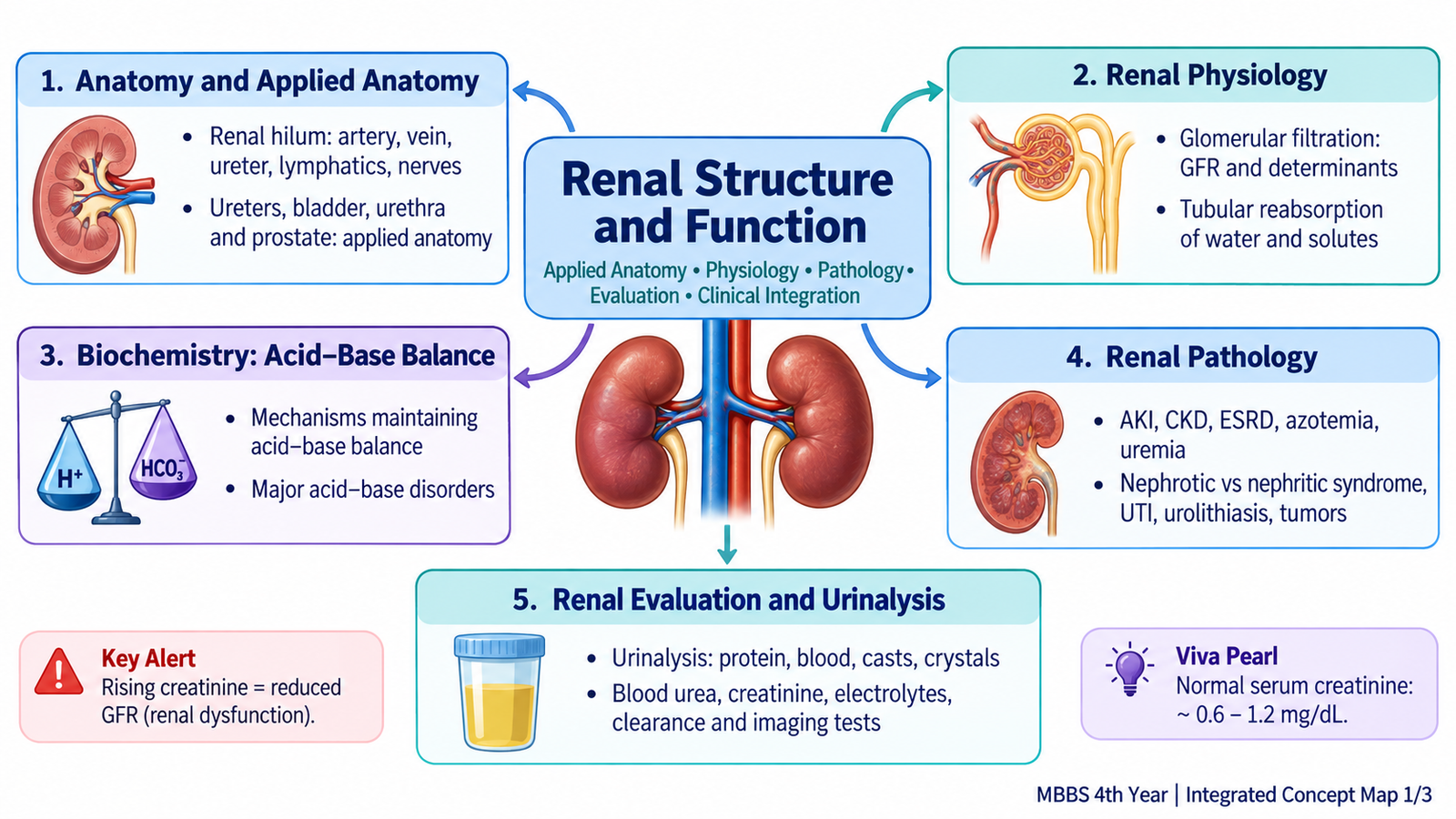
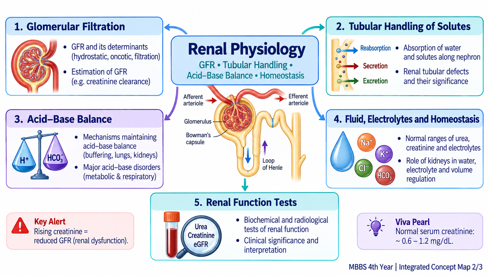
The kidneys continuously filter the blood, remove waste products and regulate water, electrolytes and acid–base balance. Understanding renal disease therefore requires more than memorizing laboratory values: a student must first understand how the kidney is organized, how glomerular filtration occurs and how each part of the nephron modifies the filtrate. This chapter connects these basic mechanisms with clinical renal evaluation. You will learn the important anatomy of the kidney and urinary tract, determinants and estimation of glomerular filtration rate, tubular handling of water and solutes, renal control of acid–base balance, common renal clinical terminology, interpretation of blood and urine tests and the role of imaging in assessing renal structure and function.
A. Applied Anatomy of the Kidney and Urinary Tract
The kidneys are paired retroperitoneal organs whose structure is closely related to their function. Blood must reach millions of nephrons for filtration, while the urine that is formed must pass through the collecting system, ureters, bladder and urethra. Important anatomical relations therefore help explain renal vascular injury, urinary obstruction and the anatomical basis of common investigations.
Gross anatomy of the kidney
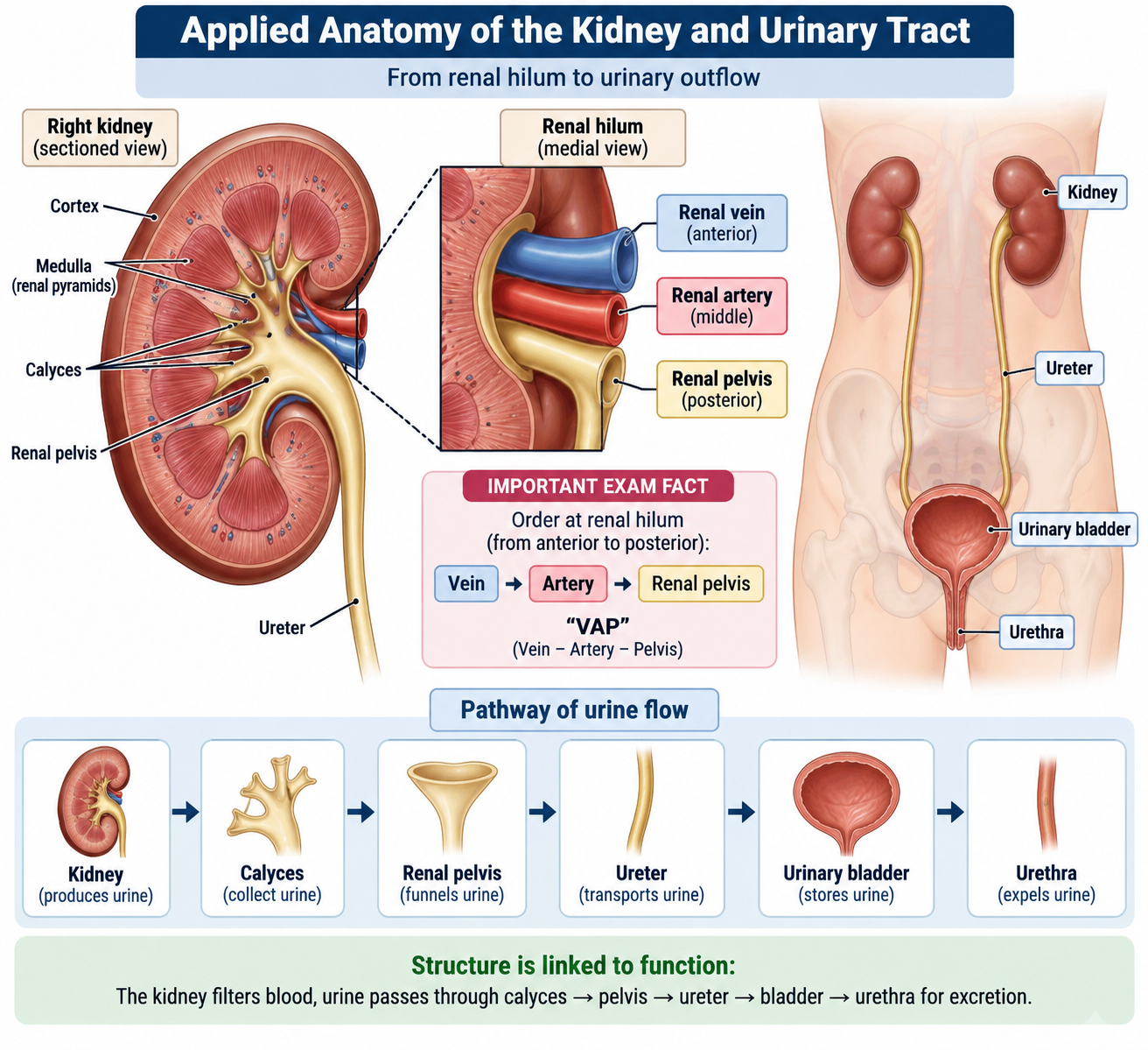
Each kidney is bean-shaped and lies on the posterior abdominal wall. The right kidney normally lies slightly lower than the left because of the liver. The kidney has an upper and lower pole, anterior and posterior surfaces, and lateral and medial borders. The medial border contains the renal hilum, which opens into the renal sinus.
When a kidney is sectioned, two major regions are visible:
- Renal cortex: the outer region containing renal corpuscles and much of the convoluted tubular system.
- Renal medulla: the inner region arranged into renal pyramids. The apex of each pyramid forms a renal papilla.
Urine flows from the papillary ducts at the renal papillae into minor calyces → major calyces → renal pelvis → ureter. This arrangement is clinically important because obstruction anywhere along this pathway may cause dilatation of the collecting system.
Renal hilum
The renal hilum provides the route through which vessels, nerves and the renal pelvis communicate with the kidney. A useful anterior-to-posterior relationship at the hilum is:
This relationship is useful when interpreting renal imaging and understanding operative or procedural anatomy.
Blood supply
Each kidney is usually supplied by a renal artery arising from the abdominal aorta. The renal artery divides into progressively smaller branches that ultimately supply individual nephron units. Segmental renal arteries behave largely as end arteries, so interruption of a significant branch may produce ischemia in the territory it supplies.
The intrarenal arterial sequence can be understood as:
Blood leaves the glomerulus through an efferent arteriole. It then enters the peritubular capillary network or, in juxtamedullary nephrons, the vasa recta. This arrangement allows filtration at the glomerulus and subsequent exchange with renal tubules.
Lymphatic drainage
Renal lymphatic vessels accompany the renal vessels and drain mainly toward the lateral aortic, or para-aortic, lymph nodes. This relationship is particularly relevant when considering spread from renal malignant disease.
Ureters, urinary bladder and urethra
The ureters are muscular tubes that transport urine from the renal pelvis to the urinary bladder by peristaltic contractions. There are three clinically important sites where the ureter is relatively narrow:
- the pelvi-ureteric junction;
- the region where the ureter crosses the pelvic brim/iliac vessels;
- the vesico-ureteric junction where it enters the bladder wall.
These sites are important because urinary calculi may become impacted there.
The urinary bladder is a muscular reservoir. Its smooth muscular wall is formed mainly by the detrusor muscle. The trigone is a relatively smooth triangular area between the two ureteric openings and the internal urethral opening.
The female urethra is short, whereas the male urethra is longer and passes through the prostate and penis. This anatomical difference contributes to important differences in urinary tract disease and catheterization.
Microscopic structure of the prostate and urethra
The prostate is composed of branching tubuloalveolar glands embedded within fibromuscular stroma. The glands drain into the prostatic urethra. Anatomically and clinically, the gland may be considered in zones; the peripheral and transition zones are especially important in prostatic disease.
The urethral epithelium changes along its length. The proximal urethra is related to the urothelium of the bladder, while more distal portions show columnar or pseudostratified columnar epithelium, becoming stratified squamous epithelium near the external opening.
B. Glomerular Filtration and Glomerular Filtration Rate
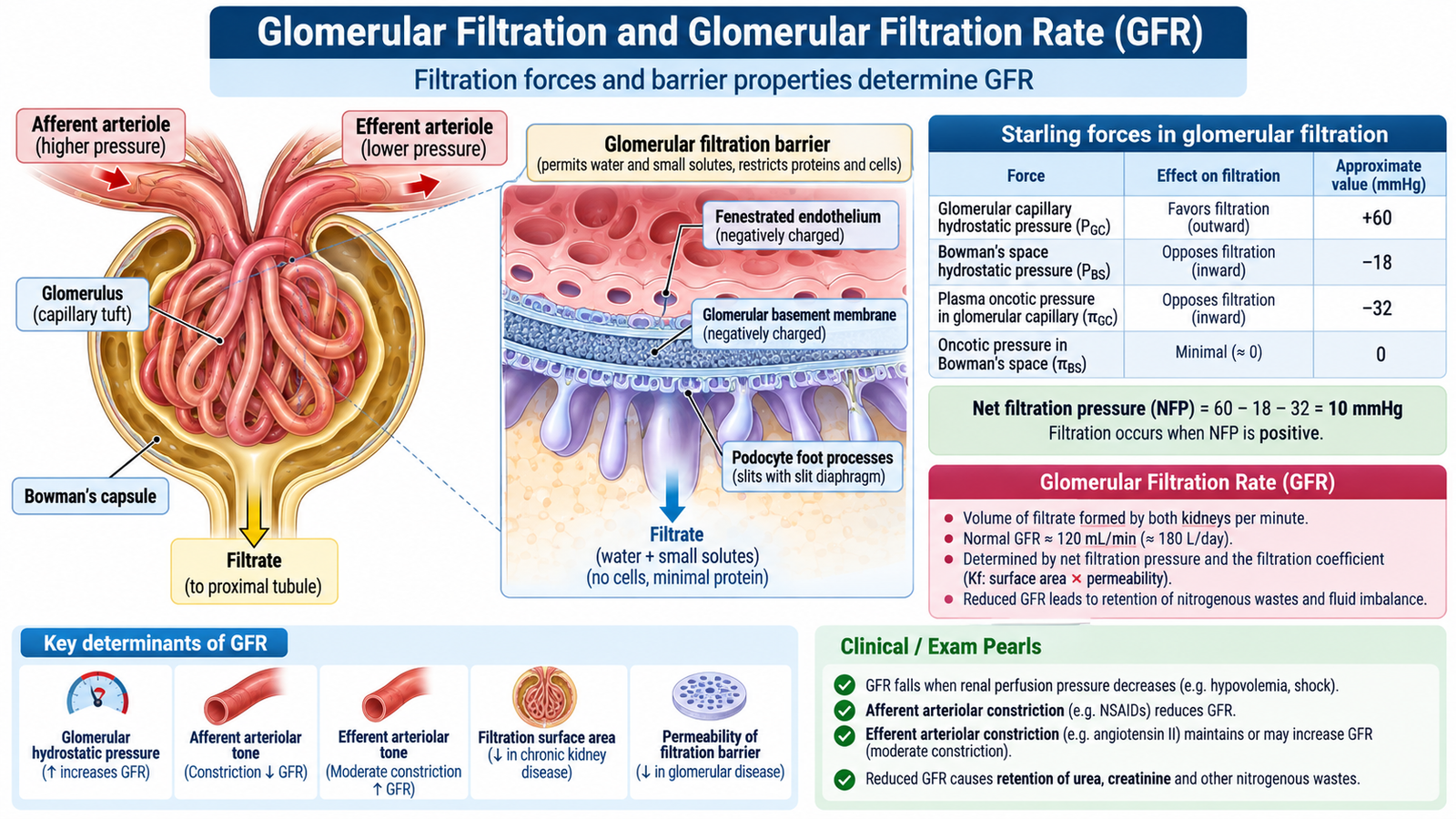
Glomerular filtration is the first major step in urine formation. Blood reaches the glomerular capillaries under pressure, and part of the plasma water and its small dissolved substances move into Bowman’s space. Blood cells and most plasma proteins normally remain within the circulation.
The filtration barrier
The glomerular filtration barrier is highly permeable to water and small solutes but restricts cells and large proteins. Its major components are:
- fenestrated glomerular capillary endothelium;
- glomerular basement membrane;
- filtration slits between podocyte foot processes.
The barrier depends on both size selectivity and physicochemical properties of molecules. Damage to this barrier may therefore allow proteins or blood to appear in urine.
What determines filtration?
Filtration depends on the balance between forces favoring movement of fluid out of glomerular capillaries and forces opposing it. Glomerular capillary hydrostatic pressure is the major force favoring filtration. Plasma oncotic pressure and pressure within Bowman’s space oppose filtration.
Renal blood flow + glomerular capillary pressure + filtration-barrier properties → amount of filtrate formed.
Glomerular filtration rate
Glomerular filtration rate (GFR) is the volume of filtrate formed by all functioning glomeruli per unit time. In a healthy young adult it is commonly around 90–120 mL/min/1.73 m², although normal GFR varies with age, body size and other physiological factors.
GFR is influenced by:
- renal perfusion and systemic arterial pressure;
- resistance of afferent and efferent arterioles;
- glomerular capillary hydrostatic pressure;
- plasma oncotic pressure;
- pressure in Bowman’s space;
- the surface area and permeability of the filtration membrane.
For example, marked urinary obstruction raises pressure proximal to the obstruction. Increased tubular and Bowman’s-space pressure opposes filtration and can therefore reduce GFR.
Renal autoregulation
Within a physiological range, the kidney can stabilize renal blood flow and GFR despite moderate changes in systemic arterial pressure. Important mechanisms include the myogenic response of the afferent arteriole and tubuloglomerular feedback involving the macula densa.
This allows filtration to remain relatively stable while systemic pressure fluctuates during normal activity.
Estimating GFR
Direct measurement of GFR is inconvenient in routine clinical practice. Instead, renal function is usually estimated using serum creatinine together with patient characteristics to obtain an estimated GFR (eGFR).
A falling GFR means that overall renal filtration capacity is decreasing. However, interpretation should not rely on a single number alone. Age, muscle mass, acute changes in kidney function and the clinical situation influence interpretation.
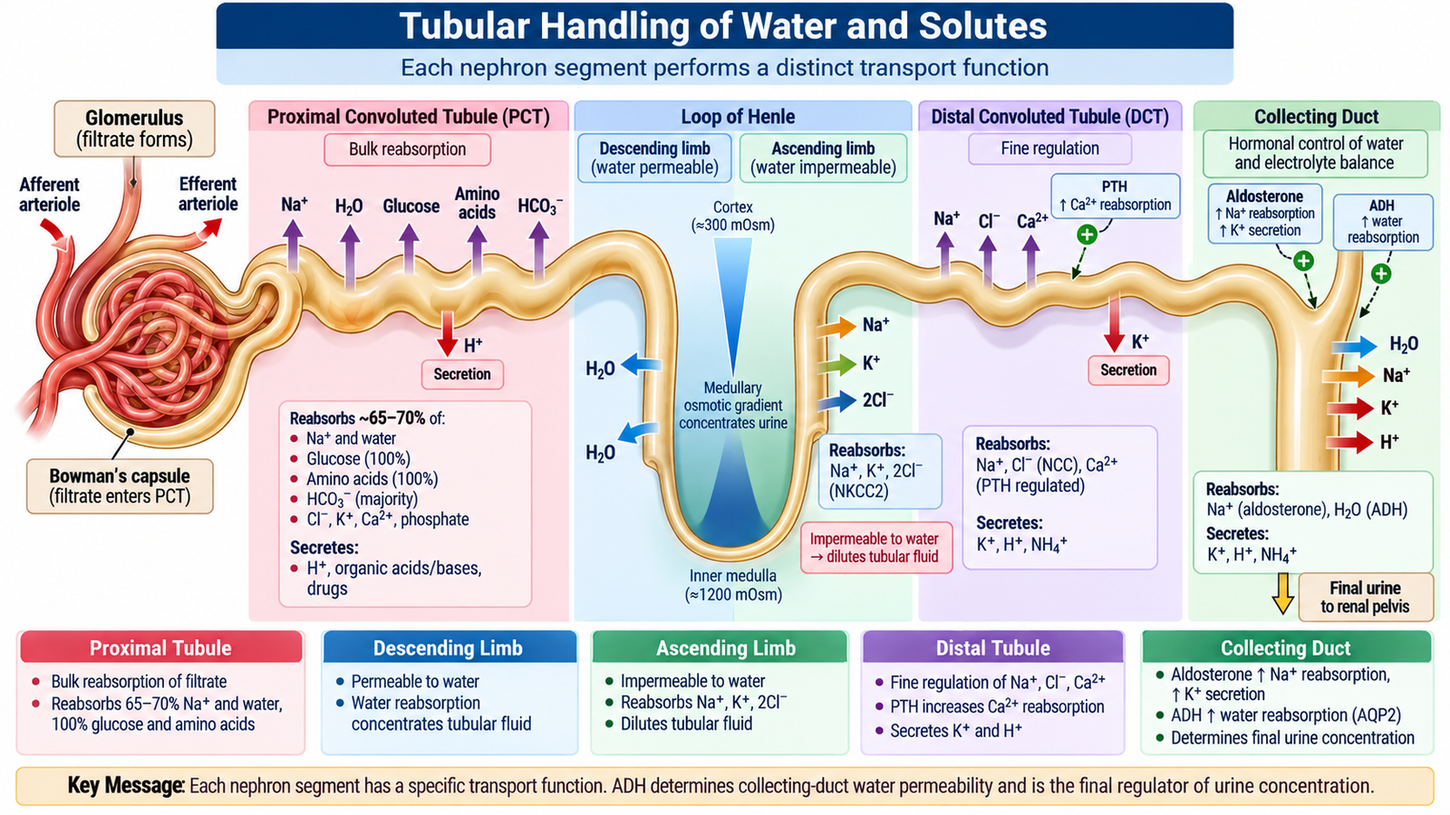
C. Tubular Handling of Water and Solutes
The glomerulus produces a large volume of filtrate, but the body cannot afford to lose all of its water and useful solutes. Renal tubules therefore selectively reabsorb substances that should be retained and secrete additional substances from blood into tubular fluid. The final urine is the result of filtration, reabsorption and secretion.
Proximal convoluted tubule
The proximal tubule performs bulk reabsorption. Approximately two-thirds of filtered sodium and water are normally reabsorbed here. Under normal circumstances, essentially all filtered glucose and amino acids are also reclaimed.
Major substances reabsorbed in the proximal tubule include:
- sodium and water;
- glucose and amino acids;
- bicarbonate;
- chloride;
- phosphate and other filtered solutes.
Because water follows the reabsorbed solutes, much proximal tubular reabsorption remains approximately isotonic.
Loop of Henle
The loop of Henle establishes an important separation between water and solute handling.
The descending limb is relatively permeable to water. Water can therefore leave the tubule as it descends into the increasingly concentrated renal medulla.
The thick ascending limb, in contrast, reabsorbs sodium, potassium and chloride but is relatively impermeable to water. Removal of solute without water makes tubular fluid more dilute and contributes to formation of the medullary concentration gradient.
Distal convoluted tubule
The distal nephron performs finer regulation rather than bulk reabsorption. Sodium chloride continues to be reabsorbed, while calcium handling is hormonally regulated, particularly by parathyroid hormone.
Collecting duct
The collecting duct determines the final composition and concentration of urine. Two hormones are especially important:
- Aldosterone increases sodium reabsorption and promotes potassium secretion in the distal nephron.
- Antidiuretic hormone (ADH) increases water permeability of collecting ducts by promoting insertion of aquaporin water channels.
When ADH is present, water is reabsorbed from the collecting duct into the hyperosmotic medullary interstitium, producing more concentrated urine. When ADH activity is low, less water is reabsorbed and a larger volume of dilute urine is formed.
Why the medullary gradient matters
The loop of Henle, particularly in juxtamedullary nephrons, helps establish a hyperosmotic medulla. The vasa recta help preserve this gradient. The collecting duct then uses the gradient, under the influence of ADH, to conserve water when required.
Glomerular filtration → bulk proximal reabsorption → loop concentration gradient → distal fine adjustment → collecting-duct control of final urine.
D. Renal Regulation of Acid–Base Balance
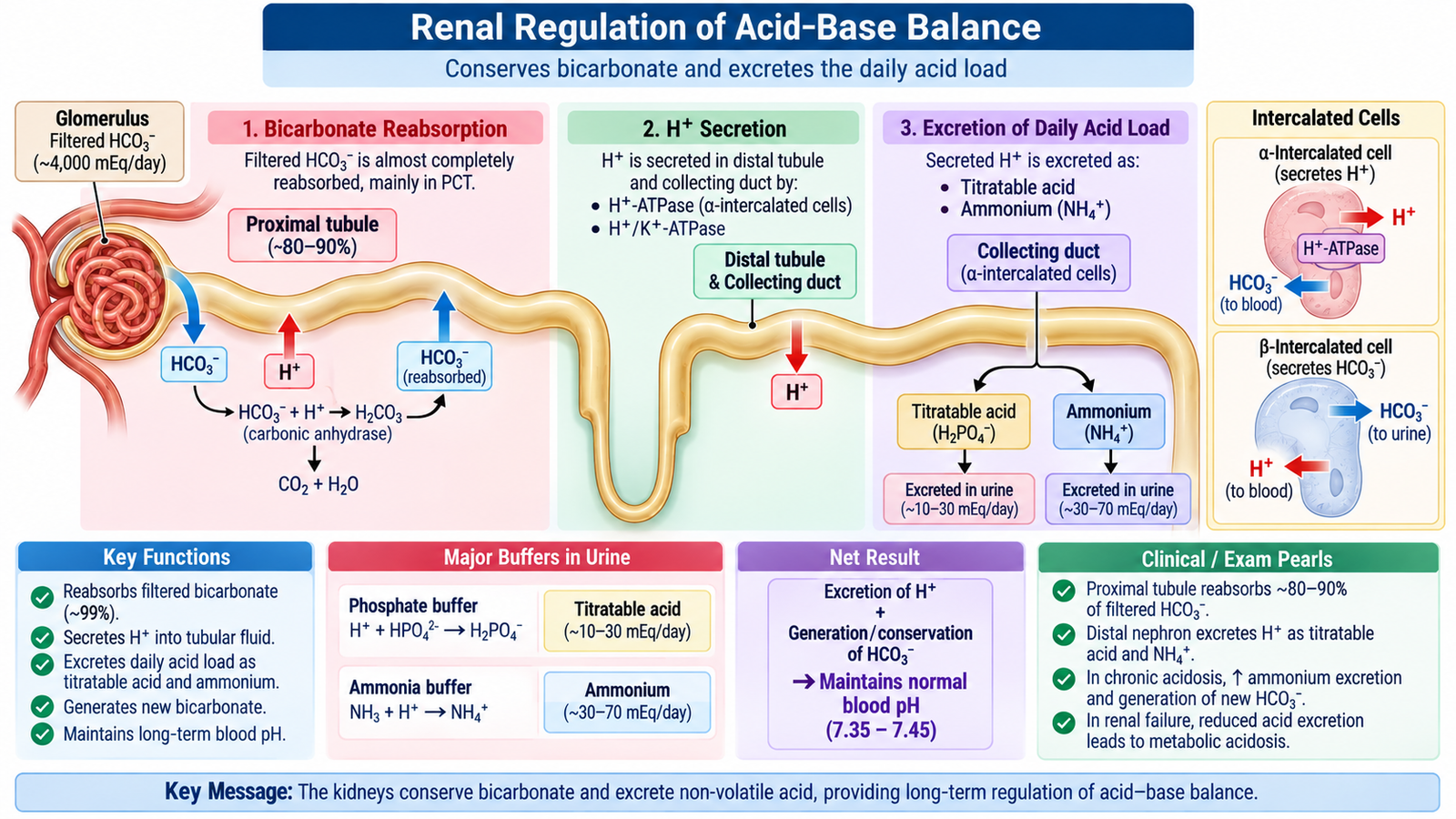
Normal cellular function requires the hydrogen-ion concentration of body fluids to remain within a narrow range. Acid is continuously generated by metabolism. Acid–base balance is maintained through coordinated action of chemical buffers, the lungs and the kidneys. The kidneys provide the slower but powerful long-term component of this regulation.
Three levels of acid–base defense
- Chemical buffers rapidly bind or release hydrogen ions.
- Lungs alter carbon dioxide elimination through ventilation.
- Kidneys regulate bicarbonate and hydrogen-ion excretion.
How the kidneys conserve bicarbonate
Filtered bicarbonate is valuable because it is a major extracellular buffer. Most filtered bicarbonate is therefore reclaimed, particularly in the proximal tubule. Tubular cells secrete hydrogen ions into the tubular lumen. These hydrogen ions interact with filtered bicarbonate, allowing bicarbonate to be effectively returned to the blood.
If bicarbonate were continually lost in urine, the body’s buffering capacity would fall and metabolic acidosis would develop.
Excretion of hydrogen ions
The kidneys cannot remove the daily acid load merely by producing very acidic free fluid. Hydrogen ions are therefore excreted after being buffered within tubular fluid.
Two important mechanisms are:
- Titratable acid: secreted hydrogen ions are buffered mainly by filtered phosphate.
- Ammonium excretion: renal tubular cells generate ammonia from glutamine. Ammonia accepts hydrogen ions to form ammonium, which can then be excreted.
Excretion of acid in these forms is linked to addition of new bicarbonate to the extracellular fluid.
Increased H+ secretion → increased titratable acid and NH4+ excretion → conservation/generation of HCO3− → restoration of extracellular buffering.
Major acid–base disorders
| Disorder | Primary disturbance | Direction of pH | Main compensation |
|---|---|---|---|
| Metabolic acidosis | Primary fall in bicarbonate | Falls | Increased ventilation lowers CO2 |
| Metabolic alkalosis | Primary rise in bicarbonate | Rises | Respiratory retention of CO2 |
| Respiratory acidosis | Primary rise in CO2 | Falls | Renal retention/generation of bicarbonate |
| Respiratory alkalosis | Primary fall in CO2 | Rises | Renal reduction of bicarbonate |
Compensation reduces the change in pH but does not remove the primary disorder. The presence of an unexpected bicarbonate or carbon dioxide pattern should therefore raise the possibility of a mixed acid–base disturbance.
E. Core Renal Terminology and Syndromic Patterns
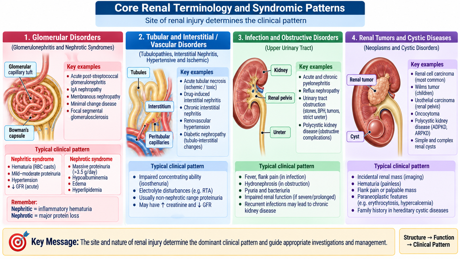
Renal terminology often describes a pattern of functional disturbance rather than one specific disease. Correct use of these terms helps the student interpret laboratory results and recognize whether the dominant problem is glomerular, tubular, vascular, infective, obstructive or neoplastic.
Azotemia and uremia
Azotemia means an abnormal accumulation of nitrogenous waste products in blood, particularly urea and creatinine. It is primarily a biochemical finding indicating reduced clearance of these substances.
Uremia is the clinical syndrome produced by severe renal dysfunction with retention of metabolic products and disturbances of fluid, electrolyte and endocrine functions. Thus, azotemia refers mainly to laboratory evidence, whereas uremia implies clinically significant systemic illness.
Acute kidney injury, chronic kidney disease and end-stage renal disease
Acute kidney injury (AKI) is a relatively rapid reduction in kidney function. GFR falls, resulting in retention of nitrogenous wastes and potentially important disturbances of fluid, potassium and acid–base balance.
Chronic kidney disease (CKD) is persistent kidney structural or functional abnormality. It develops over a prolonged period and may progressively reduce renal functional reserve.
End-stage renal disease (ESRD) describes advanced irreversible kidney failure in which residual renal function is insufficient to maintain normal internal homeostasis without renal replacement therapy.
Nephritic syndrome
Nephritic syndrome reflects inflammatory injury to glomeruli. Damage to glomerular capillary walls allows red blood cells to enter urine and reduces filtration.
The characteristic pattern includes:
- hematuria;
- red blood cell casts when glomerular bleeding is significant;
- variable proteinuria, usually less marked than in nephrotic syndrome;
- reduced GFR with azotemia in more severe cases;
- salt and water retention, which may cause edema and hypertension.
Nephrotic syndrome
Nephrotic syndrome is caused by increased permeability of the glomerular filtration barrier to plasma proteins. Large quantities of protein, particularly albumin, are therefore lost in urine.
The major pattern includes marked proteinuria, hypoalbuminemia, generalized edema and associated lipid abnormalities.
Asymptomatic hematuria
Asymptomatic hematuria means blood in urine without prominent urinary symptoms. Hematuria is a finding rather than a final diagnosis. Its source may be glomerular or may arise from another part of the urinary tract. Persistent hematuria therefore requires interpretation together with urine microscopy, protein excretion, renal function and the clinical context.
Rapidly progressive glomerulonephritis
Rapidly progressive glomerulonephritis (RPGN) is a severe clinical syndrome in which glomerular injury produces rapid deterioration in renal function over a relatively short period. It is an important pattern because delay in recognition can lead to major loss of renal function.
Renal tubular defects
Renal tubular defects occur when one or more tubular transport processes fail despite filtration occurring at the glomerulus. Depending on the affected transporter or nephron segment, abnormalities may involve glucose, electrolytes, water or acid–base regulation.
Nephrosclerosis
Nephrosclerosis refers to sclerosis and structural damage involving renal blood vessels and renal parenchyma. Progressive vascular narrowing reduces blood supply, causing chronic ischemic injury and loss of functioning nephrons.
Urinary tract infection
Urinary tract infection (UTI) means microbial infection involving part of the urinary tract. Infection may remain in the lower urinary tract or involve the kidney. Urine findings such as leukocytes, leukocyte esterase, nitrites and microorganisms may support the diagnosis when interpreted with symptoms and culture where appropriate.
Urolithiasis and hydronephrosis
Urolithiasis is formation of calculi within the urinary tract. A stone may obstruct urine flow, producing pain and hematuria and, if obstruction persists, impairing renal function.
Hydronephrosis is dilatation of the renal pelvis and calyces caused by impaired urinary drainage. Persistent obstruction increases pressure proximal to the obstruction and may compress renal parenchyma, eventually reducing renal function.
Oncocytoma and renal carcinoma
Renal oncocytoma is a generally benign renal epithelial tumor. Renal carcinoma, particularly renal cell carcinoma, is malignant. Imaging may identify a renal mass, but radiological appearance alone may not always establish the exact histological type; definitive characterization may require pathological assessment.
F. Biochemical Assessment and Renal Function Tests
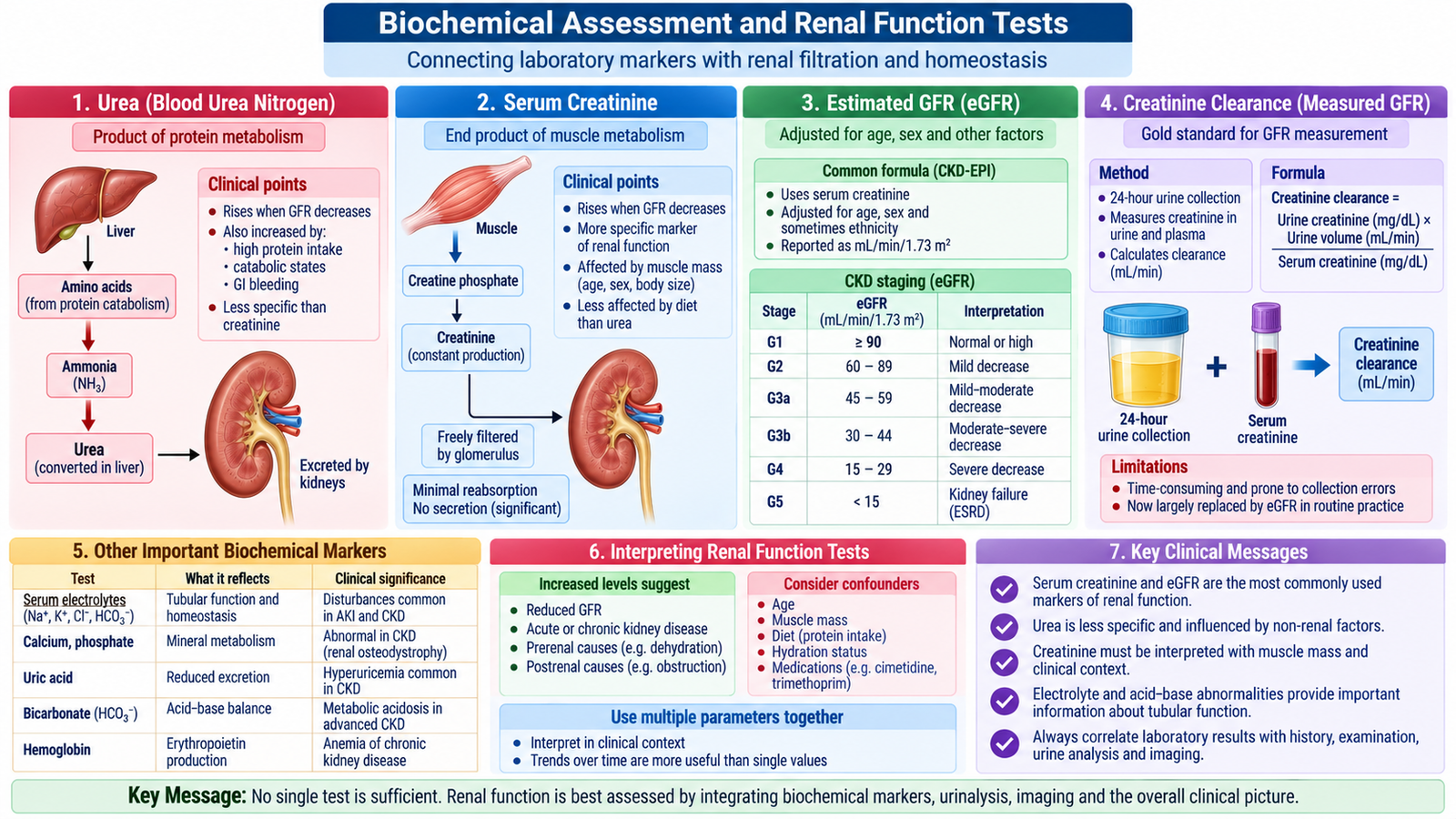
Renal function cannot be judged from a single laboratory value. Blood urea and serum creatinine mainly reflect the kidney’s ability to clear nitrogenous waste, while electrolytes show how effectively the kidneys are maintaining internal homeostasis. GFR estimates and creatinine clearance provide more direct information about filtration.
Common adult reference ranges
Reference intervals vary slightly among laboratories and analytical methods. The following are typical adult values and should always be interpreted using the reference range supplied by the reporting laboratory.
| Parameter | Typical adult reference range | Main significance |
|---|---|---|
| Serum urea | Approximately 15–45 mg/dL | Nitrogenous waste; rises when renal excretion falls but is influenced by non-renal factors |
| Serum creatinine | Approximately 0.6–1.3 mg/dL | Common endogenous marker used to assess filtration |
| Sodium | 135–145 mmol/L | Reflects water-sodium balance |
| Potassium | 3.5–5.0 mmol/L | Renal excretion is essential for potassium homeostasis |
| Chloride | Approximately 98–106 mmol/L | Closely related to sodium and acid–base balance |
| Bicarbonate | Approximately 22–28 mmol/L | Major indicator of the metabolic component of acid–base status |
Blood urea
Urea is produced mainly in the liver from nitrogen generated during protein metabolism and is excreted largely by the kidneys. Reduced renal clearance may therefore increase blood urea.
Urea is not a pure marker of GFR. Its concentration is also influenced by factors such as protein breakdown, dietary protein, gastrointestinal bleeding and hydration. A raised urea concentration must therefore be interpreted in context rather than automatically equated with intrinsic kidney disease.
Serum creatinine
Creatinine is formed from creatine in skeletal muscle and is removed mainly by glomerular filtration. It is widely used because it is convenient and changes as filtration declines.
However, serum creatinine also depends on creatinine production. A muscular individual may have a higher baseline creatinine than a person with very low muscle mass. Consequently, the same serum creatinine value does not necessarily represent exactly the same GFR in every patient.
Estimated GFR
eGFR combines serum creatinine with patient characteristics to provide an estimate of glomerular filtration. It is generally more informative than looking at creatinine alone when assessing chronic kidney function.
In rapidly changing acute kidney function, however, serum creatinine may not yet be in equilibrium. An eGFR calculated from that changing creatinine should therefore be interpreted cautiously.
Creatinine clearance
Creatinine clearance estimates the volume of plasma cleared of creatinine per unit time. It can be determined using urine creatinine concentration, urine flow and plasma creatinine concentration.
Because a small amount of creatinine is also secreted by renal tubules, creatinine clearance tends to slightly overestimate true GFR. Accurate timed urine collection is also important; incomplete collection can substantially distort the result.
Electrolytes as renal functional markers
The kidneys continually regulate sodium, potassium and bicarbonate. Advanced impairment of renal function may therefore produce electrolyte and acid–base abnormalities.
Potassium is particularly important because impaired renal potassium excretion can produce hyperkalemia. Severe disturbances of potassium may affect cardiac electrical activity. Reduced renal acid excretion may also contribute to metabolic acidosis.
G. Urinalysis and Diagnostic Significance of Urinary Abnormalities
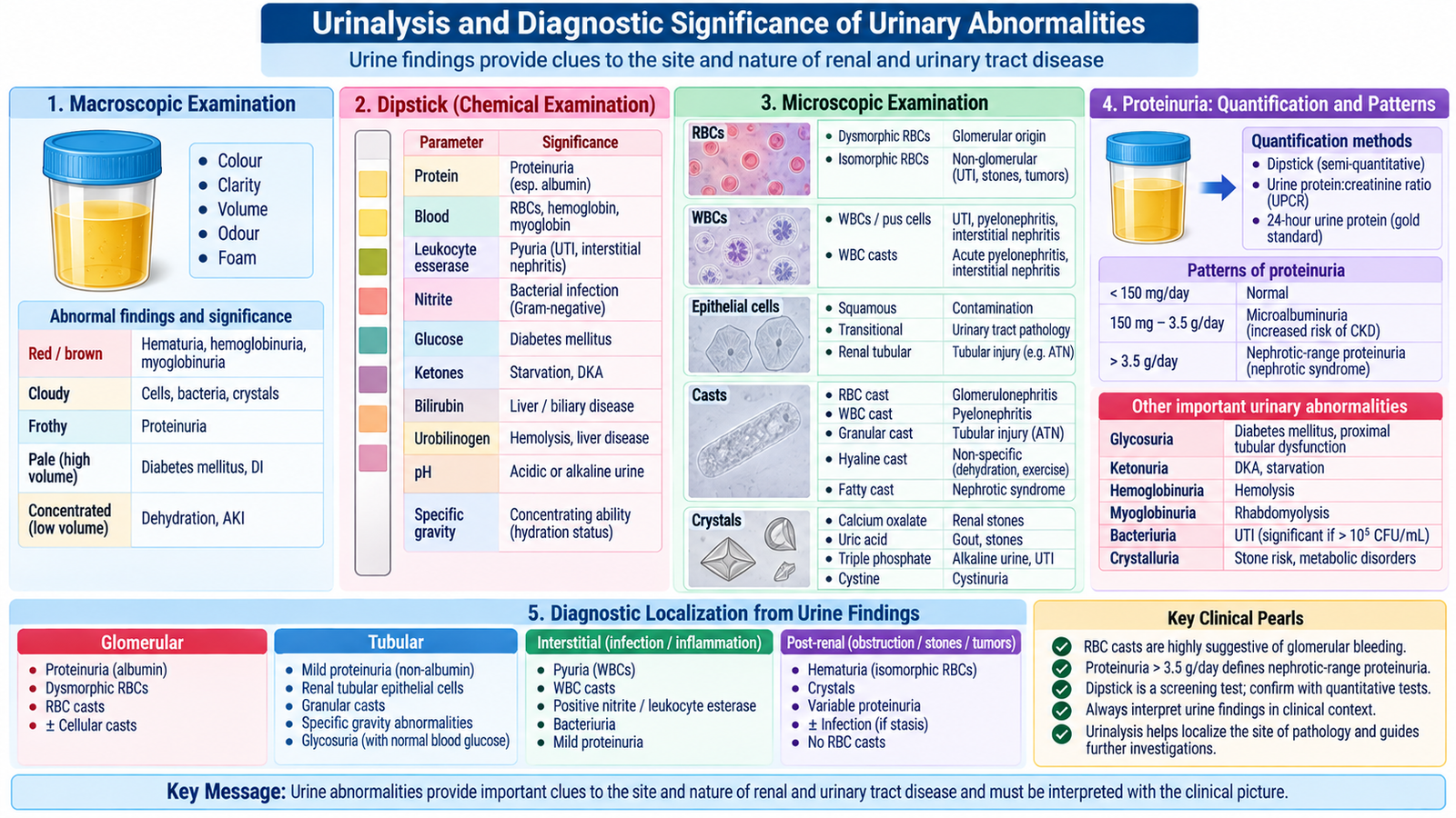
Urinalysis provides a rapid view of several processes occurring in the kidney, urinary tract and body as a whole. Correct interpretation requires the student to combine the physical appearance of urine, dipstick findings and microscopy instead of treating any single abnormality as a diagnosis.
Physical examination of urine
Important observations include color, clarity, concentration and specific gravity. Urine concentration changes according to water intake and the kidney’s ability to conserve or excrete water.
Specific gravity provides an approximate indication of urine concentration. Persistently abnormal concentration in the appropriate clinical setting may suggest impaired renal concentrating ability.
Dipstick examination
| Urine finding | What it may indicate | Interpretation point |
|---|---|---|
| Protein | Glomerular permeability abnormality; sometimes tubular disease | Persistent proteinuria requires further assessment and quantification |
| Blood | Hematuria, hemoglobin or myoglobin | Microscopy helps determine whether intact RBCs are present |
| Leukocyte esterase | White blood cells in urine | Supports urinary inflammation or infection |
| Nitrite | Certain nitrate-reducing urinary bacteria | A negative result does not exclude UTI |
| Glucose | Filtered glucose exceeds tubular reabsorptive capacity or tubular reabsorption is defective | May reflect systemic metabolic disease or tubular dysfunction |
| Ketones | Increased fat metabolism | Interpret with metabolic and nutritional context |
| pH | Urinary acidification status | Affected by acid–base state, diet and some urinary conditions |
Urine microscopy
Microscopy provides information that cannot be obtained reliably from the dipstick alone.
Red blood cells: confirm hematuria when present. Dysmorphic red cells support a glomerular source because cells become distorted while passing through an abnormal glomerular filtration barrier and nephron.
White blood cells: suggest inflammation or infection when present in significant numbers in an appropriate clinical context.
Urinary casts: are formed within renal tubules, so their presence may help localize disease to the kidney itself.
- Red blood cell casts strongly suggest glomerular bleeding.
- White blood cell casts indicate inflammation involving the renal tubules/interstitium rather than simple lower urinary contamination.
- Granular or cellular casts may accompany renal parenchymal injury.
Crystals may appear in urine depending on solute concentration and urinary conditions. Some are clinically relevant in patients with urinary stone disease, but crystals should always be interpreted in context.
Proteinuria
Proteinuria becomes clinically important when the normal filtration barrier or tubular handling of proteins is disturbed. Glomerular injury permits increased filtration of plasma proteins, whereas tubular dysfunction can reduce reabsorption of normally filtered low-molecular-weight proteins.
The amount and persistence of proteinuria provide information about the likely renal process. Marked protein loss is particularly associated with nephrotic syndromes.
Hematuria: renal versus urogenital significance
Hematuria can originate anywhere from the glomerulus to the urethra. Proteinuria, dysmorphic red blood cells and red-cell casts favor a glomerular source. In contrast, urinary stones, infection and urogenital lesions can also produce hematuria without a primary glomerular disorder.
Systemic disease reflected in urine
Urinalysis may reveal abnormalities caused by disease outside the urinary system. Examples include glycosuria or ketonuria in metabolic disease and pigment abnormalities related to hemoglobin, myoglobin or bilirubin. Thus, abnormal urine does not automatically mean that the primary disease originated in the kidney.
H. Radiological Evaluation, Integrated Renal Assessment and Disease Burden
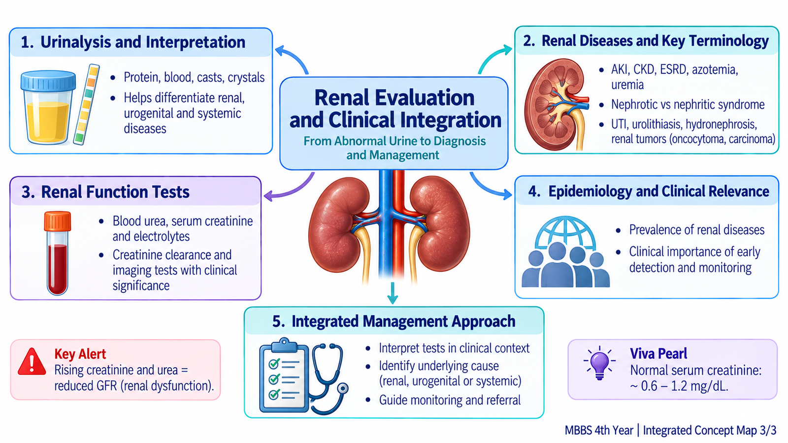
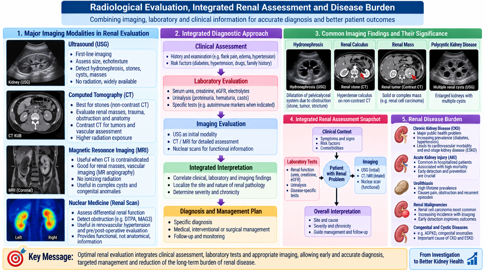
Renal evaluation becomes much more useful when biochemical tests, urinalysis and imaging are interpreted together. Blood tests describe filtration and internal homeostasis, urine provides clues to the site and nature of renal or urinary disease, and imaging shows the structure of the kidneys and urinary tract.
Ultrasonography
Ultrasound is a useful initial examination because it can assess renal size and outline without ionizing radiation. It is especially valuable for identifying:
- dilatation of the collecting system in hydronephrosis;
- gross structural abnormalities;
- renal cystic lesions;
- many renal masses;
- bladder distension and residual urine in appropriate settings.
Renal size can also provide useful context. Bilaterally small kidneys may support longstanding chronic parenchymal disease, while acute functional deterioration may occur without small kidneys.
Plain radiography
A plain abdominal radiograph may demonstrate some radiopaque urinary calculi but provides limited information about renal parenchyma and overall renal function. It has therefore largely been supplemented by more informative imaging techniques when detailed assessment is required.
Computed tomography
CT provides excellent anatomical detail and is highly useful in evaluating urinary calculi, obstruction, renal masses and many other structural abnormalities. Depending on the clinical question, CT may be performed without contrast or with contrast enhancement.
When contrast is being considered, renal function and the overall clinical context must be assessed because contrast administration may require additional caution in patients with significant renal impairment.
Contrast studies of the collecting system
Contrast imaging can demonstrate renal excretion and outline the collecting system, ureters and urinary tract. Its value lies in combining anatomical information with evidence that contrast has been filtered and excreted by the kidneys. Modern cross-sectional imaging has replaced many older applications, but the underlying diagnostic principle remains important.
Radionuclide renal studies
Radionuclide techniques can assess renal function in a way that structural imaging cannot. Depending on the tracer and technique, they may provide information about relative function of each kidney, renal perfusion and urinary drainage.
This distinction is important:
Functional renal testing asks: “How effectively are the kidneys filtering, concentrating or draining?”
How to integrate renal investigations
No single test answers every renal question. A logical interpretation proceeds from the clinical problem to the type of information required.
- Ask whether filtration is impaired: serum creatinine and eGFR provide the main initial information.
- Assess consequences of dysfunction: urea, potassium, bicarbonate and other electrolytes help identify retained waste and disturbances of homeostasis.
- Examine urine: protein, blood, leukocytes, casts and other abnormalities help identify the likely compartment involved.
- Assess structure and obstruction: imaging examines renal size, masses, stones and collecting-system dilatation.
- Assess individual renal function or drainage when necessary: appropriate functional imaging may provide additional information.
Prevalence and burden of renal disease
Renal and urinary disorders represent an important burden in clinical practice, but the frequency of individual conditions varies between populations. Chronic kidney disease becomes more common with increasing age and with common chronic conditions that damage the renal vasculature or glomeruli. Urinary tract infections are among the commonly encountered urinary disorders, while urolithiasis shows geographical and environmental variation. Glomerular diseases, hereditary tubular abnormalities and renal tumors are less frequent than many common urinary disorders but remain important because of their potential effects on renal function.
The key epidemiological principle is that renal disease is not a single entity. Its prevalence reflects the combined burden of chronic renal dysfunction, infection, glomerular disease, obstruction, stone disease and neoplasia. Population prevalence should therefore always be interpreted according to the specific renal condition being discussed rather than applying one figure to all kidney disease.
Integrated Mechanism Flow
Important Comparison — Nephritic vs Nephrotic Syndrome
| Feature | Nephritic syndrome | Nephrotic syndrome |
|---|---|---|
| Dominant injury | Inflammatory glomerular injury | Increased glomerular permeability to protein |
| Hematuria | Prominent | Not the dominant feature |
| Proteinuria | Usually less marked | Marked |
| RBC casts | Characteristic of glomerular bleeding | Not characteristic |
| Edema mechanism | Salt and water retention | Major protein loss with reduced plasma oncotic pressure |
| Renal function | GFR may fall significantly | Variable depending on underlying disease |
Renal Filtration, Reabsorption and Secretion
Use this video to reinforce glomerular filtration and the handling of water, electrolytes and other solutes along the different segments of the nephron.