
Topic 3 — Acute Kidney Injury and Renal Vascular Disorders: Tubular Injury, Electrolyte Emergencies and Acute Renal Care
1. Topic Introduction
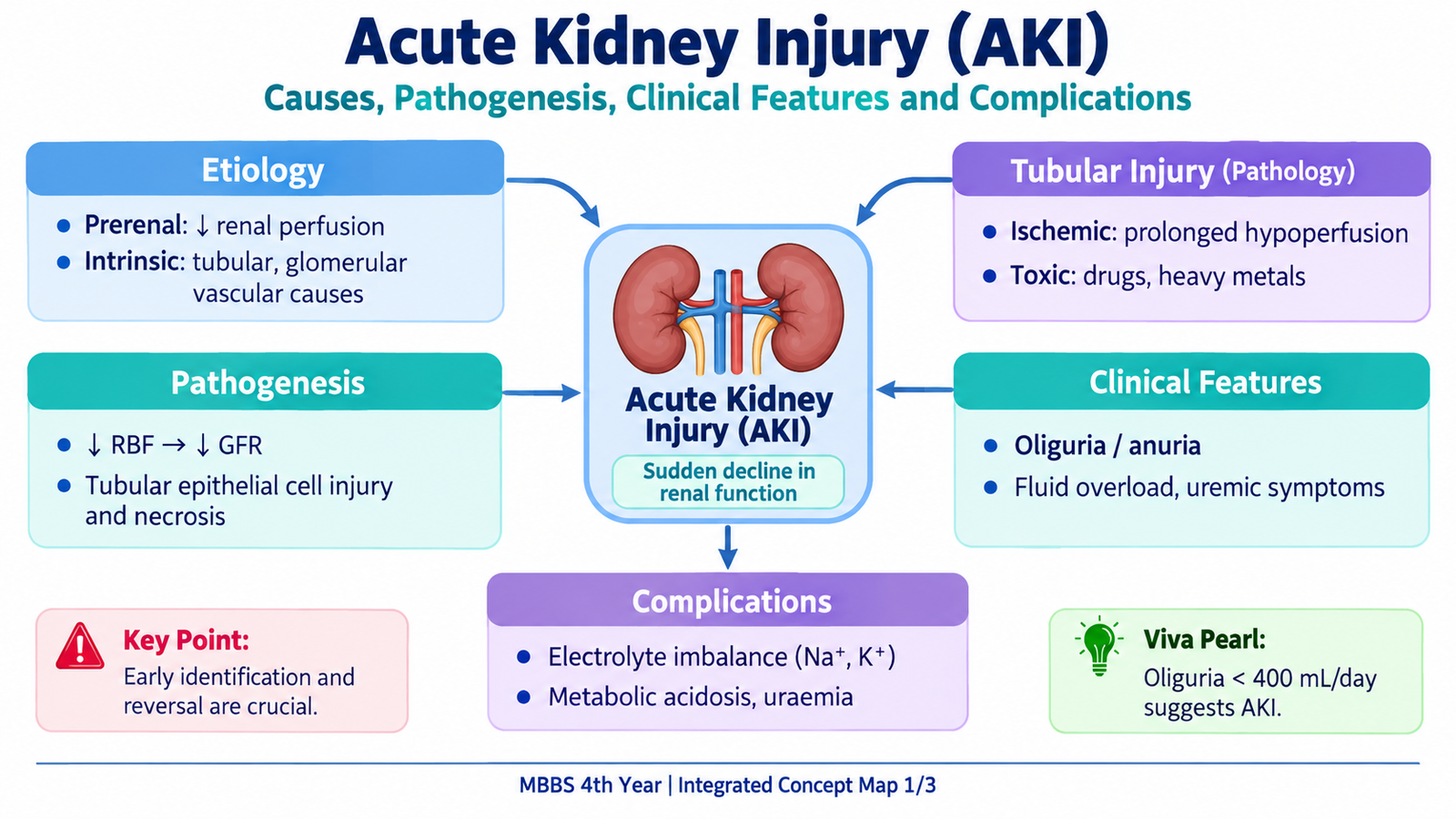
Acute kidney injury (AKI) is a sudden reduction in kidney function that causes retention of nitrogenous waste products and disturbance of fluid, electrolyte and acid–base balance. It may result from reduced renal perfusion, direct injury to kidney tissue or obstruction to urinary flow. Acute tubular injury is one of the most important intrinsic renal causes. Because AKI can rapidly produce dangerous hyperkalemia, pulmonary edema, acidosis and other complications, early recognition and correction of reversible causes are essential. This chapter also explains important sodium and potassium disorders and the renal effects of hypertension and renal vascular disease, while showing how oliguria, anuria and other renal red flags should be approached in acute and primary care.
A. Acute Kidney Injury: Definition, Classification and Diagnostic Criteria
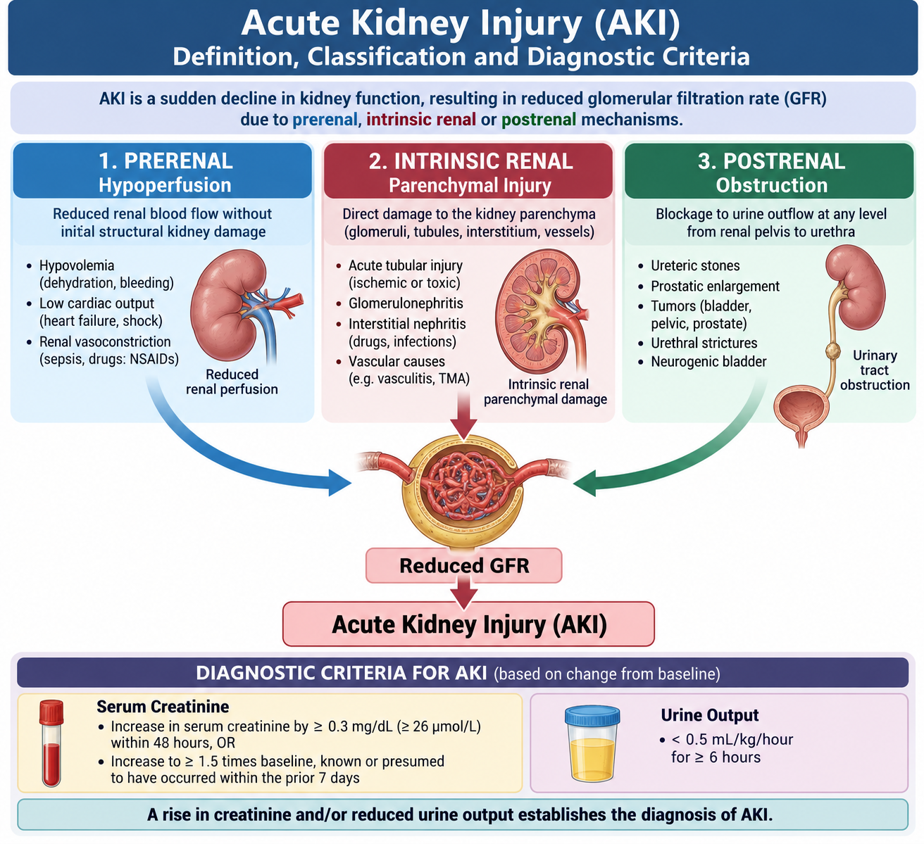
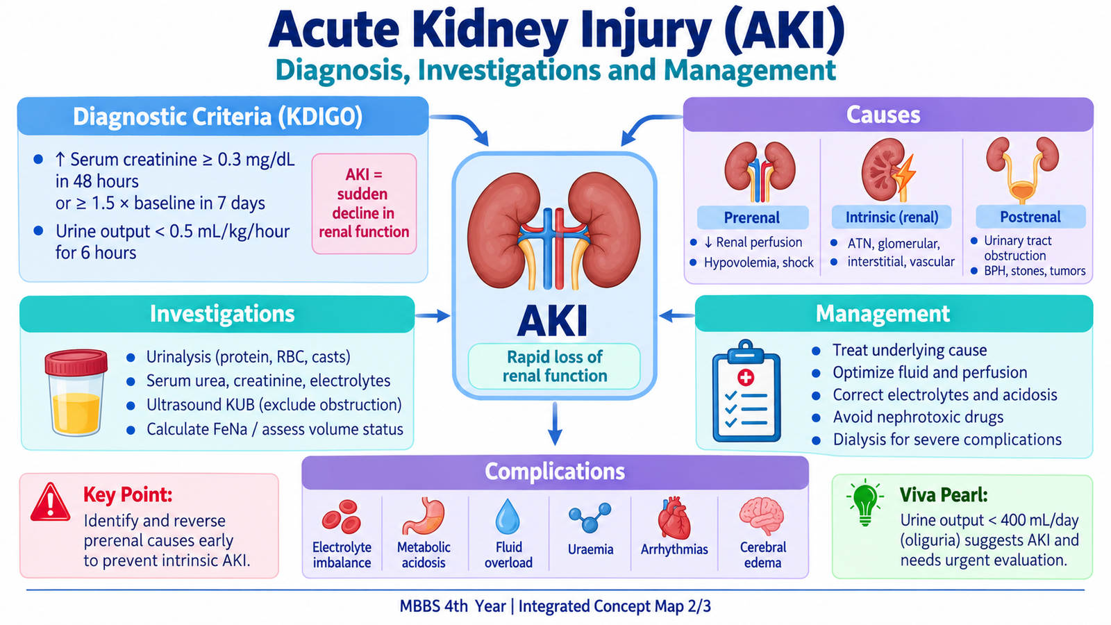
Acute kidney injury is a rapid decline in renal excretory function. The kidneys become unable to maintain normal control of waste products, water, electrolytes and acid–base balance. Serum creatinine may rise and urine output may fall, although an important point is that AKI can occur without marked oliguria. AKI is therefore a functional clinical syndrome rather than simply a low-urine-output state.
Diagnostic criteria
The commonly used diagnostic criteria identify AKI when there is an acute rise in serum creatinine or a sustained reduction in urine output. AKI is present when any one of the following occurs:
- An increase in serum creatinine of ≥0.3 mg/dL (≥26.5 µmol/L) within 48 hours.
- An increase in serum creatinine to ≥1.5 times the known or presumed baseline within the previous 7 days.
- Urine output <0.5 mL/kg/hour for at least 6 hours.
Creatinine is useful but does not rise immediately after renal injury, so early AKI may already be present before a large creatinine increase becomes visible. Urine output therefore provides important additional information.
Major etiological groups
The most useful first step is to decide whether the problem occurs before the kidney, within the kidney or after the kidney.
- Prerenal AKI: renal perfusion falls but the renal tissue is initially structurally intact. Common mechanisms include hypovolemia, reduced effective circulating volume and marked reduction in systemic blood pressure.
- Intrinsic renal AKI: the kidney itself is injured. Important categories include acute tubular injury, glomerular disease, interstitial disease and vascular injury.
- Postrenal AKI: urinary outflow is obstructed. Significant AKI usually requires bilateral obstruction or obstruction of the outflow from a solitary functioning kidney.
Reduced renal perfusion, renal tissue injury or urinary obstruction → fall in glomerular filtration rate → retention of creatinine and urea → disturbance of water, potassium and acid–base balance → clinical complications.
Prerenal AKI
In prerenal AKI, blood flow reaching the kidneys is insufficient to maintain normal filtration. Because the renal tubules are initially intact, they respond by conserving sodium and water. If severe hypoperfusion persists, ischemic tubular injury may develop and convert a functional prerenal problem into structural intrinsic AKI.
Postrenal AKI
Obstruction raises pressure proximal to the blockage. This pressure is transmitted toward the nephron and opposes glomerular filtration. Early relief can restore function, whereas prolonged obstruction may produce tubular damage and permanent renal impairment.
B. Acute Tubular Injury: Ischemic and Toxic Injury, Pathogenesis and Morphology
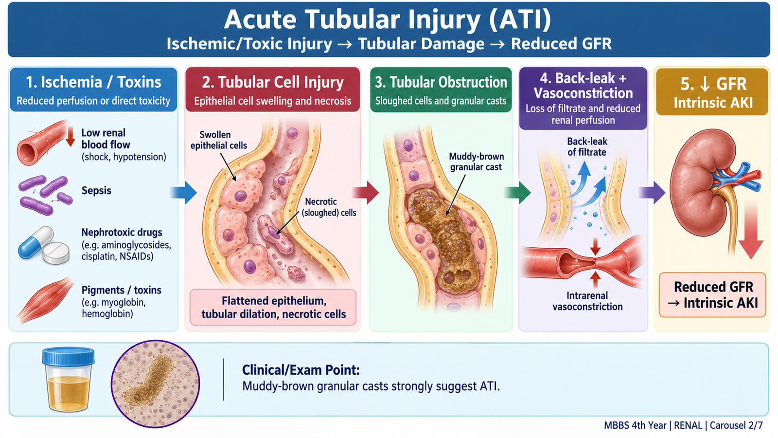
Acute tubular injury (ATI) is acute structural and functional damage to renal tubular epithelial cells and is an important cause of intrinsic AKI. The older term acute tubular necrosis is still commonly encountered, but “acute tubular injury” is more accurate because severe renal dysfunction may occur even when widespread tubular necrosis is not present.
Major causes
The two major mechanisms are ischemia and direct toxic injury. Both damage tubular cells, but their distribution and clinical setting differ.
- Ischemic ATI: follows severe or prolonged reduction of renal blood flow, such as major hypovolemia, shock or severe hemodynamic disturbance.
- Toxic ATI: follows direct tubular exposure to nephrotoxic substances, including selected drugs, pigments, chemicals or other toxins.
Pathogenesis
Tubular epithelial cells, particularly cells with high metabolic activity, are vulnerable to ischemia and toxins. Injury reduces ATP-dependent cellular functions, disturbs the cytoskeleton and causes loss of epithelial polarity and brush-border integrity. Injured cells may detach from the basement membrane and enter the tubular lumen.
Tubular obstruction increases pressure within the nephron, while disruption of the tubular epithelial barrier allows filtrate to leak back into the interstitium. At the same time, intrarenal vasoconstriction reduces glomerular blood flow. These processes combine to lower GFR.
Morphology
Gross morphology may be relatively nonspecific. The kidneys can appear swollen and pale, but the diagnostically important changes are microscopic.
Microscopic features include:
- Tubular epithelial simplification and flattening.
- Loss of the proximal tubular brush border.
- Cell swelling, degeneration and focal necrosis.
- Detachment and shedding of tubular epithelial cells.
- Dilatation of tubular lumina.
- Granular cellular debris and casts within tubules.
- Interstitial edema in injured areas.
Ischemic injury is often patchy and may affect different tubular segments, including the proximal straight tubule and thick ascending limb. Toxic injury often produces more prominent injury in the proximal tubule, because tubular cells concentrate and metabolize many filtered substances.
| Feature | Ischemic ATI | Toxic ATI |
|---|---|---|
| Main trigger | Prolonged reduction in renal perfusion | Direct nephrotoxic exposure |
| Distribution | Often patchy | May be more segment-specific |
| Commonly vulnerable area | Proximal straight tubule and thick ascending limb | Proximal tubular epithelium commonly affected |
| Shared consequence | Tubular dysfunction, casts, reduced GFR and AKI | |
Clinicopathological course
ATI is potentially reversible because surviving tubular epithelial cells can proliferate and restore the epithelial lining after the damaging stimulus is removed. Clinically, the course may pass through an initiation phase, a period of established renal dysfunction and a recovery phase. During recovery, urine output can increase before tubular concentrating ability is fully restored, so the patient may lose excessive water and electrolytes despite improving GFR.
C. Clinical Evaluation of AKI: Presentation, Investigations and Complications
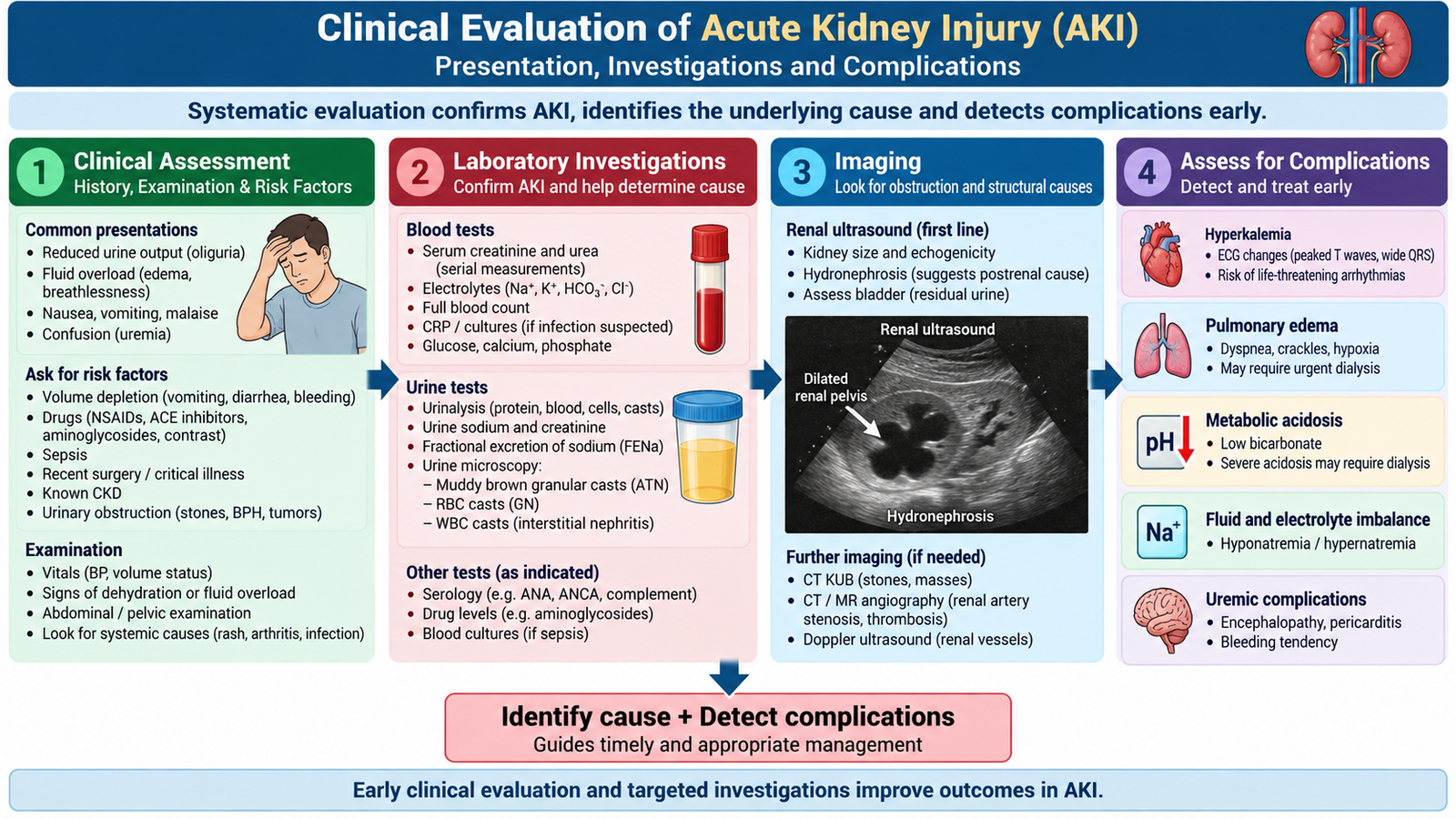
The clinical approach to AKI has two simultaneous goals: determine the severity of renal dysfunction and identify the reversible cause. The history, examination, urine findings and imaging are therefore interpreted together rather than relying on serum creatinine alone.
Clinical presentation
Patients may present because urine output has fallen, because the underlying illness is obvious, or because AKI is discovered on laboratory testing. Symptoms depend on the cause and on complications produced by loss of renal function.
- Oliguria or anuria.
- Thirst, postural symptoms or other evidence of volume depletion in prerenal states.
- Peripheral edema, raised jugular venous pressure, breathlessness or pulmonary crackles when fluid accumulates.
- Nausea, vomiting, poor appetite, lethargy or confusion with increasing uremic burden.
- Symptoms of the underlying disorder, such as infection, hemorrhage, obstruction or severe hypertension.
History
The history should focus on events capable of changing renal perfusion, directly injuring the kidney or obstructing urinary flow.
- Vomiting, diarrhea, bleeding, fever or poor fluid intake.
- Recent severe illness, hypotension, surgery or sepsis.
- Recent medicines or other potential nephrotoxic exposure.
- Urinary hesitancy, poor stream, retention, flank pain or other evidence of obstruction.
- Previous kidney disease, hypertension or known vascular disease.
Examination
Volume status is particularly important because both severe volume depletion and fluid overload can be dangerous. Blood pressure also provides clues to hypoperfusion, severe hypertension and underlying vascular disease.
- Pulse, blood pressure and peripheral perfusion.
- Dry mucosa, reduced skin turgor and postural hypotension when volume depleted.
- Peripheral edema, raised JVP and pulmonary crackles when fluid overloaded.
- Bladder distension where urinary retention is suspected.
- Features of systemic illness or severe hypertension.
Investigations
Investigation begins with confirmation of renal dysfunction and assessment of immediately dangerous consequences. Urine examination then helps localize the renal process, while imaging is particularly important when obstruction is possible.
- Serum creatinine and urea: assess renal dysfunction and trend over time.
- Electrolytes: particularly potassium, sodium and bicarbonate.
- Urinalysis: blood, protein, leukocytes and other abnormalities may suggest the underlying renal lesion.
- Urine sediment: muddy-brown granular casts and tubular epithelial cells support ATI; red-cell casts suggest glomerular injury.
- ECG: urgently required when significant hyperkalemia is present or suspected.
- Renal ultrasonography: assesses kidney size and looks for hydronephrosis or another obstructive cause.
- Additional tests are selected according to the suspected underlying disease rather than performed routinely in every patient.
Urinary indices
In uncomplicated prerenal AKI, intact tubules conserve sodium and water. Urine therefore tends to be concentrated and urine sodium is low. In established ATI, damaged tubules lose part of this concentrating and sodium-retaining ability. Fractional excretion of sodium (FENa) below about 1% supports prerenal physiology, whereas a value above about 2% is more typical of ATI. These values are supportive rather than absolute, and diuretics and several other clinical conditions can reduce their reliability.
Complications
Complications arise because the kidneys can no longer adequately remove potassium, hydrogen ions, water and nitrogenous waste products.
- Hyperkalemia: impaired potassium excretion may cause life-threatening cardiac arrhythmias.
- Metabolic acidosis: reduced acid excretion causes hydrogen-ion accumulation.
- Fluid overload: sodium and water retention may cause pulmonary edema.
- Uremic manifestations: increasing toxin retention may produce encephalopathy, pericardial inflammation, nausea and other systemic effects.
- Hyponatremia: may occur when water intake exceeds the kidney’s reduced ability to excrete water.
- Drug accumulation: medicines cleared through the kidneys may reach toxic concentrations unless doses are reviewed.
D. Management of AKI, Oliguria and Anuria: Acute Care and Primary-Care Referral
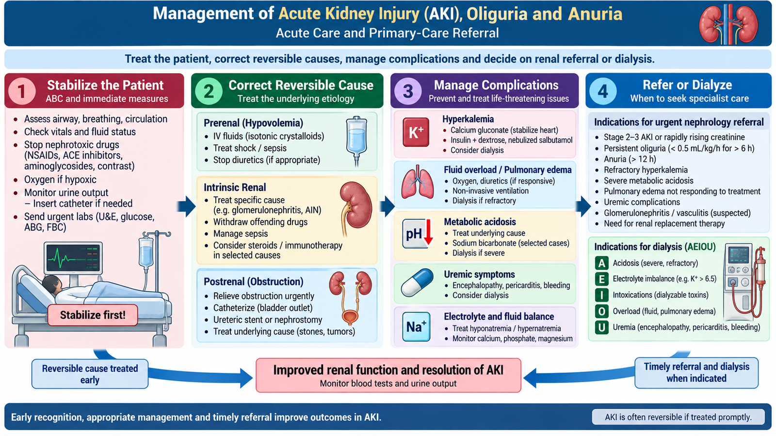
Management of AKI is based on correcting reversible causes while protecting the patient from complications until renal function recovers. There is no single treatment that reverses every form of AKI. The first questions are therefore: Is the patient hemodynamically stable? Is there obstruction? Is a dangerous electrolyte or fluid problem present? Is there a continuing renal insult?
Immediate priorities
- Assess airway, breathing and circulation in an acutely unwell patient.
- Measure blood pressure, perfusion and volume status.
- Monitor urine output and renal function.
- Measure serum potassium, sodium and bicarbonate.
- Identify and stop avoidable nephrotoxic agents where clinically appropriate.
- Review medicines whose elimination depends on kidney function.
- Search promptly for urinary obstruction.
Fluid management
A patient who is genuinely hypovolemic requires appropriate fluid replacement because restoration of effective circulating volume can reverse prerenal AKI and prevent progression to ischemic ATI. In contrast, indiscriminate fluid administration to a patient who is already euvolemic or overloaded can worsen pulmonary edema. Fluid therapy must therefore be guided by clinical volume assessment and repeated reassessment.
Oliguria and anuria
Oliguria means abnormally low urine output and is clinically important in AKI when output falls below 0.5 mL/kg/hour for a sustained period. Anuria means near-complete absence of urine production. New anuria is particularly concerning because complete urinary obstruction, severe renal vascular compromise or severe intrinsic renal failure must be considered.
When oliguria or anuria is detected:
- Confirm that urine output has truly fallen and that measurements are reliable.
- Assess circulation and hydration.
- Look for bladder distension or catheter obstruction if a catheter is present.
- Check serum creatinine, potassium and acid–base status.
- Arrange renal tract imaging when obstruction is possible.
- Do not assume that low urine output automatically means the patient needs more fluid.
Renal replacement therapy
Dialysis or another form of renal replacement therapy is considered when complications cannot be controlled adequately by medical treatment. Important indications include:
- Refractory severe hyperkalemia.
- Severe metabolic acidosis not responding to appropriate treatment.
- Pulmonary edema or severe fluid overload not responding to conservative measures.
- Important uremic complications such as encephalopathy or pericarditis.
- Selected severe toxin exposures when the substance can be removed by dialysis.
Family Medicine and primary-care recognition
In primary care, AKI may first appear as reduced urine output after vomiting, diarrhea, fever, poor intake or a recent illness. It may also be discovered after laboratory testing or after the introduction of medicines that alter renal perfusion or are nephrotoxic. Primary-care management begins with recognizing instability, assessing volume status, identifying reversible factors and deciding who needs urgent hospital assessment.
Urgent referral red flags
- New or persistent anuria.
- Severe or rapidly worsening oliguria.
- Significant or ECG-associated hyperkalemia.
- Pulmonary edema or significant breathlessness from fluid overload.
- Severe metabolic acidosis.
- Confusion, seizures, pericarditic symptoms or other major uremic manifestations.
- Rapidly increasing creatinine or rapidly worsening renal function.
- Suspected bilateral urinary obstruction.
- Severe hypertension with evidence of acute target-organ injury.
- AKI associated with major systemic illness or uncertain cause.
In chronic renal disease, urgent or specialist assessment is also important when renal function is declining rapidly, hypertension remains difficult to control, significant proteinuria or hematuria persists, recurrent hyperkalemia develops, fluid overload becomes difficult to control, or a systemic renal disease is suspected.
E. Renal Vascular Disorders: Nephrosclerosis, Malignant Hypertension and Renal Artery Stenosis
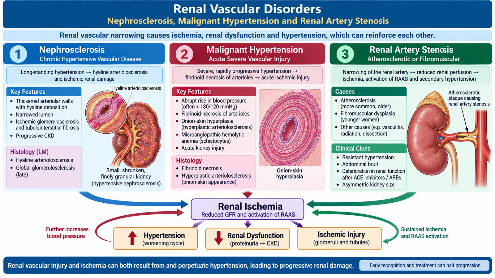
Renal blood vessels are both a target and a cause of hypertension. Long-standing hypertension gradually damages small renal arteries and arterioles, whereas severe accelerated hypertension can produce acute vascular injury. Narrowing of a major renal artery can also reduce renal perfusion and activate mechanisms that raise systemic blood pressure.
Benign nephrosclerosis
Benign nephrosclerosis is the pattern of chronic renal vascular injury associated particularly with long-standing hypertension. Persistent pressure damages small arteries and arterioles. Plasma proteins leak into vessel walls and extracellular matrix increases, producing hyaline arteriolosclerosis. The vascular lumen becomes narrower, causing chronic ischemia of the renal parenchyma.
Morphology:
- The kidneys may become symmetrically contracted in advanced disease.
- The cortical surface develops a fine, diffuse granularity.
- Microscopy shows hyaline thickening and narrowing of arterioles, with secondary ischemic changes in glomeruli and tubulointerstitium.
Malignant hypertension and malignant nephrosclerosis
Severe hypertension can cause rapid injury to the renal microcirculation. Endothelial damage increases vascular permeability and may cause fibrinoid necrosis of arterioles. In other vessels, smooth-muscle proliferation and concentric wall thickening produce hyperplastic arteriolosclerosis, classically described as an “onion-skin” appearance.
Important morphology:
- Fibrinoid necrosis of arterioles in severe acute injury.
- Concentric hyperplastic thickening of arteriolar walls.
- Narrowed vascular lumina and renal ischemia.
- Small cortical hemorrhages may produce the classic gross “flea-bitten” appearance.
Severe renal vascular injury may rapidly reduce renal function and produce hematuria, proteinuria and AKI. Because hypertension further injures already narrowed vessels, a self-amplifying cycle of ischemia and renin-mediated vasoconstriction can develop.
Renal artery stenosis
Renal artery stenosis reduces blood flow to one or both kidneys. The affected kidney interprets reduced perfusion as reduced circulating pressure and activates the renin–angiotensin–aldosterone system. Angiotensin II increases vasoconstriction and aldosterone promotes sodium retention, contributing to secondary hypertension.
Important causes include:
- Atherosclerotic renal artery disease: more common in older patients with generalized vascular disease and often affects the proximal renal artery.
- Fibromuscular dysplasia: a non-atherosclerotic vascular disorder classically affecting younger women.
Clinical clues include difficult-to-control or abruptly worsening hypertension, renal dysfunction that cannot otherwise be explained and deterioration in renal function after drugs that reduce angiotensin-II-dependent glomerular pressure, particularly when significant bilateral renal artery stenosis is present. Duplex ultrasonography, CT angiography or MR angiography may be used according to the clinical situation.
F. Sodium Disorders: Hyponatremia and Hypernatremia
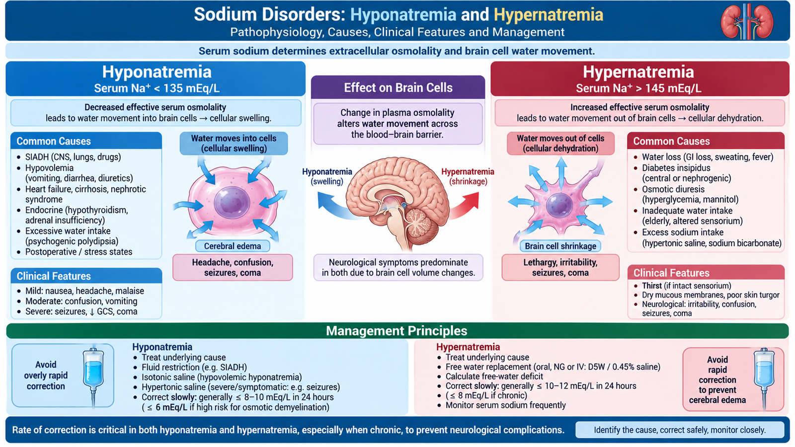
Serum sodium concentration mainly reflects the relationship between total body sodium and total body water. Therefore, a low serum sodium concentration does not always mean that the body contains too little sodium, and a high concentration usually reflects a deficit of water relative to sodium. The severity of neurological symptoms depends not only on the sodium level but also on how rapidly the abnormality developed.
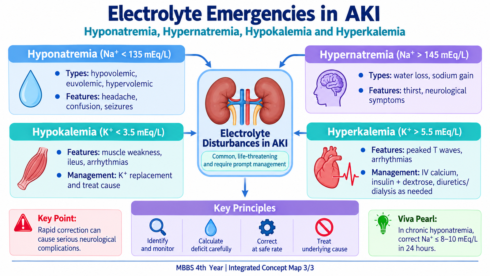
Hyponatremia
Hyponatremia is a reduced serum sodium concentration. Most clinically important cases are hypotonic, meaning plasma osmolality is also low. Water then moves into cells. Brain-cell swelling explains headache, confusion, seizures and, in severe acute cases, cerebral edema.
Types of hypotonic hyponatremia
- Hypovolemic: both sodium and water are lost, but sodium loss is proportionally greater.
- Euvolemic: body water increases without obvious edema; inappropriate antidiuretic activity is an important mechanism.
- Hypervolemic: total body sodium is increased, but total body water increases even more, producing edema and dilutional hyponatremia.
Measured hyponatremia should also be interpreted with serum osmolality and glucose because hyperglycemia can produce hypertonic hyponatremia through movement of water from cells into extracellular fluid. Some laboratory circumstances can produce pseudohyponatremia without true hypotonicity.
Clinical features
Neurological symptoms are most important. Rapidly developing hyponatremia is more dangerous because the brain has less time to adapt.
- Nausea and headache.
- Lethargy and confusion.
- Muscle cramps or weakness.
- Seizures and reduced consciousness in severe symptomatic cases.
Investigation
- Repeat serum sodium when necessary and assess the clinical context.
- Serum osmolality.
- Blood glucose.
- Urine osmolality.
- Urine sodium.
- Renal function and relevant assessment of volume status.
Urine osmolality shows whether the kidney is appropriately excreting dilute urine. Urine sodium can then help distinguish renal sodium loss from low effective circulating volume or other mechanisms, but interpretation must always be made in context.
Estimated sodium deficit
This calculation is most meaningful when a true sodium deficit is present. Hyponatremia caused mainly by water excess is not corrected simply by replacing the calculated amount of sodium.
Management and correction rate
Treatment depends on symptoms, duration and volume status. Severe neurological symptoms require urgent treatment in a monitored setting. Hypertonic saline is used for severe symptomatic hyponatremia, with the immediate aim of producing only enough sodium rise to improve dangerous cerebral symptoms.
- Severe symptomatic hyponatremia: controlled hypertonic saline therapy with close sodium monitoring; an initial rise of about 4–6 mmol/L is usually sufficient to improve acute cerebral symptoms.
- Chronic or duration-unknown hyponatremia: correction must be slow and controlled. A conservative limit of about 8 mmol/L in 24 hours is used to reduce the risk of osmotic demyelination, especially in high-risk patients.
- Hypovolemic hyponatremia: restore circulating volume appropriately.
- Euvolemic water excess: treatment commonly includes restriction of free-water intake and treatment of the cause.
- Hypervolemic hyponatremia: manage the underlying disorder and excess water/sodium balance.
Hypernatremia
Hypernatremia usually represents loss of water in excess of sodium. Increased extracellular osmolality draws water out of cells. Brain-cell dehydration produces the major neurological manifestations.
Types
- Hypovolemic hypernatremia: loss of both sodium and water, with proportionally greater water loss.
- Euvolemic hypernatremia: predominantly free-water loss.
- Hypervolemic hypernatremia: sodium gain exceeds water gain; this is less common.
Clinical features and investigations
Patients may have intense thirst, weakness, irritability, confusion, hyperreflexia, seizures or reduced consciousness. Examination should determine whether extracellular volume is depleted, normal or expanded. Serum osmolality, urine volume, urine osmolality, electrolytes and renal function help determine whether water loss is renal or extrarenal.
Free-water deficit
Total body water is commonly estimated as a fraction of body weight, with a lower proportion in older adults and in people with less lean body mass. The calculation provides an estimate; ongoing losses and clinical reassessment must also be considered.
Management and correction rate
If the patient has significant circulatory volume depletion, restoration of adequate perfusion takes priority. Once circulation is stabilized, the free-water deficit is corrected. In chronic hypernatremia or hypernatremia of uncertain duration, serum sodium is generally lowered gradually, commonly by no more than about 0.5 mmol/L per hour and approximately 10 mmol/L in 24 hours. Excessively rapid correction after cerebral adaptation may cause cerebral edema.
G. Potassium Disorders: Hypokalemia and Hyperkalemia
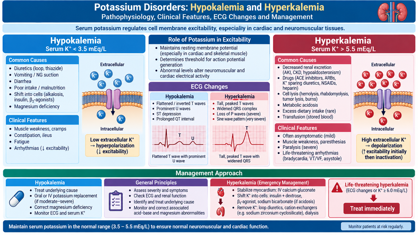
Potassium is essential for normal resting membrane potential in skeletal muscle, smooth muscle and cardiac cells. Significant changes in extracellular potassium therefore alter neuromuscular and cardiac electrical activity. In renal disease, hyperkalemia is particularly important because impaired renal excretion may allow potassium to rise rapidly.
Hypokalemia
Hypokalemia results from potassium loss, movement of potassium into cells or, less commonly, inadequate intake. Renal and gastrointestinal losses are common mechanisms. Magnesium deficiency is important because it promotes renal potassium loss and can make hypokalemia difficult to correct.
Clinical features
- Muscle weakness and fatigue.
- Muscle cramps.
- Reduced gastrointestinal motility in more marked deficiency.
- Cardiac arrhythmias, particularly when hypokalemia is severe or heart disease is present.
ECG changes
Typical changes may include ST depression, flattened or inverted T waves and prominent U waves. Electrical abnormalities become clinically important because they indicate altered myocardial repolarization and increased arrhythmic risk.
Investigation and management
Serum potassium should be interpreted together with renal function, acid–base status, magnesium and the clinical context. Urinary potassium assessment may help distinguish renal potassium wasting from extrarenal loss when the cause is uncertain.
- Treat the underlying cause.
- Replace potassium, with oral replacement preferred when clinically appropriate.
- Use intravenous potassium for selected severe or symptomatic cases under monitored conditions.
- Correct associated magnesium deficiency when present.
- Monitor potassium and ECG when the disturbance is severe.
Potassium must never be given as a rapid intravenous push because abrupt elevation of serum potassium can cause fatal arrhythmias.
Hyperkalemia
Hyperkalemia may result from reduced renal potassium excretion, release of potassium from cells or redistribution from the intracellular to extracellular compartment. AKI is an important cause because falling GFR and tubular dysfunction reduce potassium elimination.
Clinical features
Patients may have weakness or paresthesia, but dangerous hyperkalemia can also be clinically silent until a cardiac arrhythmia develops. The absence of symptoms therefore does not exclude serious risk.
ECG progression
Classically, potassium elevation may produce tall peaked T waves, progressive PR prolongation, loss of P waves and QRS widening. Severe toxicity can progress to a sine-wave pattern, ventricular arrhythmia or cardiac arrest. ECG findings are not sufficiently sensitive to safely exclude dangerous hyperkalemia when potassium is markedly elevated.
Emergency management principles
Treatment has three separate goals: protect the heart, temporarily move potassium into cells and remove potassium from the body.
Intravenous calcium is used when there are important ECG changes or other features requiring urgent membrane stabilization. Calcium does not lower serum potassium; it reduces the immediate myocardial electrical danger.
Insulin with glucose lowers extracellular potassium by stimulating cellular potassium uptake. A beta-2 agonist can provide an additional intracellular shift. Bicarbonate may be useful particularly when significant metabolic acidosis is present.
Definitive potassium removal depends on the clinical situation and may include renal excretion when adequate urine output is present, gastrointestinal potassium-removal strategies, or dialysis when hyperkalemia is severe or refractory.
Because intracellular-shift therapies are temporary, potassium must be rechecked and the underlying cause treated. Rebound hyperkalemia may occur if potassium has not actually been removed from the body.
Integrated Mechanism Flow
Important Comparison: Prerenal AKI, Acute Tubular Injury and Postrenal AKI
The distinction is clinically useful because prerenal hypoperfusion and urinary obstruction may be rapidly reversible, while ATI represents established structural tubular injury. No single laboratory result should be interpreted in isolation.
| Feature | Prerenal AKI | Acute Tubular Injury | Postrenal AKI |
|---|---|---|---|
| Main problem | Reduced renal perfusion | Tubular epithelial injury | Urinary obstruction |
| Tubular function | Initially preserved | Impaired | Variable |
| Urine sediment | Often bland | Granular “muddy-brown” casts may occur | Variable |
| FENa | Often <1% | Often >2% | Variable |
| Imaging clue | No obstructive change | Usually no obstruction | Hydronephrosis may be present |
| Key intervention | Restore effective renal perfusion | Remove insult and provide supportive care | Relieve obstruction |
⭐ AIM High-Yield Review
🎥 Video Learning — Acute Kidney Injury (AKI)
Review the causes, pathophysiology, clinical features, complications, diagnostic approach and management of acute kidney injury.