
Topic 4 — Chronic Kidney Disease, Renal Replacement Therapy and Renal Transplantation
1. Topic Introduction
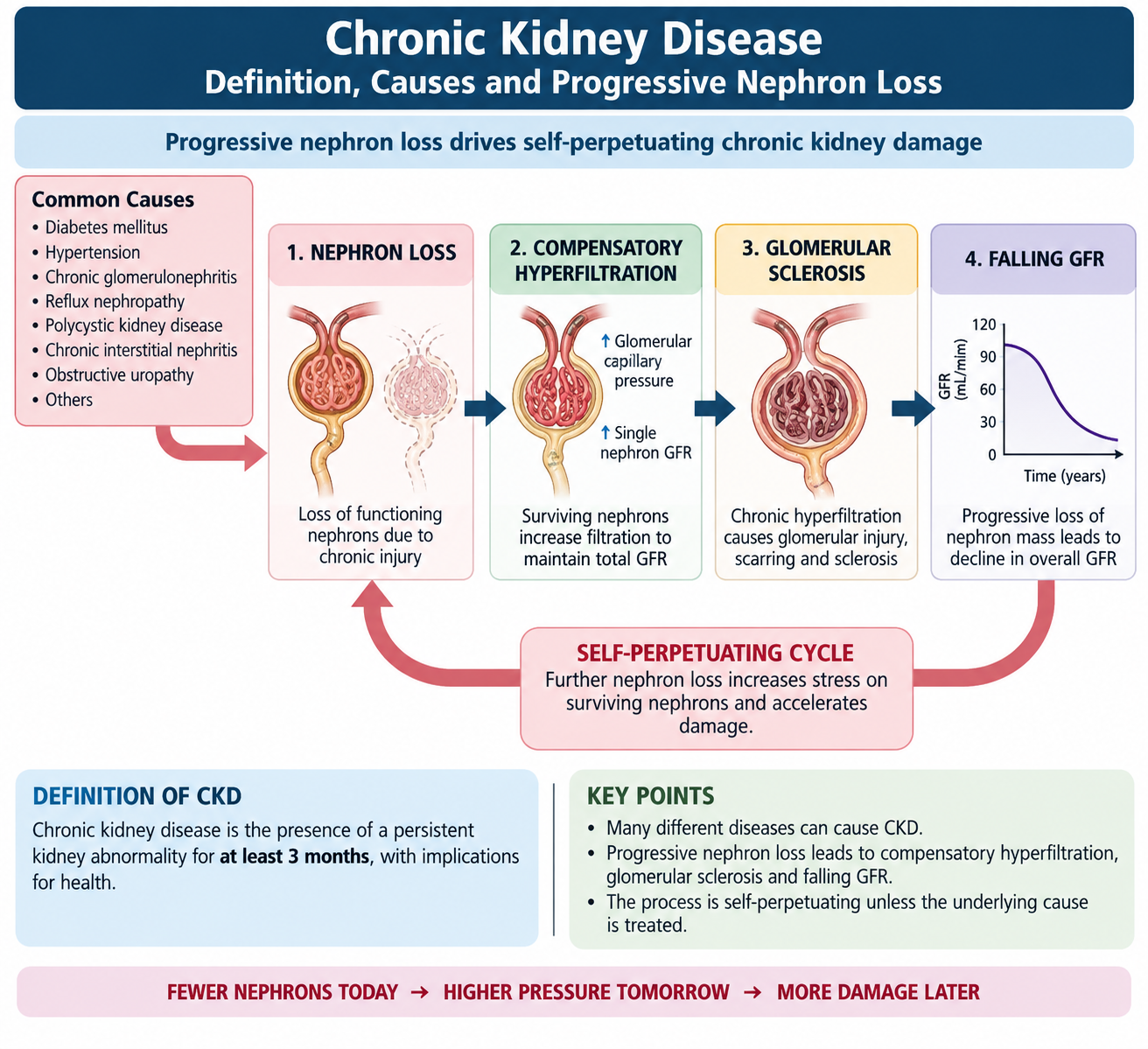
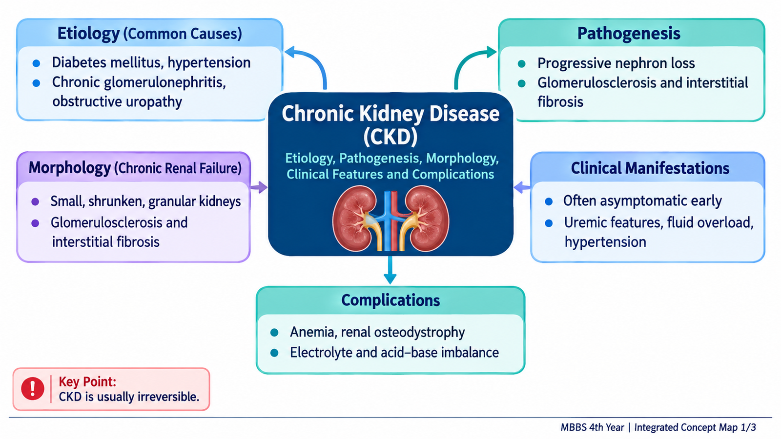
Chronic kidney disease (CKD) is a long-standing abnormality of kidney structure or function that persists for at least three months and has consequences for health. As functioning nephrons are progressively lost, the remaining nephrons initially compensate, but continued stress eventually causes further damage and a fall in glomerular filtration. CKD may therefore progress from an asymptomatic disorder detected by urine or blood tests to advanced kidney failure with fluid, electrolyte, metabolic, hematological and systemic complications. This chapter explains the causes, pathology, clinical recognition, investigations and treatment of CKD, followed by dialysis and renal transplantation. It also covers transplant rejection, immunosuppressive therapy, post-transplant care, transplantation ethics and HOTA, and the management of CKD and renal transplant recipients during pregnancy.
A. Chronic Kidney Disease: Definition, Causes and Progressive Nephron Loss
CKD is not a single disease. It is the final clinical state produced by many disorders that cause persistent structural kidney damage, persistent functional impairment, or both. The important idea is duration: a temporary fall in kidney function is not CKD unless the abnormality persists for at least three months. CKD may be present even when the glomerular filtration rate is still relatively preserved if there is convincing evidence of kidney damage.
Definition and Diagnostic Criteria
CKD is defined by abnormalities of kidney structure or function present for at least three months with implications for health. It can be diagnosed when either of the following persists:
- Reduced glomerular filtration: estimated GFR (eGFR) below 60 mL/min/1.73 m².
- Markers of kidney damage: for example persistent albuminuria or proteinuria, abnormal urinary sediment, structural abnormalities on imaging, abnormalities demonstrated by renal histology, or persistent tubular abnormalities.
A patient can therefore have CKD with an eGFR above 60 if persistent structural or urinary evidence of kidney disease is present.
GFR Categories
| Category | eGFR (mL/min/1.73 m²) | Functional Description |
|---|---|---|
| G1 | ≥90 | Normal or high GFR; CKD requires another marker of kidney damage |
| G2 | 60–89 | Mild reduction; CKD requires another marker of damage |
| G3a | 45–59 | Mild to moderate reduction |
| G3b | 30–44 | Moderate to severe reduction |
| G4 | 15–29 | Severe reduction |
| G5 | <15 | Kidney failure |
Important Causes
CKD develops when a disease causes continuing nephron injury or leaves too few functioning nephrons to maintain normal kidney function. Common etiological groups include:
- Diabetic kidney disease.
- Hypertensive renal disease.
- Chronic glomerular diseases.
- Chronic tubulointerstitial diseases.
- Chronic obstruction or reflux-related kidney damage.
- Hereditary and cystic kidney disorders such as polycystic kidney disease.
Pathogenesis: Why CKD Progresses
Different diseases may begin in different parts of the kidney, but progressive CKD tends to enter a common pathway. When some nephrons are destroyed, surviving nephrons increase their filtration to compensate. This adaptive hyperfiltration initially helps maintain total GFR, but it also increases pressure and workload within the remaining glomeruli. Persistent intraglomerular stress promotes protein leakage, glomerular injury and sclerosis. Tubular injury and interstitial inflammation then promote fibrosis, causing further irreversible nephron loss.
Nephron loss → compensatory hyperfiltration in surviving nephrons → increased intraglomerular stress → protein leakage and glomerular sclerosis → tubular/interstitial injury and fibrosis → further nephron loss → progressive fall in GFR.
This explains why CKD may continue to progress even after the original disease has become less active: damage to sufficient numbers of nephrons can itself generate further nephron stress.
B. Chronic Renal Failure: Morphology, Clinical Manifestations and Complications
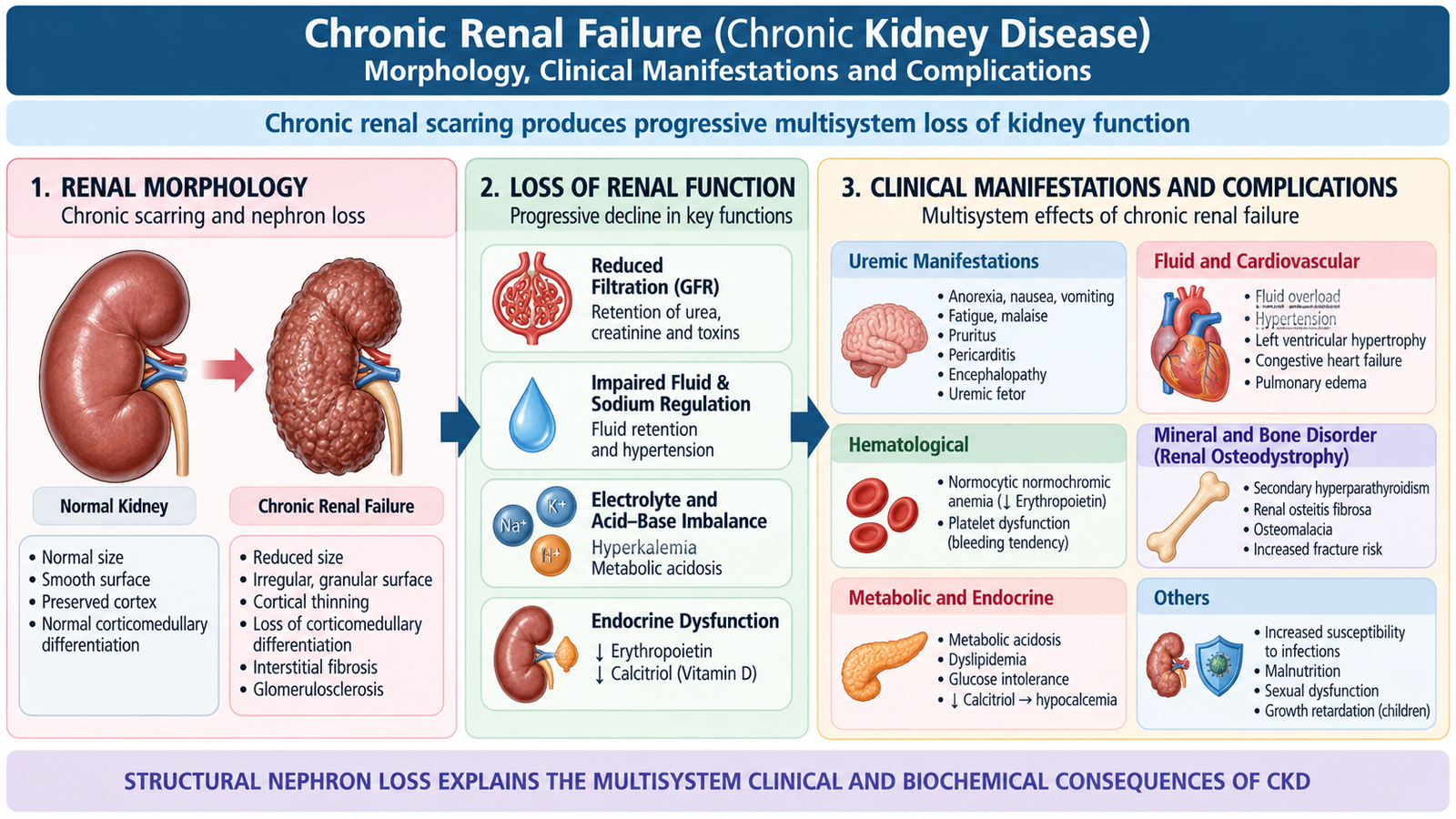
As CKD progresses, repeated nephron injury produces structural scarring together with loss of the kidney’s filtration, regulatory and endocrine functions. The pathology therefore explains many of the patient’s clinical findings: reduced filtration causes retention of waste products, impaired salt and water handling causes edema and hypertension, reduced erythropoietin contributes to anemia, and disturbed vitamin D and phosphate handling produces CKD-mineral and bone disorder.
Gross Morphology
Many causes of advanced chronic renal failure ultimately produce bilaterally small, contracted kidneys. Cortical thinning and an irregular or finely granular external surface reflect chronic loss and scarring of renal parenchyma. This appearance is not universal; kidney size may remain normal or become enlarged in some underlying diseases, so renal size must always be interpreted with the clinical context.
Microscopic Morphology
The final common microscopic pattern is chronic nephron destruction and scarring. Major changes include:
- Glomerulosclerosis: replacement of functioning glomerular capillary structures by scar tissue.
- Tubular atrophy: loss and shrinkage of renal tubules as damaged nephrons become nonfunctional.
- Interstitial fibrosis: progressive deposition of fibrous tissue between the remaining tubules.
- Chronic interstitial inflammation: may accompany ongoing tissue injury.
- Arterial and arteriolar sclerosis: vascular narrowing contributes to chronic ischemia and further nephron loss.
These changes form a self-reinforcing process: chronic vascular and glomerular injury reduces blood supply and filtration, while tubular atrophy and interstitial fibrosis reduce the amount of functioning renal tissue that remains.
Clinical Manifestations
Early CKD may be clinically silent. Symptoms become more obvious as functional renal reserve falls and complications develop.
- Fatigue and weakness: often related to anemia and accumulation of metabolic waste.
- Nocturia: damaged kidneys lose their ability to concentrate urine effectively.
- Edema and hypertension: impaired sodium and water excretion expands extracellular fluid volume.
- Anorexia, nausea and vomiting: may occur with advanced uremia.
- Pruritus: is common in advanced CKD and is associated with the uremic and mineral-bone disturbance.
- Breathlessness: may result from anemia, fluid overload or pulmonary edema.
- Neurological symptoms: advanced uremia can produce impaired concentration, neuropathy, confusion and eventually encephalopathy.
Major Complications and Why They Occur
Uremia: Severe loss of filtration causes accumulation of nitrogenous and other retained substances. Unlike simple azotemia, uremia refers to the clinical syndrome caused by advanced renal failure and may involve the gastrointestinal, neurological, cardiovascular and hematological systems.
Anemia: Diseased kidneys produce less erythropoietin, reducing bone-marrow stimulation of red-cell production. Iron deficiency, inflammation and shortened red-cell survival may further aggravate the anemia.
CKD-mineral and bone disorder: Reduced phosphate excretion causes phosphate retention, while diseased kidneys produce less active vitamin D. Calcium balance is disturbed and parathyroid hormone rises, producing secondary hyperparathyroidism and renal bone disease.
Metabolic acidosis: Progressive nephron loss reduces renal acid excretion and regeneration of bicarbonate, allowing acid to accumulate.
Hyperkalemia: Advanced renal impairment reduces potassium excretion. Severe hyperkalemia is dangerous because it may disturb cardiac conduction and cause life-threatening arrhythmia.
Fluid overload: Failure to excrete sodium and water produces peripheral edema, worsening hypertension and, in severe cases, pulmonary edema.
Cardiovascular disease: Hypertension, volume overload, anemia, metabolic disturbance and vascular disease together contribute substantially to cardiovascular morbidity in CKD.
Uremic pericarditis and encephalopathy: These are manifestations of severe uremia and are important indications that renal replacement therapy may be required urgently.
C. Clinical Evaluation, Investigations and Treatment of CKD
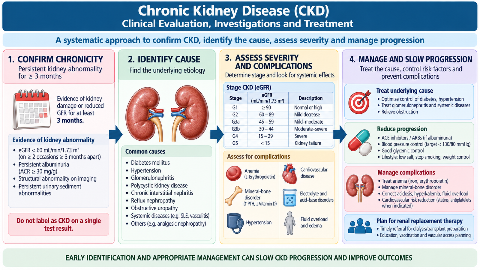
Clinical evaluation of CKD has four practical aims: confirm that kidney disease is chronic, identify its cause, determine the degree of functional impairment, and detect complications that require treatment. A single elevated creatinine value does not establish CKD; previous laboratory results, persistent urine abnormalities and imaging findings help demonstrate chronicity.
History and Examination
History should look for diabetes, hypertension, previous glomerular or urinary disease, recurrent obstruction, inherited kidney disease, previous episodes of kidney injury, and exposure to potentially nephrotoxic drugs. The clinician should also ask about edema, change in urine pattern, fatigue, anorexia, nausea, pruritus, breathlessness and neurological symptoms.
Examination should assess blood pressure, hydration and volume status, peripheral edema, signs of anemia and evidence of systemic disease. Breathlessness with basal crepitations may indicate pulmonary fluid overload, while confusion or a pericardial rub in advanced renal failure suggests serious uremic complications.
Core Investigations
- Serum creatinine and eGFR: estimate the degree of filtration impairment and allow assessment of progression over time.
- Urinalysis: detects protein, blood and other abnormalities that may suggest the underlying renal lesion.
- Urinary albumin or protein assessment: persistent protein loss is both evidence of kidney damage and a marker of progression risk.
- Serum electrolytes and bicarbonate: detect hyperkalemia, acid-base disturbance and other metabolic consequences.
- Complete blood count: identifies anemia.
- Calcium, phosphate and related mineral assessment: helps evaluate CKD-mineral and bone disorder.
- Renal ultrasonography: assesses kidney size and architecture and looks for obstruction or structural disease.
Small echogenic kidneys with cortical thinning support a chronic process. However, a normal or enlarged kidney does not exclude CKD because some underlying disorders do not produce contracted kidneys.
Renal biopsy is considered when identifying the renal lesion would change diagnosis or management. It is not routinely useful in an end-stage, markedly scarred kidney where the histology may show only nonspecific chronic damage.
Treatment Principles
CKD management aims to treat the underlying disease where possible, slow additional nephron loss, control complications and prepare appropriately for renal replacement therapy if kidney failure progresses.
Control the underlying cause and progression. Good blood-pressure control reduces further vascular and glomerular injury. In proteinuric kidney disease, renin-angiotensin system blockade with an ACE inhibitor or angiotensin receptor blocker may reduce intraglomerular pressure and proteinuria when clinically appropriate. Good glycemic management is important in diabetic kidney disease, and suitable patients may benefit from kidney-protective therapies such as SGLT2 inhibitors.
Avoid additional renal injury. Potentially nephrotoxic medicines should be avoided or used cautiously, and doses of renally eliminated drugs may need adjustment as renal function declines.
Control fluid and salt retention. Dietary sodium moderation and diuretics, particularly loop diuretics when appropriate, can help control edema and volume-related hypertension. Diuretics can improve fluid balance but do not restore lost nephrons.
Treat anemia. Iron deficiency should be identified and corrected. Erythropoiesis-stimulating therapy may be used in selected patients when anemia is primarily related to reduced renal erythropoietin production.
Treat mineral and bone abnormalities. Management may include control of phosphate balance, phosphate-binding therapy and vitamin D-related treatment according to the patient’s biochemical abnormalities.
Correct significant metabolic acidosis. Alkali therapy may be used in suitable patients with persistent metabolic acidosis.
Plan ahead for advanced kidney failure. A patient with progressive advanced CKD should be referred early enough to discuss dialysis options, vascular or peritoneal access planning and possible kidney transplantation. Renal replacement should not be delayed until a life-threatening emergency develops.
D. Renal Replacement Therapy and Dialysis
Renal replacement therapy (RRT) provides essential functions that the patient’s kidneys can no longer perform adequately. The major forms are hemodialysis, peritoneal dialysis and kidney transplantation. Dialysis mainly replaces excretory and fluid-regulatory functions; it does not reproduce every endocrine and metabolic function of a normal kidney. Kidney transplantation provides the most complete form of renal replacement in a suitable recipient.
Hemodialysis
In hemodialysis, blood is circulated through an extracorporeal dialyzer. A semipermeable membrane separates blood from dialysis fluid. Small waste solutes move mainly by diffusion down concentration gradients, while excess water is removed by ultrafiltration.
Reliable vascular access is essential. An arteriovenous fistula is generally preferred for long-term hemodialysis because it provides durable high-flow access and has a lower infection risk than a long-term central venous catheter. A catheter may nevertheless be necessary when dialysis is required before permanent access is available.
Peritoneal Dialysis
Peritoneal dialysis uses the patient’s peritoneal membrane as the dialysis surface. Dialysis fluid is introduced into the peritoneal cavity, where solutes diffuse between peritoneal capillary blood and dialysate. An osmotic gradient promotes removal of excess water. The fluid is then drained and replaced.
This treatment avoids extracorporeal circulation and can be performed repeatedly outside a dialysis unit, but successful therapy requires careful catheter care and attention to infection prevention.
When Dialysis Is Indicated
The decision to start dialysis is based on the patient’s clinical condition rather than on a creatinine value alone. Important indications include complications that cannot be controlled adequately by conservative treatment:
- Refractory hyperkalemia.
- Severe or refractory metabolic acidosis.
- Fluid overload causing pulmonary edema despite appropriate medical treatment.
- Uremic complications such as encephalopathy or pericarditis.
- Progressive symptomatic uremia with major deterioration in general condition.
- Selected dialyzable toxic substances in appropriate poisoning situations.
Complications of Hemodialysis
Rapid movement of water and solutes during hemodialysis explains several important complications.
- Hypotension: excessive or rapid intravascular volume removal reduces circulating volume.
- Muscle cramps: may accompany rapid fluid and electrolyte shifts.
- Vascular-access infection: particularly important with dialysis catheters.
- Access thrombosis or stenosis: reduces effective blood flow through the access.
- Dialysis disequilibrium: rapid changes in plasma osmolality can cause cerebral symptoms, particularly during initial or very intensive dialysis.
Complications of Peritoneal Dialysis
- Peritonitis: presents characteristically with abdominal symptoms and cloudy dialysis effluent and requires prompt treatment.
- Catheter exit-site or tunnel infection.
- Protein loss: proteins can be lost into dialysate.
- Hernias and abdominal-wall problems: repeated dialysate filling increases intra-abdominal pressure.
- Metabolic effects: glucose-containing dialysate may contribute to weight gain and disturbances of glucose control.


E. Renal Transplantation: Indications, Evaluation and Prerequisites
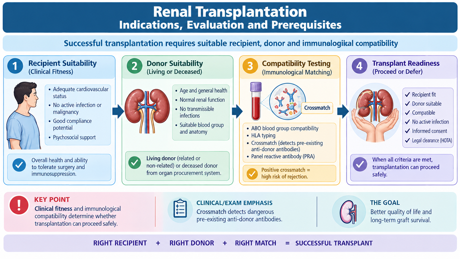
Kidney transplantation places a functioning donor kidney into a patient with irreversible advanced kidney failure. In a suitable patient it can restore filtration and many regulatory and endocrine functions more completely than dialysis. The transplant process begins well before surgery because successful transplantation depends on careful evaluation of both the recipient and donor.
Indication and Diagnostic Considerations
The major indication is irreversible advanced kidney failure in a patient who is medically and surgically suitable for transplantation. Transplant evaluation may begin before dialysis is required when progressive kidney failure is clearly approaching the stage at which renal replacement will be necessary.
The underlying renal diagnosis remains important because some diseases may recur in the transplanted kidney and because active systemic disease may influence transplant timing or prognosis.
Recipient Assessment
The objective is to determine whether transplantation is likely to provide meaningful benefit without an unacceptable risk. Important areas include:
- Confirmation that kidney failure is advanced and irreversible.
- Assessment of cardiovascular and general surgical fitness.
- Identification and treatment of active infection.
- Assessment for active malignancy or other conditions that may be worsened by immunosuppression.
- Evaluation of the urinary tract when structural or urological disease may affect the graft.
- Assessment of the patient’s ability to adhere to lifelong medication and follow-up.
Donor-Recipient Compatibility
A successful graft requires immunological assessment because the recipient’s immune system can recognize donor antigens as foreign. Blood-group compatibility is normally assessed, and tissue compatibility testing helps estimate immunological risk.
A particularly important investigation is the crossmatch. Recipient serum is tested against donor lymphocytes to detect clinically important pre-existing antibodies directed against donor antigens. A positive crossmatch indicates a high risk of immediate antibody-mediated graft injury and usually prevents routine transplantation with that donor.
Donor antigens + pre-existing recipient anti-donor antibodies → immediate antibody binding and vascular injury → graft thrombosis and failure. Therefore, detecting these antibodies before transplantation is crucial.
Prerequisites for Successful Transplantation
- A suitable recipient with irreversible kidney failure.
- A medically suitable donor and properly evaluated donor organ.
- Acceptable immunological compatibility and appropriate crossmatching.
- Control of active infection before transplantation.
- Adequate cardiovascular and surgical fitness.
- A functioning lower urinary tract or a correctable urological problem.
- A plan for effective immunosuppression.
- Reliable lifelong follow-up and treatment adherence.
Transplantation is therefore not simply a surgical procedure. It is a coordinated process involving nephrology, transplant surgery/urology, immunology, infection assessment and long-term patient care.
F. Transplant Rejection, Immunosuppression and Post-Transplant Care
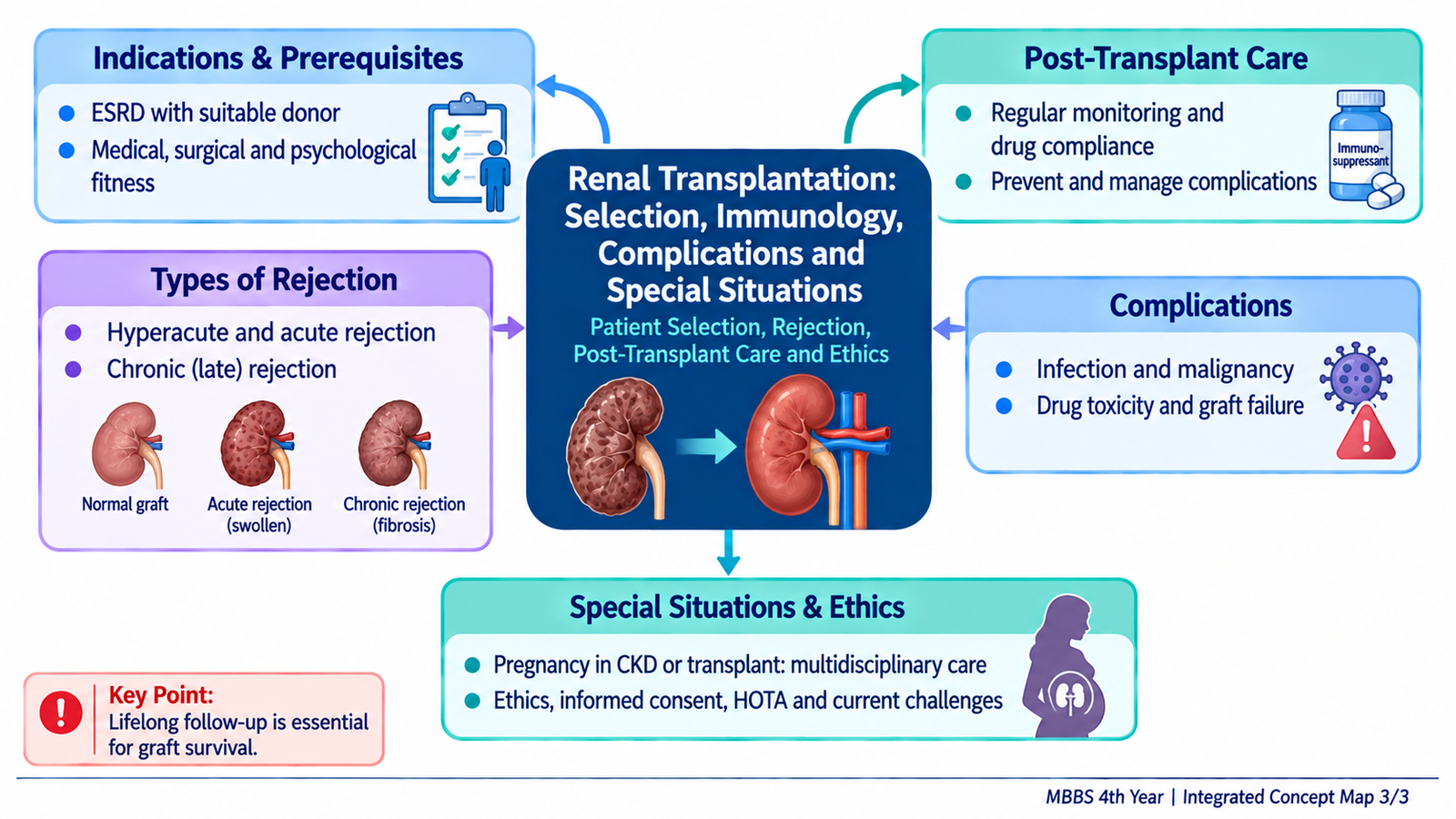
A transplanted kidney contains donor antigens that can be recognized as foreign by the recipient’s immune system. Without immunosuppression, cellular and antibody-mediated responses can injure graft vessels and renal tissue. Successful transplantation therefore requires a balance: immune activity must be suppressed enough to prevent rejection without producing excessive infection, malignancy or drug toxicity.
Types of Transplant Rejection
Hyperacute rejection occurs when the recipient already has circulating antibodies against donor antigens. Antibodies bind rapidly to graft vascular endothelium, activate complement and promote thrombosis. The graft becomes ischemic and may fail almost immediately. Appropriate pre-transplant crossmatching is designed to prevent this form of rejection.
Acute rejection may occur after transplantation when recipient immune responses attack the graft. T-cell-mediated rejection causes lymphocytic inflammation and tubular or vascular injury. Acute antibody-mediated rejection results from antibodies directed against donor antigens and particularly affects the graft microvasculature. Acute rejection may be reversible if recognized and treated promptly.
Chronic rejection develops gradually. Persistent immune-mediated vascular and tissue injury leads to progressive arterial intimal thickening, interstitial fibrosis, tubular atrophy and loss of functioning renal tissue. The result is slowly declining graft function, and established chronic scarring is difficult to reverse.
Immunosuppressive Drugs
Immunosuppressive therapy usually combines agents acting at different points in lymphocyte activation and proliferation. This improves rejection prevention while allowing the intensity of each individual drug to be limited.
| Drug Class | Examples | Core Action | Important Problems |
|---|---|---|---|
| Calcineurin inhibitors | Tacrolimus, cyclosporine | Reduce T-cell activation and IL-2-mediated immune responses | Nephrotoxicity, hypertension; tacrolimus may cause diabetes, while cyclosporine may cause gingival hyperplasia and hirsutism |
| Antimetabolites | Mycophenolate, azathioprine | Reduce lymphocyte proliferation | Bone-marrow suppression; mycophenolate also causes gastrointestinal effects and is teratogenic |
| Corticosteroids | Prednisolone and related agents | Suppress inflammatory gene transcription and lymphocyte responses | Infection, diabetes, osteoporosis, weight gain and other metabolic effects |
| mTOR inhibitors | Sirolimus, everolimus | Reduce lymphocyte proliferation downstream of growth signals | Dyslipidemia, cytopenias and impaired wound healing |
| Biologic induction agents | Basiliximab, antithymocyte globulin | Reduce or inhibit T-cell immune responses around transplantation | Greater immunosuppression increases infection-related risk |
A common maintenance strategy combines a calcineurin inhibitor, an antimetabolite and corticosteroid therapy, modified according to the patient’s immunological risk, adverse effects and graft function.
Post-Transplant Patient Care
Early post-transplant care focuses on determining whether the graft is functioning, recognizing surgical complications and maintaining an appropriate level of immunosuppression.
- Monitor urine output and serum creatinine for evidence of graft function.
- Monitor fluid balance, electrolytes and blood pressure.
- Monitor immunosuppressive therapy and drug toxicity.
- Look for infection because immunosuppression may reduce the usual inflammatory response.
- Investigate an unexpected deterioration in graft function for rejection, obstruction, vascular problems, infection, dehydration or drug toxicity.
- Maintain long-term adherence to immunosuppressive treatment and transplant follow-up.
Complications of Renal Transplant Surgery and Follow-Up
Complications may arise from the operation itself, from impaired graft function, or from the immunosuppressed state.
- Bleeding: may occur around the operative site.
- Renal artery or vein thrombosis: compromises graft blood supply and may rapidly threaten graft survival.
- Urinary leak: usually relates to the urinary tract anastomosis.
- Ureteric obstruction: impairs urinary drainage and can cause graft dysfunction.
- Lymphocele: lymphatic fluid may accumulate around the graft and occasionally compress adjacent structures.
- Wound or systemic infection: risk is increased by surgery and immunosuppression.
- Delayed graft function: the transplanted kidney may not begin effective filtration immediately.
- Rejection: must always be considered when graft function deteriorates.
- Drug toxicity: particularly calcineurin-inhibitor nephrotoxicity, may mimic or contribute to graft dysfunction.

G. Ethics of Organ Transplantation and HOTA
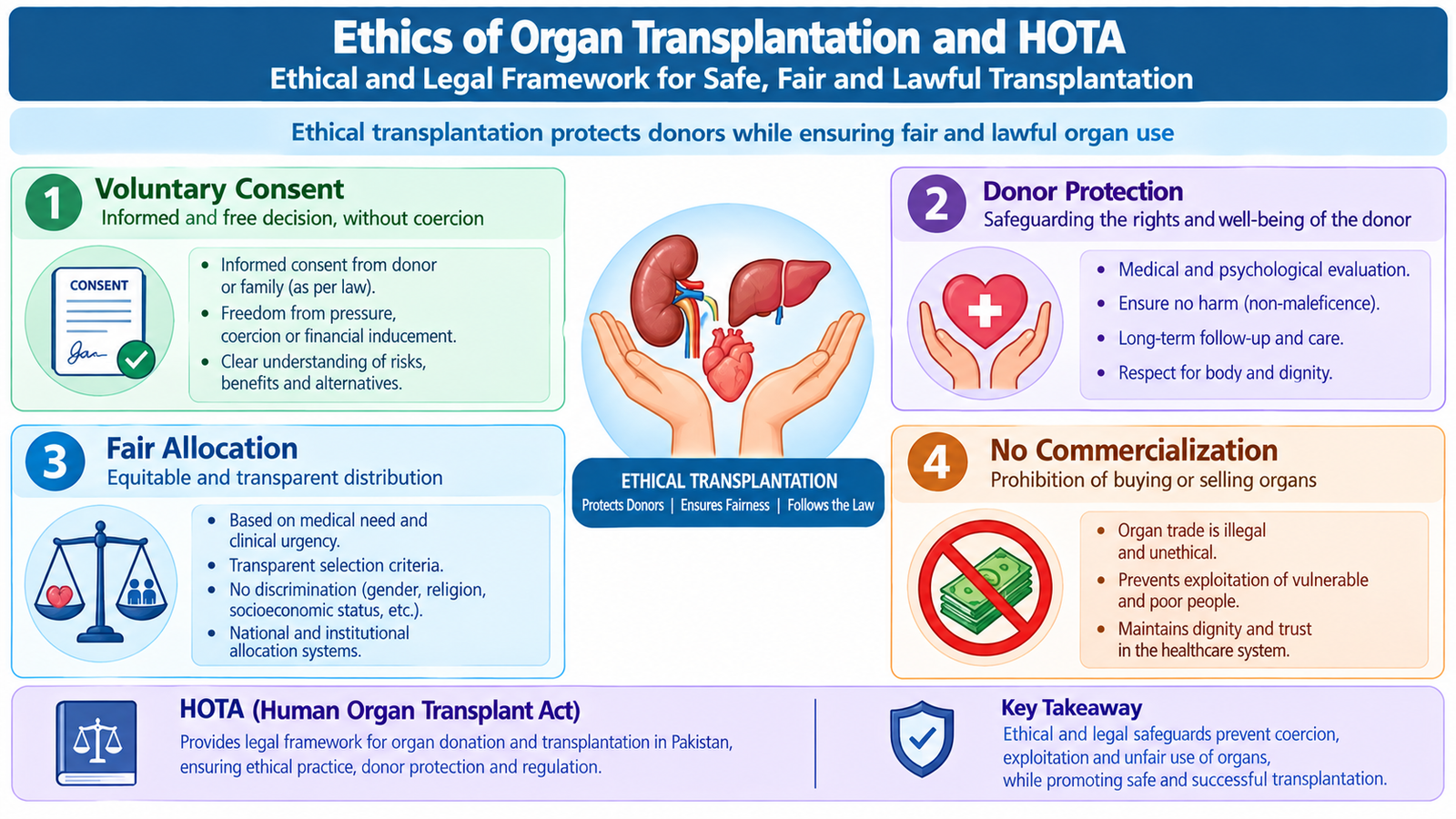
Organ transplantation can save or greatly improve a recipient’s life, but obtaining an organ also involves another human being and a scarce medical resource. Ethical and legal safeguards are therefore essential. Their purpose is to protect donors from harm or exploitation, ensure valid consent, protect recipients, and maintain fairness and public trust in the transplant system.
Core Ethical Principles
Autonomy and informed consent: A competent donor must understand the nature of donation, the expected benefits to the recipient, the risks to the donor and the available alternatives. Consent must be voluntary.
Freedom from coercion: Family pressure, financial dependence or other forms of coercion can undermine genuine consent. This is particularly important in living donation because a healthy person is accepting medical risk for another person’s benefit.
Beneficence and non-maleficence: The transplant team should seek substantial benefit for the recipient while minimizing preventable harm to both donor and recipient. A donor must therefore undergo appropriate medical and psychosocial assessment.
Justice: Organs are scarce. Allocation should therefore be based on ethically defensible medical principles rather than wealth, social influence or the ability to purchase an organ.
Non-commercialization: Treating organs as commodities creates a major risk of exploitation of economically vulnerable individuals. Ethical transplantation separates legitimate medical donation from commercial organ trade.
Human Organ Transplant Act (HOTA): Undergraduate Concept
The KMU learning outcome refers to the Human Organ Transplant Act (HOTA). At undergraduate level, it should be understood as the legal framework intended to regulate human organ transplantation, protect donors and recipients, require valid consent and appropriate oversight, and prevent unauthorized or commercial transplantation.
The central medico-legal principles are that organ retrieval and transplantation must occur through an authorized and ethically accountable process; the donor’s consent must be genuine; donor and recipient evaluation must be documented; and financial exploitation or sale of human organs must not replace voluntary, lawful donation.
Problems and Vulnerabilities in the Organ Transplantation System
Even where a legal framework exists, transplantation can face practical and ethical difficulties. Important problems include:
- Shortage of legally donated organs compared with the number of patients who need transplantation.
- Commercial pressure and illegal organ trade.
- Coercion or exploitation of poor and otherwise vulnerable potential donors.
- Difficulty proving that consent is genuinely free from pressure or financial inducement.
- Fraudulent documentation or attempts to bypass donor safeguards.
- Inequitable access to transplantation because of socioeconomic differences.
- Insufficient development of deceased-donor systems and public awareness.
- Weak monitoring, enforcement or long-term donor follow-up within poorly functioning systems.
These problems explain why transplant ethics cannot be reduced to obtaining a signature on a consent form. A trustworthy system must protect voluntariness, prevent exploitation and maintain transparent oversight from donor assessment through long-term follow-up.
H. CKD and Renal Transplant Recipients During Pregnancy
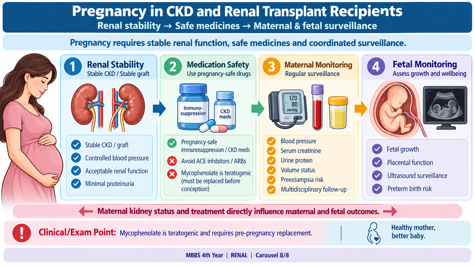
Pregnancy places additional physiological demands on the kidneys. A woman with CKD therefore requires assessment of renal function, proteinuria, blood pressure, medications and the severity of underlying disease before and throughout pregnancy. Pregnancy is also possible after successful kidney transplantation, but the pregnancy remains high risk and should be managed jointly by obstetric and renal/transplant teams.
Why CKD Increases Pregnancy Risk
The degree of maternal risk depends mainly on the severity of renal impairment, the amount of proteinuria and the presence of hypertension. More advanced disease reduces the kidney’s ability to adapt to the increased physiological demands of pregnancy.
Maternal complications can include worsening hypertension, pre-eclampsia, increasing proteinuria, deterioration in renal function, anemia and fluid-balance problems. Fetal risks include impaired fetal growth, prematurity and fetal loss, particularly when maternal kidney disease is severe.
Management of Pregnancy in CKD
Pre-pregnancy counseling is important where possible. Renal function, proteinuria and blood pressure should be assessed, the underlying kidney disease reviewed, and medications checked for pregnancy safety.
During pregnancy, management includes:
- Regular blood-pressure monitoring.
- Serial assessment of renal function and proteinuria.
- Monitoring fluid balance and signs of worsening renal disease.
- Assessment and treatment of anemia when present.
- Fetal growth and obstetric surveillance.
- Joint follow-up by obstetric and renal teams in significant CKD.
ACE inhibitors and angiotensin receptor blockers are not used during pregnancy because of fetal toxicity. When antihypertensive treatment is required, agents with established pregnancy use such as labetalol, nifedipine or methyldopa may be selected according to the clinical situation.
A woman already receiving dialysis who becomes pregnant requires specialist management. Dialysis often needs to be intensified, with close attention to maternal volume status, blood pressure, biochemical control and fetal growth.
Pregnancy After Kidney Transplantation
Successful transplantation frequently improves general health and reproductive function. Pregnancy should nevertheless be planned when graft function is stable, blood pressure is adequately controlled, there has been no recent significant rejection, and the immunosuppressive regimen has been reviewed for pregnancy safety.
The transplanted kidney must be monitored throughout pregnancy because physiological changes, infection, hypertension, rejection and drug toxicity can all affect graft function.
Immunosuppression During Pregnancy
Immunosuppression must not simply be stopped because this could precipitate graft rejection. Instead, treatment is adjusted to drugs with an acceptable pregnancy safety profile.
- Tacrolimus or cyclosporine: may be continued with specialist monitoring when required for graft maintenance.
- Azathioprine: is commonly used when an antimetabolite is required during pregnancy.
- Corticosteroids: may be continued when clinically required.
- Mycophenolate: is teratogenic and must be replaced with an appropriate alternative before a planned pregnancy under transplant-specialist supervision.
- mTOR inhibitors: are generally avoided when planning and managing pregnancy because of limited safety and fetal concerns.
Mode and timing of delivery are primarily determined by obstetric indications together with maternal and graft condition. The presence of a functioning renal transplant by itself does not automatically require operative delivery.
3. Integrated Mechanism Flow — CKD to Renal Replacement
Persistent nephron injury
Surviving nephrons hyperfilter
Glomerulosclerosis and fibrosis
Loss of renal reserve
Uremia, fluid, electrolyte, endocrine effects
Dialysis or transplantation when indicated
4. Important Comparison — Types of Renal Transplant Rejection
| Feature | Hyperacute | Acute | Chronic |
|---|---|---|---|
| Typical timing | Minutes to hours | Usually days to months, but may occur later | Months to years |
| Main mechanism | Pre-existing anti-donor antibodies | T-cell-mediated and/or antibody-mediated immune injury | Persistent immune-mediated vascular and parenchymal injury |
| Characteristic damage | Vascular thrombosis and ischemic necrosis | Tubular, interstitial and/or vascular inflammation | Vascular narrowing, interstitial fibrosis and tubular atrophy |
| Clinical pattern | Immediate graft failure | Acute deterioration in graft function | Gradual progressive graft dysfunction |
| Key principle | Prevent with compatibility testing and crossmatch | May respond to appropriate anti-rejection treatment | Established structural scarring is difficult to reverse |