
Topic 5 — Tubulointerstitial Disease and Urinary Tract Infection: Pathology, Antimicrobial Therapy and Special Populations
Topic Introduction
Tubulointerstitial disease primarily affects the renal tubules and the surrounding interstitium, while urinary tract infection (UTI) occurs when microorganisms produce infection within the urinary tract. Infection may remain confined to the bladder as cystitis or ascend to involve the kidney as pyelonephritis. This topic connects the pathological changes produced by infection and interstitial inflammation with the way UTI presents, is investigated and is treated. You will also learn the clinical pharmacology of important UTI drugs and understand why children and pregnant women require special attention. The overall aim is to recognize the site and severity of disease, identify important complications and select appropriate management principles without losing sight of the underlying pathology.
A. Understanding UTI: Definition, Classification and Diagnostic Approach
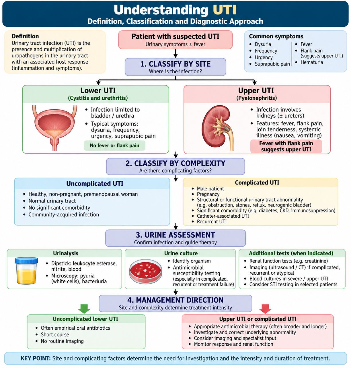
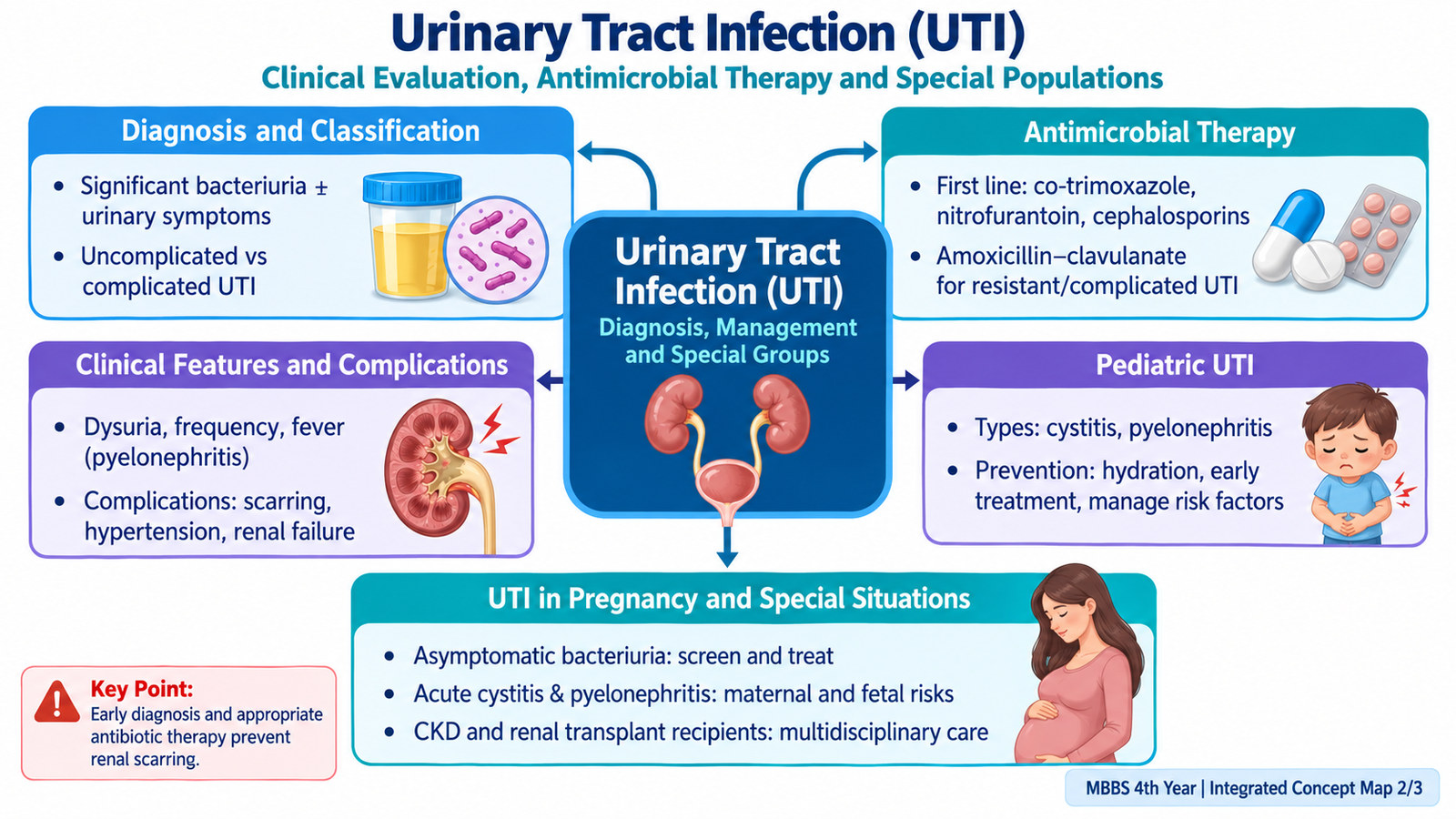
Urinary tract infection means microbial infection involving one or more parts of the urinary tract. The most useful first step is to decide where the infection is located and whether factors are present that increase the risk of treatment failure or complications. Lower UTI mainly involves the bladder, whereas upper UTI involves the renal pelvis and kidney. This distinction matters because renal infection produces more systemic illness and requires antimicrobial drugs that achieve effective concentrations in renal tissue.
Clinical classification
- Lower UTI: primarily cystitis, producing dysuria, frequency, urgency and suprapubic discomfort.
- Upper UTI: pyelonephritis, in which infection reaches the kidney and commonly produces fever, flank pain and systemic illness.
- Uncomplicated UTI: infection occurring without important structural or functional factors that substantially increase the risk of persistence or complications.
- Complicated UTI: infection associated with circumstances such as urinary obstruction, anatomical or functional urinary abnormalities, instrumentation, important comorbidity or another factor that makes eradication more difficult or complications more likely.
Most bacterial UTIs develop through an ascending route. Organisms gain access to the lower urinary tract, multiply in the bladder and may ascend through the ureters toward the kidney when host defenses are inadequate or urinary flow is disturbed. Urinary stasis is important because normal urine flow helps remove bacteria mechanically.
Diagnostic approach
Diagnosis combines the clinical pattern with urine findings. Symptoms establish whether infection is clinically suspected, while urinalysis and, where appropriate, urine culture provide objective evidence.
- Pyuria indicates leukocytes in urine and supports urinary tract inflammation.
- Bacteriuria supports bacterial infection when interpreted with symptoms and specimen quality.
- Leukocyte esterase is an indirect marker of urinary leukocytes.
- Nitrite positivity supports infection with organisms capable of converting urinary nitrate to nitrite, but a negative result does not exclude UTI.
- Urine culture identifies the organism and provides antimicrobial susceptibility information. It is especially useful when infection is complicated, recurrent, atypical, involves the upper tract, occurs during pregnancy or occurs in children.
Imaging is not required for every simple lower UTI. It becomes more relevant when obstruction, stones, structural abnormality, recurrent infection, an atypical course or another complication is suspected.
| Feature | Uncomplicated UTI | Complicated UTI |
|---|---|---|
| Urinary tract | No major complicating abnormality | Structural, functional or important host factor may be present |
| Risk | Usually lower risk of persistence or severe complications | Greater risk of persistence, recurrence or progression |
| Evaluation | Often relatively simple | Culture and assessment of the complicating factor are more important |
| Management principle | Treat infection appropriately | Treat infection and correct the underlying factor where possible |

B. Interstitial Nephritis: Etiology, Pathogenesis and Drug-Induced Disease
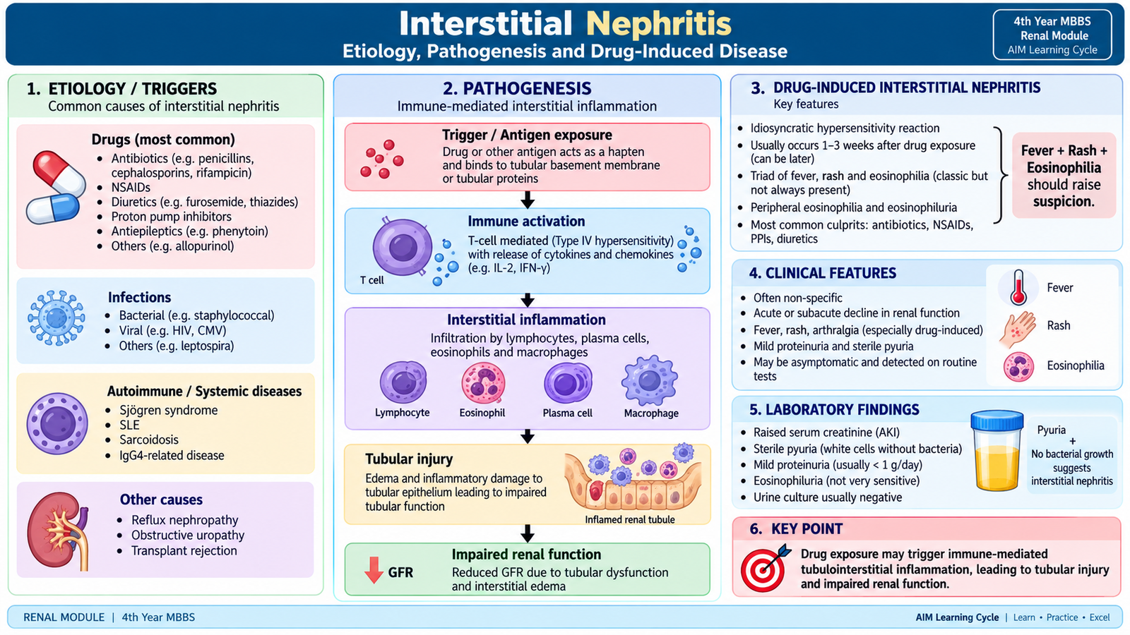
Interstitial nephritis is inflammation involving the renal interstitium and tubules rather than primarily affecting the glomeruli. It may develop acutely or become chronic. In an acute process, inflammatory cells and interstitial edema disturb tubular function and may reduce renal function. Drugs are an important cause of acute interstitial nephritis, but infections and immune-mediated processes may also produce interstitial inflammation.
Pathogenesis
Drug-induced acute interstitial nephritis is usually an immune-mediated reaction rather than direct dose-dependent toxicity. A drug or drug-related antigen triggers an immune response within the interstitium. Inflammation then produces edema and tubular injury, reducing the kidney’s ability to maintain normal filtration and tubular function.
Morphology
The key microscopic abnormality is expansion of the interstitium by edema and inflammatory cells. The inflammatory infiltrate may contain lymphocytes and other leukocytes. Eosinophils may be prominent in some drug-induced cases, although they are not required for the diagnosis. Tubular epithelial injury may accompany the interstitial inflammation.
Clinical and diagnostic correlation
Renal dysfunction may appear after exposure to a causative drug. Patients may have nonspecific illness or features of hypersensitivity. Fever, rash and peripheral eosinophilia are classically associated with drug-induced disease but are not present in every patient. Urinalysis may show pyuria and other evidence of tubulointerstitial inflammation.
The central management principle in drug-induced disease is recognition and withdrawal of the offending drug. Renal function and fluid-electrolyte status are then monitored while supportive management is provided. Persistent or severe cases require specialist assessment.
C. Acute Pyelonephritis: Pathogenesis, Morphology and Clinical Recognition
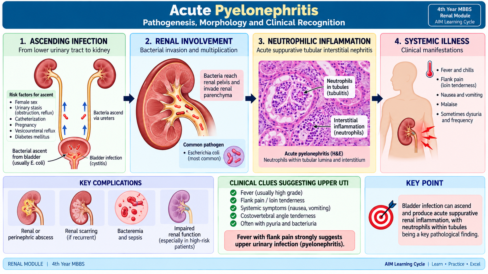
Acute pyelonephritis is an acute suppurative infection of the renal pelvis, tubules and interstitium. Infection most commonly reaches the kidney by ascending from the lower urinary tract. Conditions that interfere with normal urinary flow or facilitate backward movement of urine increase the opportunity for organisms to reach renal tissue.
Pathogenesis
Once organisms reach the kidney, they provoke an acute neutrophilic inflammatory response. Neutrophils enter the interstitium and renal tubules, producing focal suppuration and tubular injury. The patchy nature of infection explains why lesions may be unevenly distributed through the renal parenchyma.
Gross morphology
The affected kidney may contain small yellow-white suppurative areas or abscesses within the parenchyma. Inflammation may extend from the pelvis into the medulla and cortex. Severe infection may produce larger areas of suppuration.
Microscopic morphology
- Prominent neutrophilic inflammation in the interstitium.
- Neutrophils within tubular lumina.
- Tubular epithelial injury and focal necrosis in severe inflammation.
- Microabscess formation where suppuration is marked.
Clinical features
Acute renal infection produces both urinary and systemic manifestations. Fever and flank pain suggest upper urinary involvement because inflammation is no longer confined to the bladder.
- Fever, chills and systemic malaise.
- Flank or loin pain.
- Costovertebral-angle tenderness.
- Dysuria, frequency or urgency may coexist if lower UTI is also present.
- Nausea or vomiting may occur with significant systemic illness.
Investigations and complications
Urinalysis commonly shows pyuria and bacteriuria. Urine culture is important because identifying the organism and its susceptibility assists antimicrobial selection. Systemic investigations assess the severity of illness and renal function when clinically indicated.
Severe or inadequately treated infection may produce renal or perinephric abscess formation, bloodstream infection and sepsis. Persistent obstruction is particularly important because antimicrobial therapy alone may be insufficient when infected urine cannot drain normally.
D. Chronic Pyelonephritis: Recurrent Injury, Renal Scarring and Complications
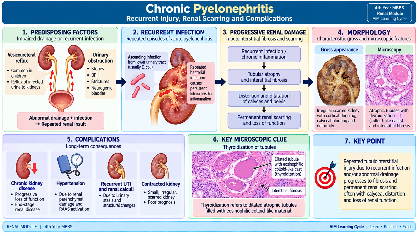
Chronic pyelonephritis is a chronic tubulointerstitial disease characterized by renal scarring and deformity related to repeated or persistent urinary infection, usually in the presence of an important predisposing urinary abnormality. Reflux or obstruction allows repeated renal injury, so chronic pyelonephritis is more than simply a prolonged episode of uncomplicated cystitis.
How chronic damage develops
Repeated infection and abnormal urine flow injure tubules and interstitium. Acute inflammation gradually gives way to tubular atrophy, chronic inflammatory infiltration and interstitial fibrosis. As fibrosis contracts, the renal surface becomes irregularly scarred and the underlying calyces may become deformed.
Morphology
Grossly, the kidneys may show coarse, irregular and often asymmetric scars associated with deformity or blunting of the underlying calyces. The relationship between cortical scarring and calyceal distortion is an important pathological clue.
Microscopically, there is interstitial fibrosis with chronic inflammatory cells and tubular atrophy. Some atrophic tubules become dilated and contain eosinophilic proteinaceous material, producing an appearance resembling thyroid follicles. This is called thyroidization of tubules.
Clinical significance and complications
Because renal scarring is permanent, extensive bilateral disease may progressively reduce renal function. Patients may also experience recurrent episodes of UTI. The important principle is that preventing further infection is not enough when a correctable obstructive or reflux-related abnormality remains untreated.
E. Acute and Chronic Cystitis: Pathology, Presentation and Complications
Cystitis is inflammation of the urinary bladder, most commonly caused by infection. Acute bacterial cystitis is primarily a lower urinary tract process. Because inflammation remains within the bladder rather than the renal parenchyma, local urinary symptoms predominate and major systemic features are usually absent. Chronic cystitis develops when inflammation persists or recurs, often because the underlying predisposing factor has not been removed.
Acute cystitis
Microbial growth within the bladder stimulates mucosal inflammation. The inflamed bladder becomes more sensitive, producing urgency and frequency, while irritation of the urethra and bladder mucosa causes dysuria.
Morphology: the mucosa may be congested and edematous. Microscopically, acute inflammation is characterized by neutrophilic infiltration of the mucosa and submucosa. Severe inflammation may cause mucosal hemorrhage or ulceration.
Typical clinical features include:
- Dysuria.
- Urinary frequency and urgency.
- Suprapubic discomfort.
- Cloudy urine or hematuria may occur.
- High fever and marked flank pain suggest that infection may have extended beyond simple cystitis.
Chronic cystitis
Repeated or persistent inflammation leads to chronic inflammatory infiltration and reactive mucosal changes. Fibrosis may develop in prolonged disease. The key clinical task is therefore to look for a cause of persistence, such as impaired bladder emptying, obstruction or another factor promoting recurrent infection.
Complications
An untreated lower UTI may ascend and cause pyelonephritis. Recurrent infection should prompt assessment for an underlying structural or functional abnormality rather than repeated treatment without considering the cause.

F. Clinical Management of UTI: From Presentation to Treatment
Management becomes easier when approached in a fixed sequence. First decide whether infection is confined to the lower urinary tract or involves the kidney. Next determine whether the patient is clinically stable and whether complicating factors are present. Investigations then confirm infection and guide antimicrobial therapy. Finally, any factor that allows infection to persist—particularly obstruction or abnormal urinary drainage—must be addressed.
History and clinical assessment
- Dysuria, frequency, urgency and suprapubic pain suggest lower UTI.
- Fever, rigors, flank pain and vomiting raise concern for pyelonephritis.
- Previous UTI, recent antimicrobial exposure and recurrent symptoms influence investigation and drug selection.
- Ask about urinary obstruction, stones, instrumentation and other relevant complicating factors.
Investigations
Urinalysis provides rapid supportive evidence, while urine culture identifies the organism and susceptibility pattern. Culture assumes greater importance in upper, recurrent, complicated, pediatric and pregnancy-associated infection. Renal function is assessed when illness is significant or renal impairment is possible. Imaging is selected when a structural problem, obstruction or complication is suspected.
Management principles
- Choose an antimicrobial appropriate for the likely site of infection and patient characteristics.
- Use local susceptibility information and previous culture results when available.
- Modify therapy when culture and susceptibility results become available.
- Ensure adequate hydration when clinically appropriate.
- Treat pain, fever and other symptoms appropriately.
- Correct urinary obstruction or another important predisposing factor when present.
- Reassess patients whose symptoms persist or worsen despite treatment.

G. Antimicrobial Pharmacology in UTI
Successful UTI treatment depends not only on whether a drug inhibits the organism but also on whether the drug reaches the site of infection. A drug that becomes highly concentrated in urine may work well for bladder infection but may be unsuitable for renal parenchymal infection if tissue concentrations are inadequate. Culture and susceptibility testing help refine treatment, particularly when resistance or complicated infection is possible.
Co-trimoxazole
Co-trimoxazole combines trimethoprim and sulfamethoxazole. The two components inhibit successive steps in bacterial folate synthesis. Sequential blockade reduces production of folate-dependent bacterial metabolites required for nucleic-acid synthesis.
It may be useful for susceptible urinary pathogens. Important adverse effects include gastrointestinal disturbance, allergic skin reactions, bone-marrow suppression and hyperkalemia. Severe hypersensitivity reactions can occur. Renal function, interacting medicines and pregnancy status influence its safe use. Because the components affect folate metabolism and have gestation-specific concerns, use during pregnancy requires careful clinical selection.
Nitrofurantoin
Nitrofurantoin is reduced by bacterial enzymes to reactive intermediates that damage multiple bacterial cellular components, including DNA. It is excreted into urine and achieves useful urinary concentrations, making it particularly suited to lower urinary infection when the organism is susceptible.
Its major limitation is that urinary concentration does not equal adequate renal tissue concentration. Therefore, nitrofurantoin should not be selected to treat pyelonephritis. Common adverse effects include gastrointestinal symptoms. Important but less common toxicity includes pulmonary and hepatic reactions. Hemolysis may occur in susceptible patients with glucose-6-phosphate dehydrogenase deficiency.
Cephalosporins
Cephalosporins are β-lactam antibiotics. They bind bacterial penicillin-binding proteins and interfere with peptidoglycan cross-linking, weakening the bacterial cell wall and producing bacterial death in susceptible organisms.
Different cephalosporins may be used in urinary infection depending on susceptibility, disease severity and the need for oral or parenteral therapy. Important adverse effects include gastrointestinal disturbance and hypersensitivity. A history of serious β-lactam allergy requires careful assessment.
Amoxicillin–clavulanate
Amoxicillin inhibits bacterial cell-wall synthesis by binding penicillin-binding proteins. Clavulanate inhibits several bacterial β-lactamases and therefore protects amoxicillin from enzymatic destruction by susceptible β-lactamase-producing organisms.
It may be used for susceptible urinary pathogens. Gastrointestinal disturbance, especially diarrhea, is common. Hypersensitivity reactions may occur, and hepatic adverse effects are important though less frequent.
Drug-selection logic
| Drug | Main pharmacological idea | Important UTI point | Important caution |
|---|---|---|---|
| Co-trimoxazole | Sequential folate inhibition | Useful when organism is susceptible | Hypersensitivity, marrow effects, hyperkalemia, pregnancy considerations |
| Nitrofurantoin | Reactive bacterial metabolites damage cellular components | Strong lower-UTI role | Not suitable for pyelonephritis |
| Cephalosporins | β-lactam cell-wall inhibition | Options for lower or systemic UTI depending on agent | β-lactam hypersensitivity |
| Amoxicillin–clavulanate | Cell-wall inhibition plus β-lactamase inhibition | Use when susceptibility supports activity | GI effects, allergy, hepatic adverse effects |

H. Urinary Tract Infection in Children
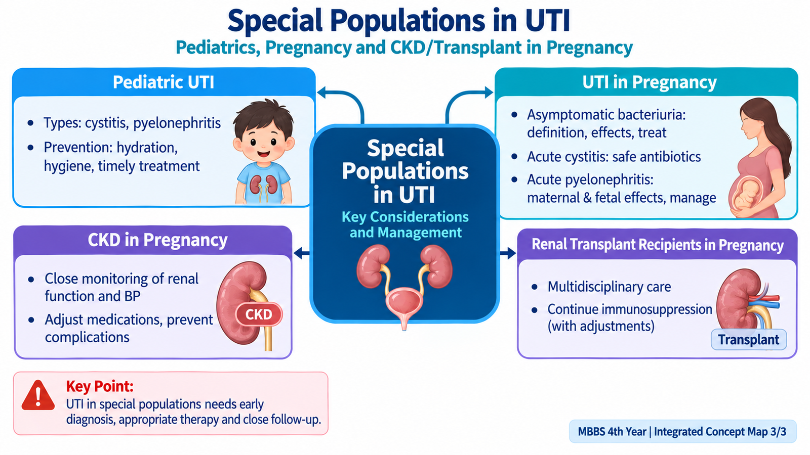
UTI in children is important because presentation varies with age and recurrent or febrile infection may reveal an underlying urinary tract abnormality. Older children may describe typical urinary symptoms, whereas infants and young children can present with nonspecific illness. Recognition therefore depends on combining age-appropriate clinical suspicion with a properly obtained urine specimen.
Types of pediatric UTI
- Lower UTI: primarily bladder infection or cystitis.
- Upper UTI: pyelonephritis involving renal tissue.
- Febrile UTI: fever associated with UTI raises concern for upper-tract involvement, especially in younger children.
- Recurrent UTI: repeated episodes require assessment for factors allowing infection to recur.
Age-related presentation
Infants may not localize urinary symptoms. Fever, irritability, poor feeding or vomiting may therefore be the main clues. Older children are more likely to report dysuria, frequency, urgency, suprapubic pain or flank pain.
Investigation and management
A reliable urine specimen is essential because contamination can lead to an incorrect diagnosis. Urinalysis provides rapid supportive evidence, while culture confirms the causative organism and guides antibiotic therapy. The child’s age, clinical condition and whether infection is lower or upper influence treatment.
A febrile or recurrent UTI may require assessment for congenital or functional urinary abnormalities. The purpose is not merely to treat the present infection but to prevent repeated renal injury when an underlying abnormality is present.
Prevention
- Encourage adequate fluid intake when appropriate.
- Encourage regular bladder emptying and avoid prolonged holding of urine.
- Address constipation when present because bowel dysfunction may interfere with normal bladder emptying.
- Teach appropriate perineal hygiene.
- Investigate recurrent or atypical infection rather than repeatedly treating episodes without identifying the cause.

I. UTI During Pregnancy: Asymptomatic Bacteriuria, Cystitis and Pyelonephritis
Pregnancy increases susceptibility to urinary infection because physiological changes promote urinary stasis. Dilatation of the collecting system and reduced efficiency of urinary drainage make bacterial persistence and ascent more likely. For this reason, bacteriuria that might remain clinically silent in another setting is important during pregnancy because it can progress to symptomatic infection and pyelonephritis.
Asymptomatic bacteriuria
Asymptomatic bacteriuria means significant bacteriuria in a properly collected urine specimen without symptoms attributable to UTI. During pregnancy it is clinically important because untreated bacteriuria may progress to symptomatic infection, particularly pyelonephritis.
Maternal infection may also adversely affect pregnancy. Therefore, detection by urine culture and treatment with an antimicrobial appropriate for pregnancy and the susceptibility pattern are important management principles. Follow-up assessment is used to ensure that infection has been adequately addressed when clinically indicated.
Acute cystitis during pregnancy
Cystitis presents with dysuria, frequency, urgency and suprapubic discomfort without the systemic features expected in pyelonephritis. Urine culture is important because drug selection must consider both bacterial susceptibility and maternal-fetal safety. Lower-tract drugs may be suitable when appropriate, but the pregnancy stage and patient-specific cautions must be considered.
Acute pyelonephritis during pregnancy
Fever, flank pain, costovertebral-angle tenderness and systemic illness suggest renal involvement. Pyelonephritis is more serious than cystitis because maternal complications can include severe systemic infection and deterioration, while significant maternal illness can also affect pregnancy.
Management requires prompt clinical assessment, urine culture, an antimicrobial that achieves adequate renal tissue concentrations and is suitable for pregnancy, supportive care and close monitoring. Significant systemic illness may require hospital-based care. Obstruction or another complicating factor should be considered when improvement is unsatisfactory.

Integrated Mechanism Flow
Organisms reach lower urinary tract
Cystitis develops
Favored by stasis, reflux or obstruction
Acute pyelonephritis
Tubular damage and interstitial inflammation
Fibrosis, renal scarring and functional loss
Important Comparison — Acute versus Chronic Pyelonephritis
| Feature | Acute Pyelonephritis | Chronic Pyelonephritis |
|---|---|---|
| Main process | Acute suppurative infection | Repeated/persistent tubulointerstitial injury with scarring |
| Dominant inflammatory cell | Neutrophils | Chronic inflammatory cells with fibrosis |
| Characteristic morphology | Suppurative foci, neutrophils in tubules/interstitium | Coarse scars, calyceal deformity, tubular atrophy and thyroidization |
| Clinical pattern | Fever, flank pain, systemic illness | Recurrent infection and progressive renal damage may dominate |
| Major concern | Sepsis, abscess, acute complication | Permanent scarring and loss of renal function |