
Topic 6 — Cystic and Congenital Renal Disorders: Polycystic Kidney Disease, PUJ Obstruction and Urinary Tract Anomalies
Topic Introduction
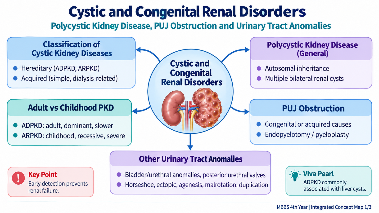
Cystic and congenital renal disorders are abnormalities in which the kidney or urinary tract develops an altered structure that may interfere with normal renal function or urine drainage. Some conditions, such as polycystic kidney disease, are inherited and progressively replace normal renal tissue with cysts. Others arise because the kidneys do not ascend, rotate or fuse normally during development. Congenital obstruction may occur at the pelviureteric junction or within the urethra and can lead to hydronephrosis and progressive renal damage. In this chapter, you will learn how these abnormalities develop, how they appear clinically and on imaging, and the basic principles used to diagnose and manage them.
A. Cystic Diseases of the Kidney: Classification and Core Concepts
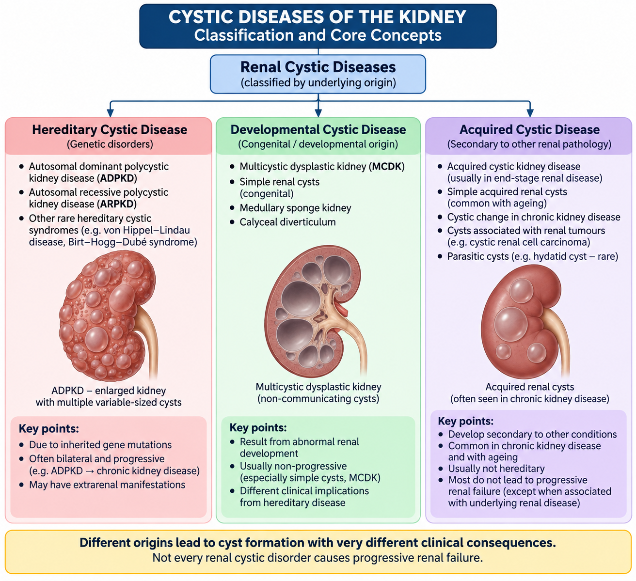
A renal cyst is a fluid-filled or occasionally material-filled cavity within the kidney. Cysts may arise because of inherited abnormalities of tubular epithelial cells, abnormal renal development, or acquired changes occurring later in life. Their clinical importance varies greatly: a solitary simple renal cyst may have little effect on renal function, whereas inherited polycystic kidney disease may involve both kidneys extensively and eventually cause chronic kidney disease.
Useful Classification
For undergraduate learning, cystic renal diseases are most easily understood by grouping them according to their origin.
- Hereditary cystic diseases: autosomal dominant polycystic kidney disease and autosomal recessive polycystic kidney disease are the most important examples in this topic.
- Developmental cystic disorders: these include multicystic dysplastic kidney and medullary sponge kidney.
- Other inherited tubulointerstitial cystic disorders: nephronophthisis and related inherited disorders may produce corticomedullary cysts and progressive renal impairment.
- Acquired/non-hereditary cysts: simple renal cysts are common incidental lesions, while acquired cystic kidney disease may develop in patients with longstanding advanced renal failure.
Why Cysts Can Damage the Kidney
A cyst begins within or from a renal tubular structure. As it enlarges, it can compress surrounding nephrons and blood vessels. Extensive bilateral cyst formation therefore reduces the amount of functioning renal parenchyma and promotes interstitial fibrosis. The result may be progressive loss of renal function.
Abnormal tubular development or epithelial function → cyst formation and enlargement → compression of normal nephrons → interstitial damage and fibrosis → progressive loss of renal function.
B. Autosomal Dominant Polycystic Kidney Disease: Pathogenesis and Pathology
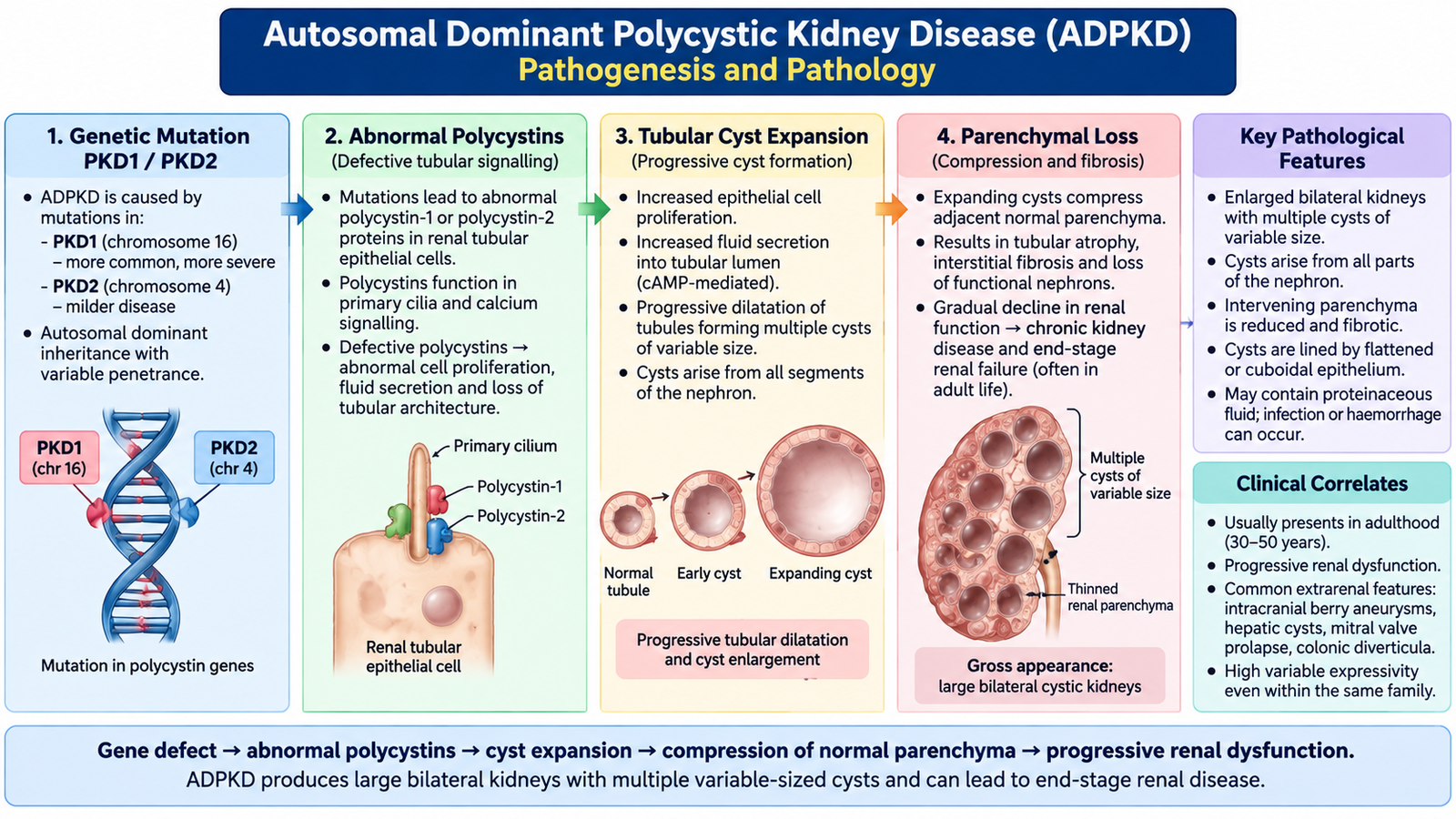
Autosomal dominant polycystic kidney disease, or ADPKD, is an inherited disorder characterized by progressive formation of numerous cysts in both kidneys. It is the typical form of “adult polycystic kidney disease,” although structural abnormalities begin much earlier than the appearance of symptoms. The kidneys gradually enlarge as cysts develop from different parts of the nephron.
Genetic Basis
ADPKD is inherited in an autosomal dominant pattern. The major genes are PKD1 and PKD2. They encode the proteins polycystin-1 and polycystin-2, which participate in signaling and regulation of tubular epithelial cells. Abnormal polycystin function alters tubular cell behavior, fluid secretion and tubular architecture, allowing cysts to develop and expand.
Disease associated with PKD1 is generally more severe and tends to produce renal failure earlier than disease associated with PKD2, although considerable variation occurs between individuals.
Pathogenesis
Early cysts remain connected with the tubule from which they originate. With growth, many cysts lose that connection and continue to enlarge because their epithelial lining proliferates and secretes fluid. Expansion of multiple cysts explains the greatly increased kidney size and the gradual destruction of normal parenchyma.
Gross Morphology
- Both kidneys are usually enlarged.
- The external surface is distorted by numerous cysts.
- Cysts vary considerably in size and may involve the cortex and medulla.
- Cyst contents may be clear, turbid or hemorrhagic.
- Residual normal renal tissue becomes compressed between expanding cysts.
Microscopic Morphology
Cysts are lined by flattened or cuboidal epithelial cells. With progression, the intervening renal parenchyma may show tubular atrophy, vascular changes and interstitial fibrosis. These chronic changes contribute to the gradual decline in glomerular filtration.
Extrarenal Manifestations
The genetic defect affects tissues outside the kidney as well. Therefore, ADPKD is not simply a renal cystic disease.
- Hepatic cysts are an important extrarenal manifestation.
- Intracranial berry aneurysms may occur and can rupture to produce subarachnoid hemorrhage.
- Cardiac valve abnormalities, including mitral valve prolapse, may occur.
- Cysts can also occur in some other organs.
C. ADPKD in Clinical Practice and Childhood Polycystic Kidney Disease
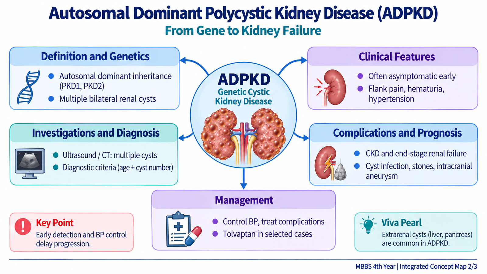
ADPKD often remains clinically silent for years because functioning nephrons initially compensate for the cystic change. As cysts enlarge, patients develop manifestations related to renal enlargement, vascular compression, cyst complications and progressive nephron loss. In contrast, autosomal recessive polycystic kidney disease usually presents much earlier and has a different pathological pattern.
Clinical Manifestations of ADPKD
- Hypertension: may appear before major reduction in renal function. Cyst expansion can compress renal vessels and activate mechanisms that increase blood pressure.
- Flank or abdominal discomfort: enlarged kidneys and expanding cysts may produce pressure or pain.
- Hematuria: may result from bleeding into a cyst or communication of cyst bleeding with the urinary tract.
- Urinary tract infection or cyst infection: abnormal renal structure predisposes to infection.
- Renal calculi: altered urinary drainage and metabolic factors increase stone risk.
- Progressive chronic kidney disease: continuing cyst expansion and interstitial fibrosis gradually reduce functioning nephron mass.
Diagnostic Approach
Diagnosis is based mainly on the combination of family history and renal imaging. Ultrasonography is commonly used because it can demonstrate renal cysts without radiation. CT and MRI can show smaller cysts and give more detailed anatomical information.
In a person at risk because a first-degree relative has ADPKD, commonly used unified ultrasound criteria are age dependent:
| Age | Ultrasound Finding Supporting Diagnosis in an At-Risk Individual |
|---|---|
| 15–39 years | At least 3 renal cysts in total, involving one or both kidneys |
| 40–59 years | At least 2 cysts in each kidney |
| 60 years or older | At least 4 cysts in each kidney |
These imaging criteria are most useful when there is an established family history. When the family history is absent or the imaging result is uncertain, the diagnosis requires broader clinical assessment. Genetic testing may be useful in selected situations, particularly when imaging is equivocal or a definite diagnosis is important for family or transplant-related decisions.
Investigations
- Blood pressure measurement: detects an important early complication.
- Serum creatinine and estimated GFR: assess renal function.
- Urinalysis: may show hematuria or evidence of infection.
- Ultrasonography: demonstrates renal cysts and kidney enlargement.
- CT or MRI: provides additional anatomical detail when required.
- Evaluation for extrarenal disease: is directed by the patient’s history, examination and individual risk rather than performed identically in every patient.
Management Principles of ADPKD
Management aims to preserve renal function, control complications and identify patients who need specialist treatment. The inherited structural abnormality itself cannot simply be removed, so long-term care is important.
- Control hypertension carefully; blockade of the renin–angiotensin system is commonly used when appropriate.
- Treat urinary infection promptly and assess suspected cyst infection appropriately.
- Evaluate hematuria, renal stones and persistent pain rather than assuming every symptom is caused by uncomplicated cysts.
- Monitor renal function and progression of chronic kidney disease.
- Avoid unnecessary nephrotoxic exposure where possible.
- Specialist disease-modifying therapy such as the vasopressin V2-receptor antagonist tolvaptan may be considered in selected adults at risk of rapid progression.
- Advanced renal failure may ultimately require dialysis or renal transplantation.
- Genetic counseling is useful because each child of an affected individual has a significant inherited risk in an autosomal dominant disorder.
Autosomal Recessive Polycystic Kidney Disease
Autosomal recessive polycystic kidney disease (ARPKD) is the major inherited childhood form of polycystic kidney disease. It is caused by abnormalities of the PKHD1 gene, which encodes fibrocystin. The renal lesion mainly affects collecting ducts. These ducts become elongated and dilated, producing numerous small cystic spaces arranged in a radial pattern through the kidney.
Morphology and Clinical Outcome
- Both kidneys are enlarged but usually retain a relatively smooth external outline.
- Numerous elongated cystic channels involve the collecting ducts.
- Severe disease presenting around birth may produce major renal dysfunction.
- Severe fetal renal dysfunction may reduce amniotic fluid and contribute to pulmonary hypoplasia.
- Surviving children may develop hypertension and chronic kidney disease.
- Congenital hepatic fibrosis is an important associated abnormality and may cause portal hypertension in older children.
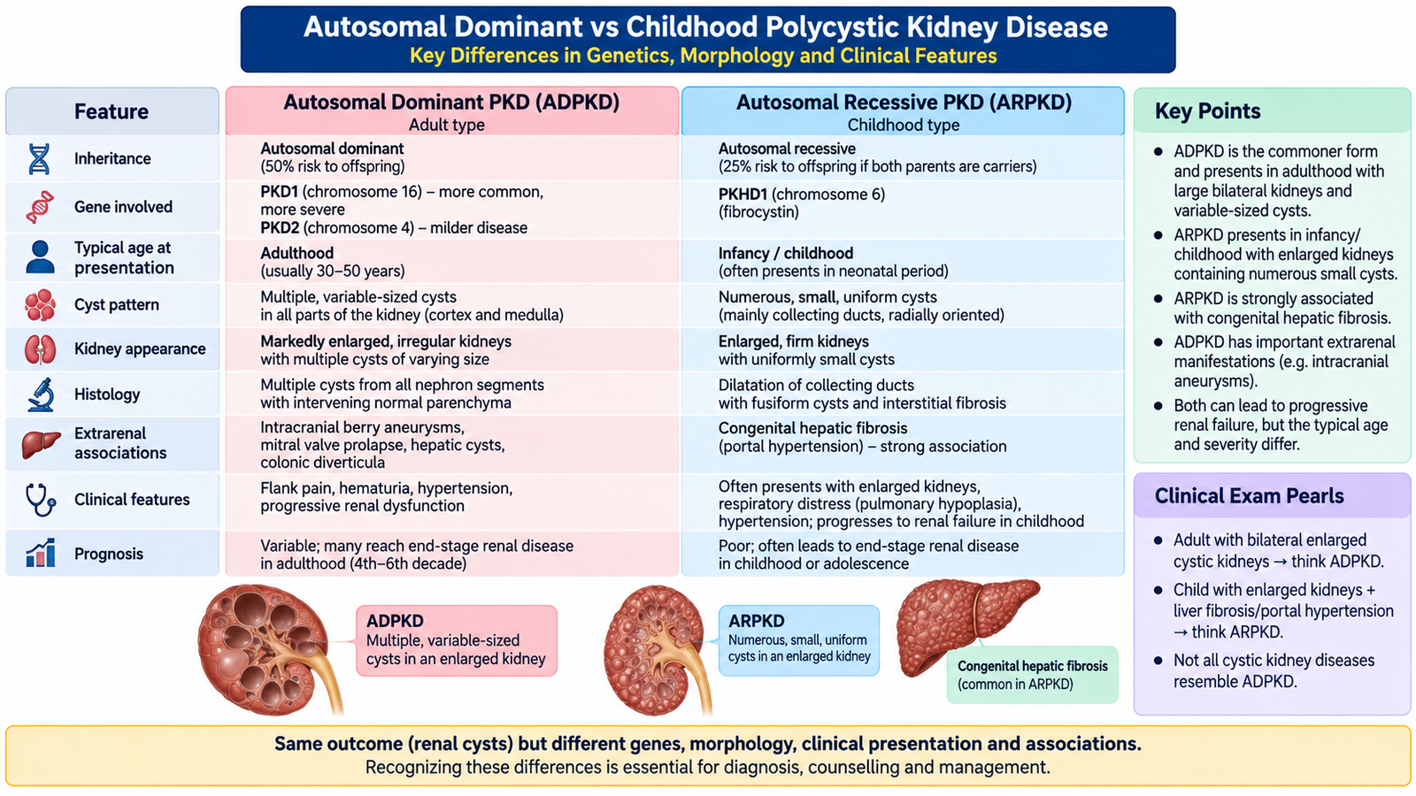
Important Comparison: ADPKD versus ARPKD
| Feature | ADPKD | ARPKD |
|---|---|---|
| Inheritance | Autosomal dominant | Autosomal recessive |
| Major genes | PKD1, PKD2 | PKHD1 |
| Typical presentation | Usually later life, although disease begins earlier | Infancy or childhood |
| Cyst pattern | Variable-sized cysts involving different nephron segments | Dilated collecting ducts, usually small and elongated |
| Kidney appearance | Massively enlarged and externally distorted by cysts | Enlarged with relatively smooth external surface |
| Important extrarenal association | Hepatic cysts and intracranial aneurysms | Congenital hepatic fibrosis and portal hypertension |
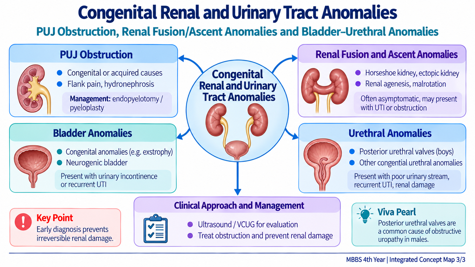
D. Pelviureteric Junction Obstruction
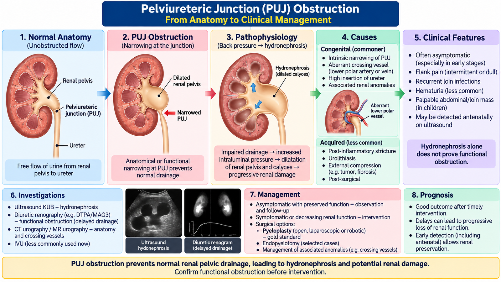
Pelviureteric junction (PUJ) obstruction is impaired drainage of urine from the renal pelvis into the proximal ureter. Persistent obstruction raises pressure within the collecting system and produces hydronephrosis. If significant obstruction continues, the expanding renal pelvis and altered intrarenal pressure can progressively damage renal parenchyma and reduce renal function.
Causes
PUJ obstruction may be congenital or acquired. Congenital disease is more common and can result from an abnormal segment of the junction that fails to conduct normal ureteric peristalsis. The obstruction may also be influenced by abnormal insertion of the ureter or compression by a crossing vessel.
- Congenital intrinsic obstruction: abnormal muscular arrangement or an aperistaltic/narrow PUJ segment.
- Congenital anatomical factors: high ureteric insertion or an associated crossing lower-pole vessel.
- Acquired causes: scarring, calculi, previous intervention or other lesions that narrow or distort the PUJ region.
How Obstruction Damages the Kidney
Clinical Presentation
Presentation depends on age and severity. Many congenital cases are first recognized during antenatal or childhood ultrasonography. Older children and adults may present when increased urine production distends an obstructed collecting system.
- Intermittent flank or loin pain, sometimes worse after a large fluid intake.
- Recurrent urinary tract infection.
- Hematuria.
- Renal calculi caused partly by urinary stasis.
- Antenatally detected hydronephrosis in infants.
- Occasionally reduced renal function when obstruction is severe or longstanding.
Investigations
Imaging must answer two questions: Is the collecting system dilated? and is that dilatation functionally obstructive?
- Ultrasonography: shows hydronephrosis and can assess renal parenchymal thickness.
- Urinalysis and urine culture: assess hematuria and associated infection.
- Serum creatinine and renal function tests: are particularly important when bilateral disease or significant renal impairment is suspected.
- Diuretic renography: evaluates drainage and differential function of the affected kidney and helps determine whether dilatation represents significant functional obstruction.
- CT urography or other anatomical imaging: may be useful in selected patients to define anatomy, identify stones or demonstrate an external anatomical cause.
Management
Management depends on symptoms, renal function and evidence of significant obstruction. Mild stable dilatation with preserved function can sometimes be observed with appropriate follow-up. Intervention is considered when obstruction causes symptoms, repeated infection, stones or deterioration of renal function.
Pyeloplasty
Pyeloplasty reconstructs the PUJ to create a wide, dependent and freely draining connection between the renal pelvis and ureter. Dismembered pyeloplasty is the standard reconstructive principle in many patients. The undergraduate focus is the purpose of the operation rather than its detailed operative steps.
Endopyelotomy
Endopyelotomy is an endoscopic treatment in which the narrowed PUJ is incised to improve drainage. It is less invasive but is suitable only for selected patients and is generally less universally applicable than pyeloplasty.
E. Renal Fusion, Ascent, Rotation and Duplication Anomalies
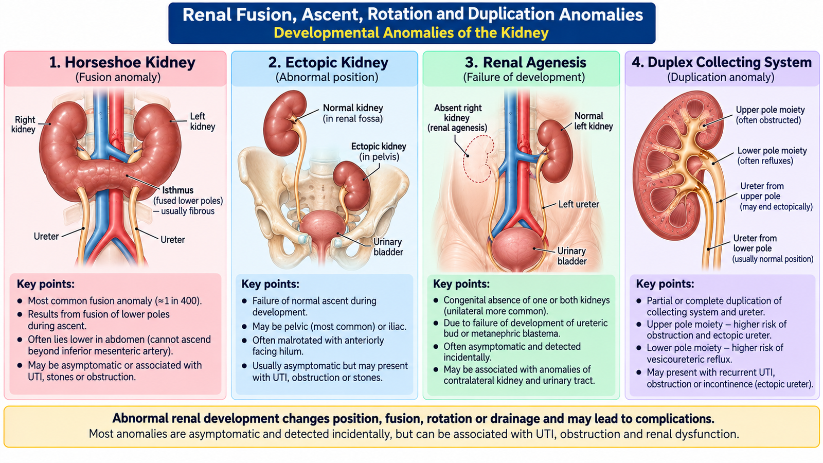
During normal development, the kidneys form in the pelvis and ascend to the upper posterior abdomen while changing their orientation. Abnormal ascent, fusion, rotation or ureteric development therefore produces predictable congenital anomalies. Many are discovered incidentally, but abnormal anatomy may disturb urinary drainage and predispose to obstruction, infection or stone formation.
Horseshoe Kidney
A horseshoe kidney results when the two developing kidneys become fused, usually at their lower poles. The fused tissue forms an isthmus across the midline. During ascent, the fused kidney is unable to rise normally and therefore remains lower than usual.
- The kidneys are joined across the midline, usually at their lower poles.
- Ascent is restricted, classically when the fused organ reaches the region of the inferior mesenteric artery.
- Many patients remain asymptomatic.
- Abnormal position and drainage may predispose to PUJ obstruction, urinary infection and renal calculi.
Ectopic Kidney
An ectopic kidney lies outside its normal renal fossa because normal ascent has failed. A pelvic kidney is a common pattern. Because its blood supply and ureteric course develop in an unusual position, drainage may sometimes be impaired, although many ectopic kidneys function normally and are discovered incidentally.
Renal Agenesis
Renal agenesis means complete absence of renal development. The consequence depends greatly on whether one or both kidneys are absent.
- Unilateral renal agenesis: may be clinically silent because the opposite kidney undergoes compensatory enlargement and maintains renal function.
- Bilateral renal agenesis: causes profound fetal renal failure and severe reduction of amniotic fluid. This interferes with normal fetal lung development and is incompatible with sustained postnatal survival.
Renal Malrotation
During ascent, the kidney normally rotates so that the renal pelvis is directed medially. In malrotation, this process is incomplete or abnormal. Malrotation may be an incidental finding but can accompany other renal positional abnormalities and occasionally contribute to impaired drainage.
Duplication of the Collecting System
Duplication occurs when ureteric development produces two drainage systems. Duplication may be incomplete, with the ureters joining before reaching the bladder, or complete, with two ureters entering the lower urinary tract separately.
Many duplications cause no symptoms. Clinically important cases may be associated with abnormal ureteric insertion, vesicoureteric reflux, obstruction, ureterocele or recurrent urinary infection.
F. Congenital Bladder and Urethral Anomalies with Posterior Urethral Valves
Congenital abnormalities of the lower urinary tract are important because they may interfere with storage or outflow of urine. When obstruction is severe, pressure can be transmitted backward from the bladder to the ureters and kidneys. Consequently, a lesion that originates in the urethra can eventually produce hydronephrosis, infection and chronic renal damage.
Important Congenital Bladder Anomalies
Urachal Abnormalities
The urachus is an embryological connection between the developing bladder and the umbilical region that normally disappears. Failure of complete closure may produce a patent urachus, urachal sinus or urachal cyst. A completely patent urachus may allow urine to discharge from the umbilicus.
Bladder Exstrophy
Bladder exstrophy is a major congenital defect in which the anterior bladder wall and overlying lower abdominal wall fail to develop normally, leaving the bladder mucosa exposed. It requires specialized reconstructive management.
Congenital Urethral Abnormalities
Congenital urethral abnormalities include abnormal development of the urethral lumen or its external opening. Examples include posterior urethral valves, congenital stenosis or atresia, and abnormalities such as hypospadias or epispadias. Within the renal module, posterior urethral valves are particularly important because they can produce severe urinary obstruction and renal damage.
Posterior Urethral Valves
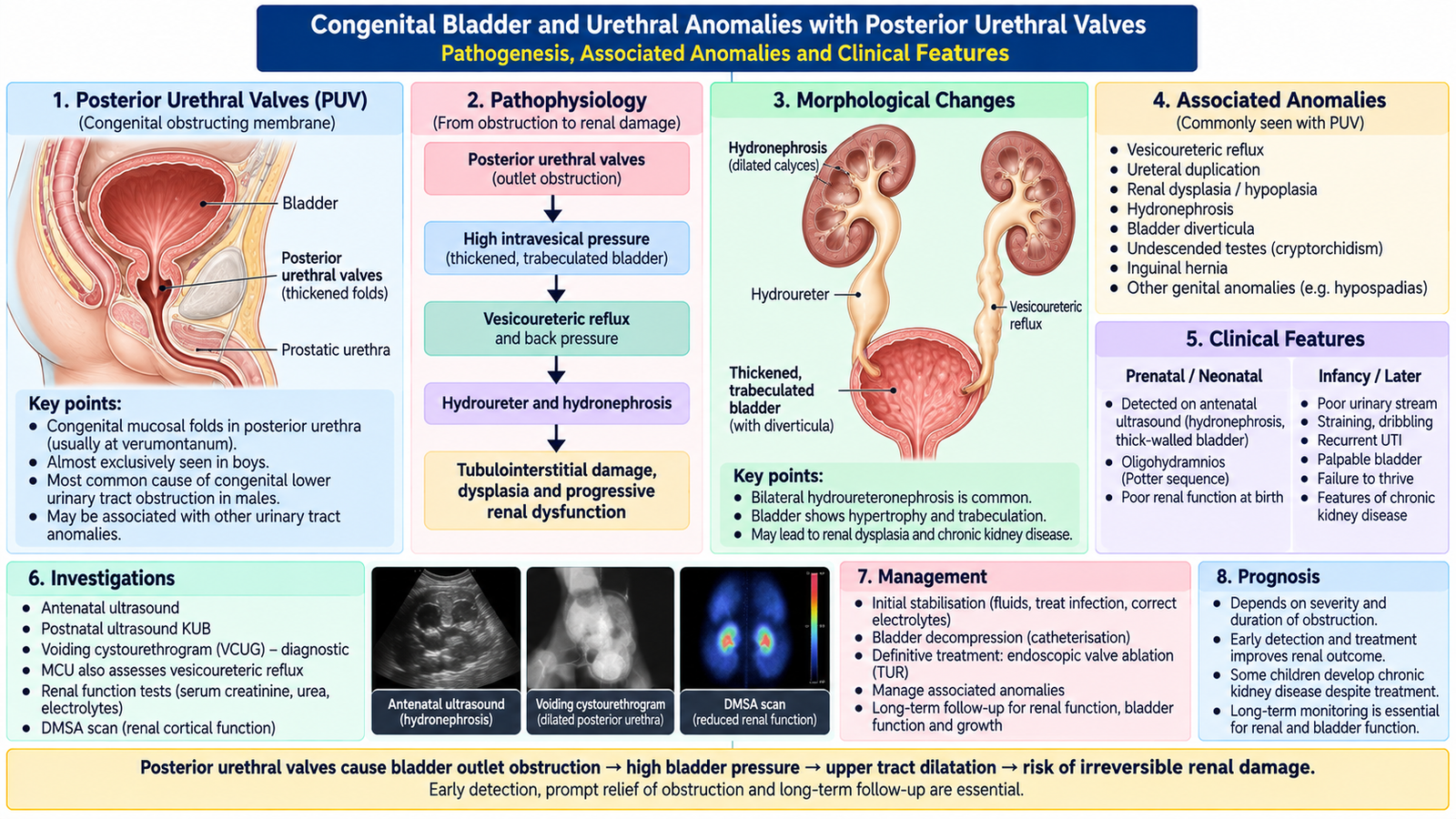
Posterior urethral valves (PUV) are congenital obstructing membranous folds within the posterior urethra of male infants and children. They interfere with bladder emptying. The bladder must generate higher pressure to overcome the obstruction, and that pressure can eventually be transmitted to the ureters and kidneys.
Clinical Presentation
Severe disease may be recognized antenatally because urinary obstruction produces a large bladder and upper urinary tract dilatation. After birth, the presentation depends on the degree of obstruction and existing renal damage.
- Antenatal bilateral hydronephrosis or a distended fetal bladder.
- Poor urinary stream or difficulty passing urine.
- Urinary retention or a palpable distended bladder.
- Recurrent urinary tract infection.
- Failure to thrive in a child with significant chronic disease.
- Raised serum creatinine or electrolyte disturbances when renal function is impaired.
- Severe fetal obstruction may produce oligohydramnios and impaired pulmonary development.
Investigations
- Ultrasonography: assesses the bladder, hydronephrosis, ureters and renal parenchyma.
- Serum creatinine and electrolytes: evaluate renal dysfunction.
- Urinalysis and urine culture: detect associated urinary infection.
- Micturating cystourethrogram / voiding cystourethrogram: demonstrates the posterior urethral obstruction and may also show vesicoureteric reflux.
Management
The immediate objective is to relieve urinary obstruction and stabilize the child. Definitive treatment then removes the obstructing valve tissue while renal function and bladder behavior are followed over time.
- Initial bladder drainage: urethral catheterization is used when feasible to decompress the urinary tract.
- Correct dehydration, electrolyte disturbance and infection when present.
- Endoscopic valve ablation: is the usual definitive treatment when the infant or child is stable and the urethra can be safely instrumented.
- Temporary urinary diversion, such as vesicostomy, may be required in selected cases when adequate decompression cannot otherwise be achieved.
- Long-term follow-up is essential because bladder dysfunction, reflux and chronic kidney disease may persist despite successful relief of the original obstruction.
Integrated Mechanism Flow: From Structural Abnormality to Renal Damage
The initiating abnormality differs between ADPKD, PUJ obstruction and posterior urethral valves, but each can ultimately reduce functioning renal tissue when structural distortion or obstruction becomes severe.
⭐ AIM High-Yield Review
Cystic and Congenital Renal Disorders
Use these videos after reading the AIM learning material to reinforce polycystic kidney disease, PUJ obstruction, congenital renal anomalies and posterior urethral valves.
1. Cystic Kidney Disease — ADPKD & ARPKD
Recommended for cyst formation, ADPKD, ARPKD, complications, diagnosis and management.
2. Pelviureteric Junction (PUJ) Obstruction
Focus on presentation, obstruction, hydronephrosis, pyeloplasty and endopyelotomy.
3. Congenital Renal Anomalies
Reinforces renal agenesis, ectopic kidney, horseshoe kidney and other congenital urinary tract abnormalities.
4. Posterior Urethral Valves
Covers pathogenesis, clinical presentation, investigations, treatment and follow-up of posterior urethral valves.