
Topic 9 — Obstructive Uropathy and Lower Urinary Tract Dysfunction
Topic Introduction
Obstructive uropathy occurs when urine cannot flow normally through the urinary tract. The obstruction may occur from the renal pelvis down to the urethra, and its effects depend on the level, severity and duration of blockage. Persistent obstruction produces back-pressure, progressive dilatation of the collecting system and, eventually, loss of renal tissue and function. In the lower urinary tract, benign prostatic hyperplasia, urinary incontinence and urethral stricture are important causes of urinary symptoms and dysfunction. In this chapter, you will understand how urinary obstruction damages the kidney, how BPH produces lower urinary tract symptoms, how BPH is assessed and treated, how major forms of urinary incontinence are recognized and managed, and how urethral stricture presents and is investigated.
A. Obstructive Uropathy and Hydronephrosis
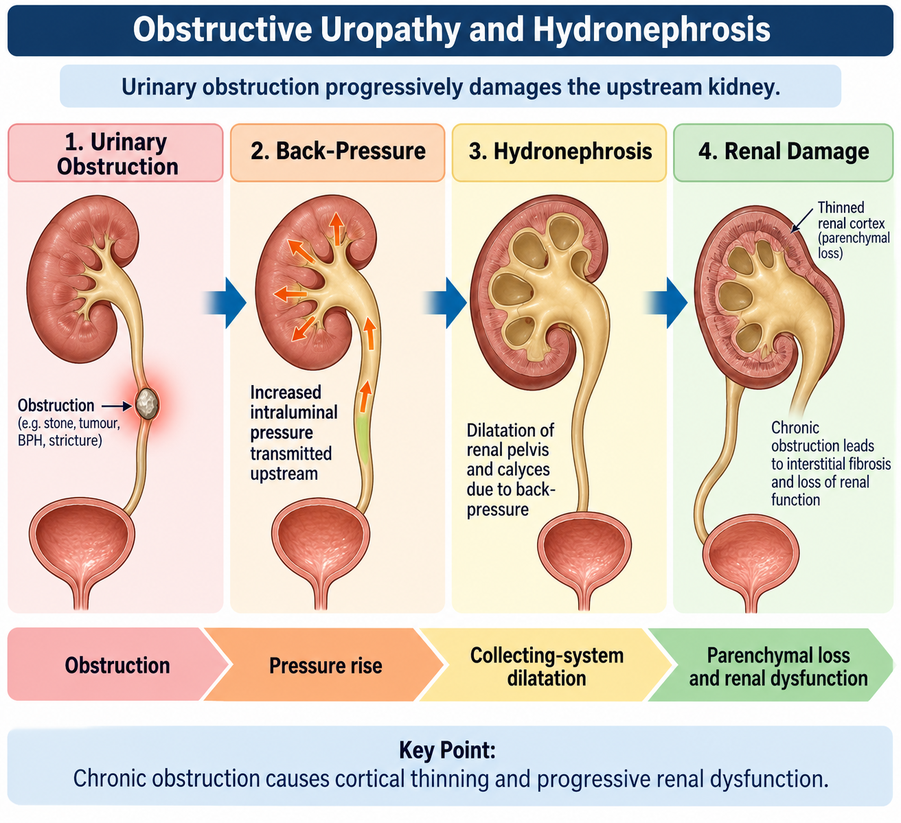
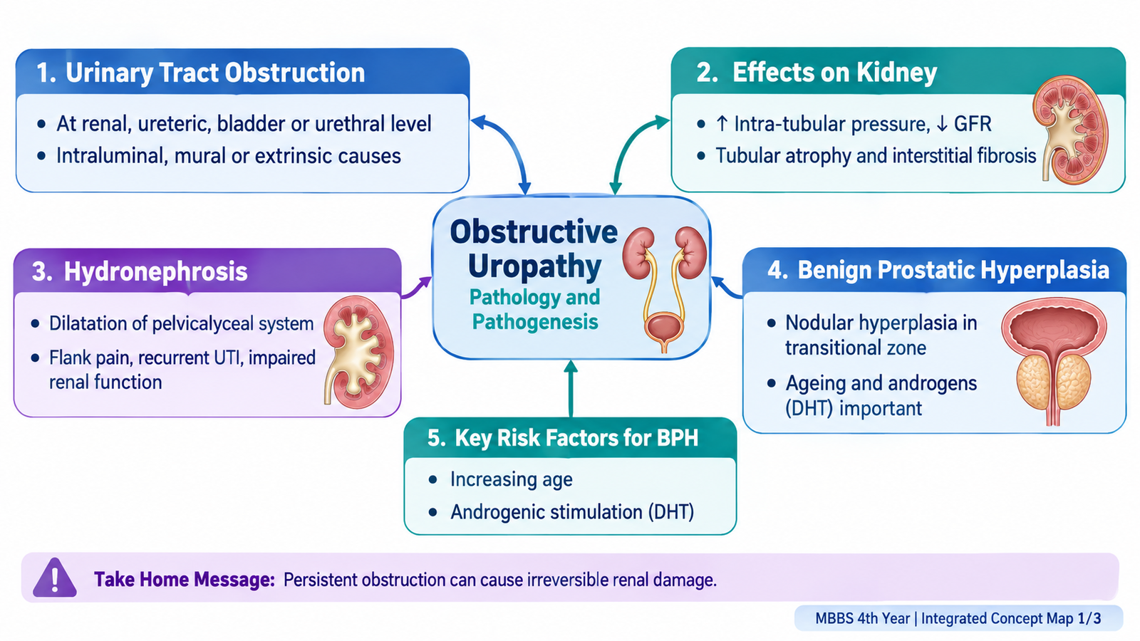
Obstructive uropathy means structural or functional interference with urine flow anywhere from the renal collecting system to the urethral meatus. When urine cannot pass freely, pressure rises proximal to the obstruction. Initially this disturbs filtration and tubular function. If the obstruction persists, progressive pressure-related injury causes tubular atrophy, interstitial fibrosis and loss of renal parenchyma.
Anatomical Levels of Urinary Tract Obstruction
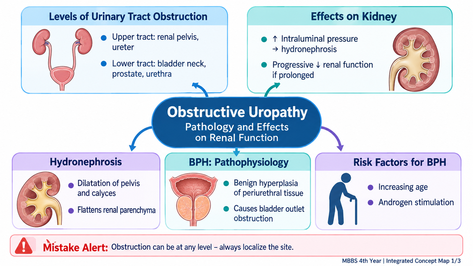
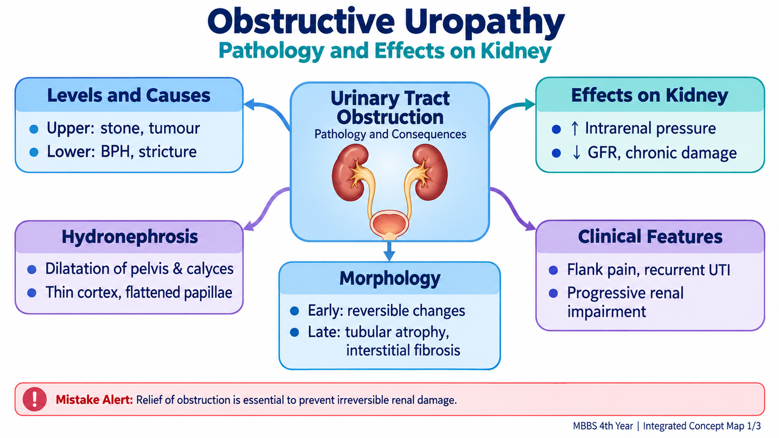
The consequences of obstruction are closely related to its anatomical level. A unilateral upper-tract obstruction usually affects one kidney, whereas obstruction at the bladder outlet or urethra can affect both kidneys because both ureters drain into the same bladder.
- Renal pelvis / pelvi-ureteric junction: narrowing at the junction between the renal pelvis and ureter obstructs drainage from that kidney.
- Ureter: stones, strictures, tumors or external compression may cause unilateral or bilateral obstruction.
- Bladder outlet: BPH is an important acquired cause in older men.
- Urethra: urethral stricture can obstruct bladder emptying.
Effects on Renal Function
Obstruction raises hydrostatic pressure in the collecting system and renal tubules. This pressure is transmitted backward toward the nephron and opposes glomerular filtration. Renal blood flow and tubular handling of water and electrolytes also become abnormal. If obstruction is complete and prolonged, nephron function progressively declines.
Urinary obstruction → increased upstream pressure → reduced filtration and tubular dysfunction → tubular atrophy and interstitial fibrosis → progressive loss of renal function.
With unilateral obstruction, serum creatinine may remain near normal because the opposite kidney continues to function. Bilateral obstruction, or obstruction in a patient with only one functioning kidney, can produce marked renal impairment and acute kidney injury.
Hydronephrosis
Hydronephrosis is dilatation of the renal pelvis and calyces caused by impaired urinary drainage. It is a structural consequence of obstruction rather than a specific disease. The associated renal damage depends mainly on how severe and prolonged the obstruction is.
Gross Morphology
- The renal pelvis and calyces become progressively dilated.
- Renal papillae become flattened as pressure increases.
- With longstanding obstruction, the renal cortex becomes thin because functioning parenchyma is lost.
- Severe chronic disease may leave a markedly dilated collecting system surrounded by only a thin rim of renal tissue.
Microscopic Morphology
Persistent pressure produces tubular dilatation followed by tubular atrophy. Chronic interstitial inflammation and fibrosis develop, while glomeruli may remain relatively preserved early in the process. Advanced disease ultimately produces widespread parenchymal loss.
Clinical Consequences
The presentation depends on the cause and rate of obstruction. Acute obstruction may produce pain, whereas slowly developing obstruction may remain relatively silent until significant renal damage has occurred. Urinary stasis also increases susceptibility to infection.
- Flank or loin pain may occur with acute upper-tract obstruction.
- Reduced urinary flow or retention suggests lower-tract obstruction.
- Urinary stasis predisposes to infection.
- Prolonged bilateral obstruction may cause renal failure.
- Relief of obstruction before irreversible fibrosis develops may allow substantial recovery of function.
B. Benign Prostatic Hyperplasia: Pathophysiology and Lower Urinary Tract Dysfunction
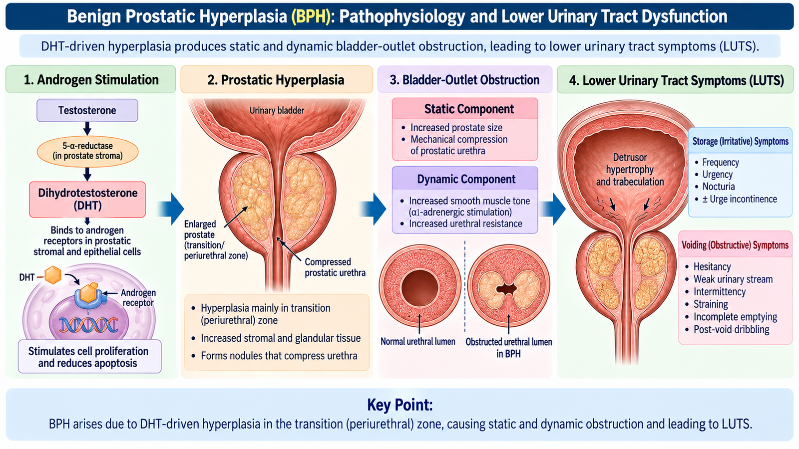
Benign prostatic hyperplasia (BPH) is a non-malignant proliferation of prostatic glandular and stromal tissue. It develops mainly in the periurethral or transition-zone region of the prostate. Because this tissue surrounds the proximal urethra, progressive enlargement can compress the urethral lumen and increase resistance to urinary flow.
Pathophysiology
Prostatic growth is androgen dependent. Within prostatic tissue, testosterone is converted by 5-alpha-reductase to dihydrotestosterone (DHT). DHT binds androgen receptors and supports growth and survival of stromal and epithelial cells. With increasing age, this hormonal environment favors nodular hyperplasia.
BPH causes obstruction through two related mechanisms. The static component results from the physical bulk of enlarged prostatic tissue. The dynamic component results from alpha1-adrenergic-mediated smooth-muscle tone in the prostate and bladder neck. This distinction explains why alpha blockers can improve symptoms rapidly without actually shrinking the prostate.
Risk Factors and Predisposing Factors
- Increasing age is the major clinical association.
- Androgenic stimulation is required for the development and maintenance of prostatic hyperplasia.
- Family or genetic susceptibility may increase risk in some men.
- Metabolic factors such as obesity and metabolic syndrome are associated with greater risk in some populations.
Lower Urinary Tract Symptoms
The symptoms produced by BPH are collectively called lower urinary tract symptoms (LUTS). They are commonly divided into storage and voiding symptoms. Symptoms do not always correlate perfectly with prostate size because smooth-muscle tone and bladder function also contribute.
- Weak urinary stream
- Hesitancy
- Intermittency
- Straining
- Feeling of incomplete emptying
- Frequency
- Urgency
- Nocturia
- Urgency incontinence in some patients
Effects on the Bladder and Upper Urinary Tract
To overcome increased outlet resistance, the detrusor muscle initially contracts more strongly and undergoes hypertrophy. Prolonged obstruction may produce bladder trabeculation, increased residual urine and eventually impaired detrusor function. Severe longstanding obstruction can transmit pressure to the ureters and kidneys, producing hydroureter, hydronephrosis and renal dysfunction.
C. Clinical Assessment of BPH and the IPSS
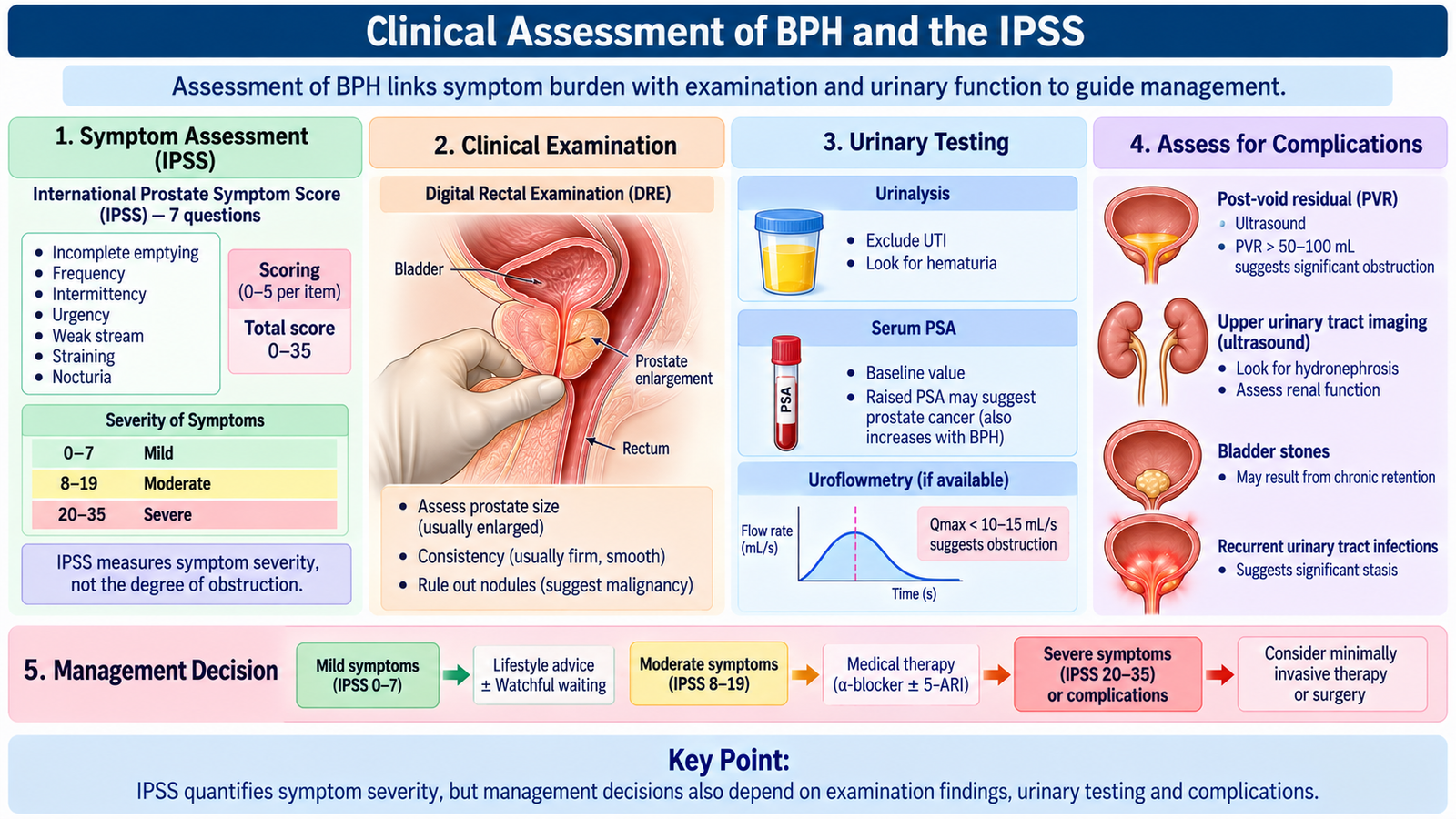
Assessment of a patient with an enlarged prostate should determine three things: the severity of symptoms, whether significant urinary obstruction or complications are present, and whether another disorder could explain the symptoms. BPH should therefore not be diagnosed from prostate size alone.
History
Ask about both storage and voiding symptoms, their duration, their effect on sleep and daily activity, episodes of retention, visible hematuria, recurrent urinary infection and current medications. Neurological disease, diabetes and previous urinary tract procedures may suggest alternative or additional causes of dysfunction.
International Prostate Symptom Score — IPSS
The IPSS provides a standardized method for measuring the severity of male LUTS. It contains seven symptom questions, each scored from 0 to 5, producing a total score from 0 to 35.
Mild
Moderate
Severe
The questions assess incomplete emptying, frequency, intermittency, urgency, weak stream, straining and nocturia. A separate quality-of-life question asks how bothered the patient is by his urinary condition.
Examination
General examination looks for complications and alternative causes. Abdominal examination may reveal a palpable distended bladder. A focused neurological examination is useful when neurogenic dysfunction is suspected.
On digital rectal examination (DRE), uncomplicated BPH classically produces a smooth, firm or rubbery, symmetrically enlarged prostate. A hard, irregular or nodular prostate requires assessment for malignancy rather than being attributed automatically to BPH.
Investigations
- Urinalysis: looks for infection, blood and other urinary abnormalities.
- Renal function testing: is useful where renal impairment or significant obstruction is suspected.
- Post-void residual urine: estimates how much urine remains after voiding and helps assess emptying.
- Uroflowmetry: provides an objective measure of urinary flow; a reduced flow suggests impaired emptying but is not specific for BPH.
- Ultrasonography: may assess bladder residual urine, prostate size and the upper urinary tract when clinically indicated.
- PSA: may be considered when assessment for prostate cancer would influence clinical management. PSA is not a specific diagnostic test for BPH.
Watchful Waiting
Men with mild symptoms, or symptoms that cause little bother and are not associated with important complications, may be managed initially by watchful waiting. This means active observation rather than neglect. The patient is reviewed for symptom progression or complications.
Simple measures may include reducing excessive evening fluid intake, moderating caffeine where it worsens urgency or frequency, reviewing medicines that affect urination and avoiding unnecessary delay in voiding.
D. Pharmacological Treatment of BPH
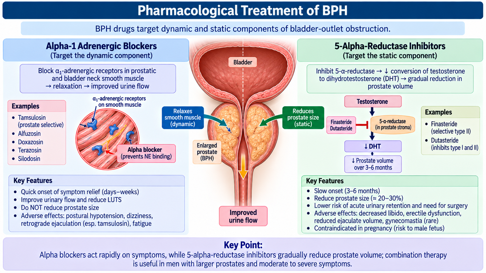
Drug treatment for BPH targets either the dynamic component of obstruction, caused by smooth-muscle tone, or the static component, caused by enlarged prostatic tissue. Understanding this difference makes the drug classification easy: alpha1-adrenergic blockers relax smooth muscle, while 5-alpha-reductase inhibitors reduce DHT formation and gradually reduce the glandular component of enlargement.
Classification
- Alpha1-adrenergic blockers
- Less prostate-selective examples: doxazosin, terazosin.
- Uroselective / prostate-selective agents: tamsulosin, silodosin; alfuzosin is also used for LUTS with relatively greater urinary selectivity.
- 5-alpha-reductase inhibitors: finasteride and dutasteride.
- Combination therapy: an alpha blocker plus a 5-alpha-reductase inhibitor in appropriately selected patients.
Alpha1-Adrenergic Blockers
Alpha1 receptors are present in smooth muscle within the prostate and bladder neck. Their activation increases smooth-muscle tone. Blocking these receptors reduces this tone and lowers resistance to urinary flow.
Because these drugs act on muscle tone rather than tissue growth, symptomatic improvement occurs more rapidly than with 5-alpha-reductase inhibition. They do not significantly reduce prostate volume.
Adverse Effects
- Dizziness and postural hypotension, especially with less uroselective agents.
- Weakness or fatigue.
- Nasal congestion.
- Ejaculatory dysfunction, particularly with highly alpha1A-selective drugs.
- Tamsulosin is associated with intraoperative floppy iris syndrome, so a history of its use is relevant before cataract surgery.
5-Alpha-Reductase Inhibitors
Finasteride inhibits 5-alpha-reductase and therefore reduces conversion of testosterone to DHT. Dutasteride acts on more than one isoenzyme of 5-alpha-reductase. Reduced intraprostatic DHT gradually decreases androgen-dependent prostatic growth.
These drugs work more slowly than alpha blockers but can be particularly useful when significant prostatic enlargement contributes to symptoms and risk of progression.
Adverse Effects
- Reduced libido.
- Erectile dysfunction.
- Ejaculatory disturbance or reduced ejaculate volume.
- Gynecomastia may occur.
- Serum PSA is reduced during treatment, which must be considered when PSA results are interpreted.
Combination Therapy
Combination therapy targets both components of BPH. The alpha blocker provides relatively rapid relaxation of prostatic smooth muscle, while the 5-alpha-reductase inhibitor acts more slowly on the enlarged prostate. The potential benefit must be balanced against exposure to adverse effects from both classes.
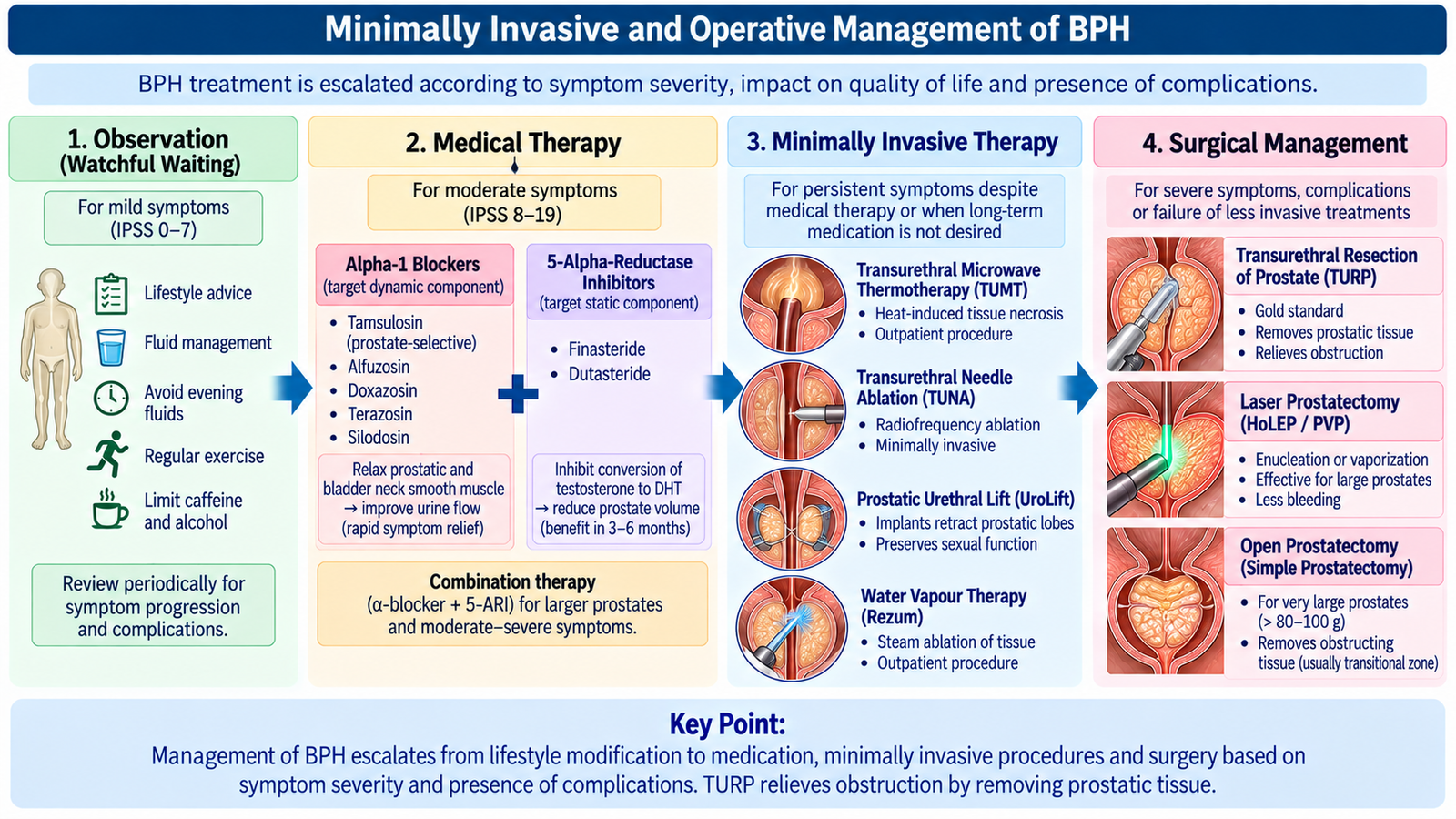
E. Minimally Invasive and Operative Management of BPH
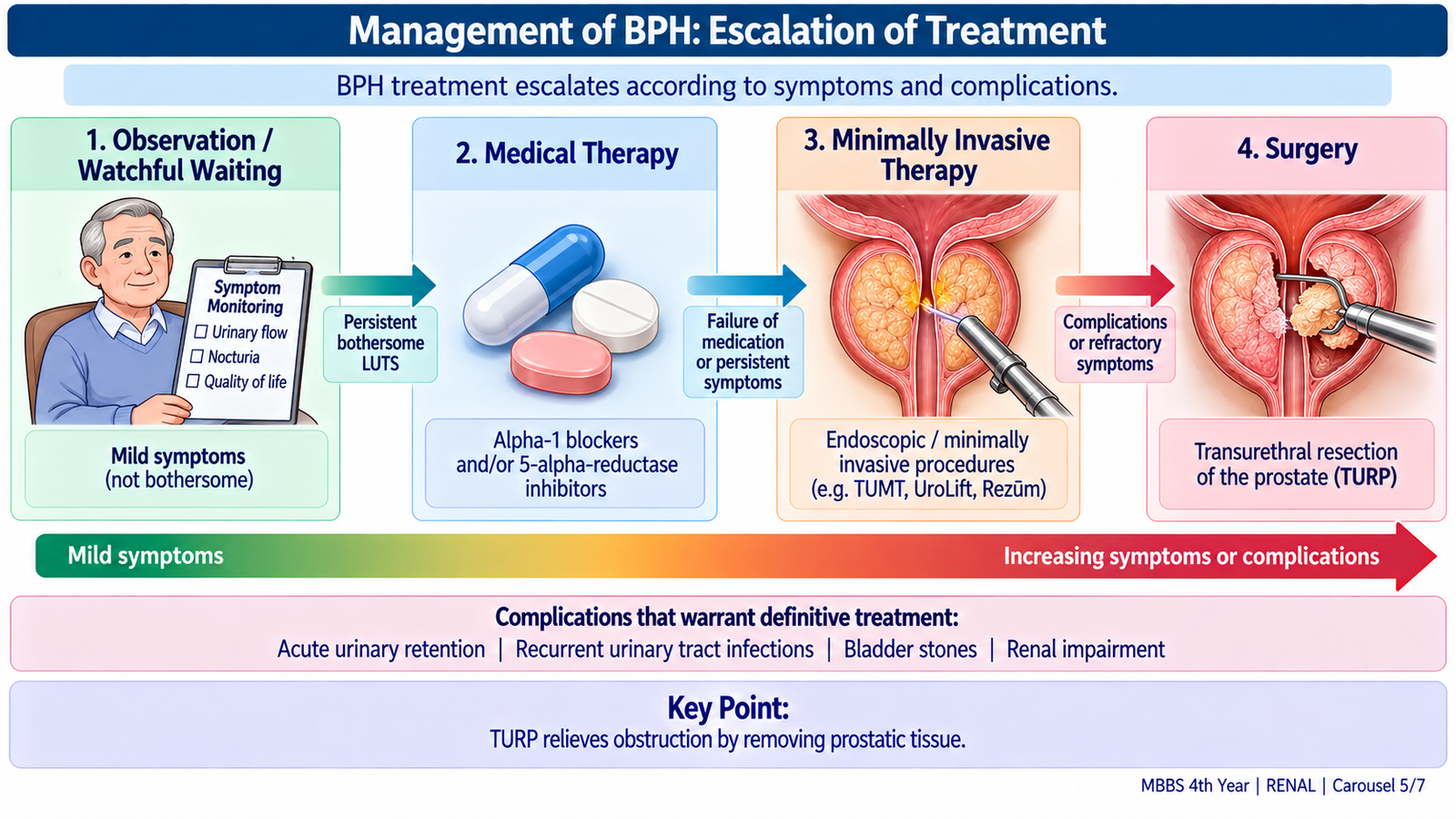
Procedural treatment is considered when symptoms remain troublesome despite appropriate conservative or medical therapy, when the patient prefers intervention, or when clinically important complications of obstruction develop. The aim is to reduce resistance at the prostatic urethra while avoiding unnecessary operative morbidity.
When Intervention Becomes Important
The decision is based on symptoms, quality of life, prostate characteristics, bladder function and complications rather than prostate size alone. Important complications of significant outlet obstruction include recurrent retention, recurrent infection, bladder stones and upper-tract or renal deterioration.
Minimally Invasive Treatment
Minimally invasive procedures attempt to improve the prostatic urethral channel using less invasive techniques than conventional resection. Depending on the technique, this may involve mechanically widening the prostatic urethra or destroying obstructing tissue with thermal or other energy.
Examples include prostatic urethral lift and selected thermal ablation techniques. Their suitability depends on prostate anatomy, severity of symptoms, patient preference and availability. Detailed procedural technique is beyond the undergraduate requirement.
Transurethral Resection of the Prostate — TURP
TURP is a standard transurethral operation in which obstructing prostatic tissue is resected through the urethra. By removing tissue that compresses the prostatic urethra, it reduces the static component of bladder-outlet obstruction and improves urinary flow.
Important complications to recognize at undergraduate level include bleeding, urinary infection, retrograde ejaculation, urethral stricture and, less commonly with modern techniques, fluid-related complications historically associated with TURP.
Open Simple Prostatectomy
Open prostatectomy for BPH is a simple prostatectomy, meaning that obstructing adenomatous prostatic tissue is removed rather than the entire prostate as in radical prostatectomy for cancer. It may be considered when the benign gland is very large or when an open approach is appropriate because of associated pathology.
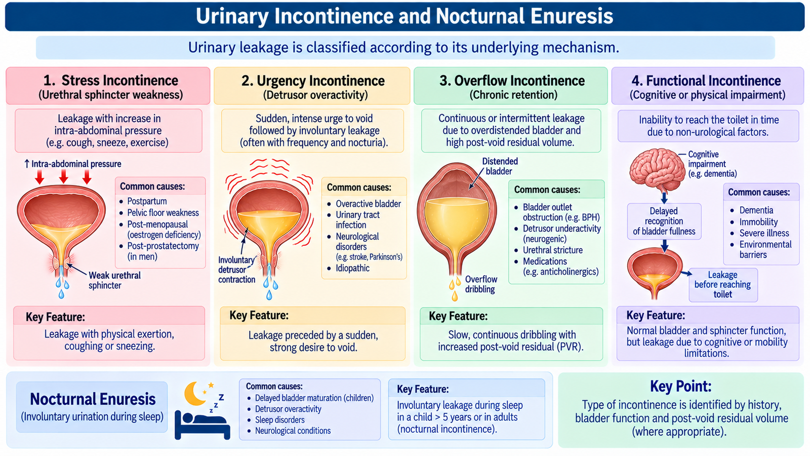
F. Urinary Incontinence and Nocturnal Enuresis
Urinary incontinence is involuntary leakage of urine. It is a symptom with several different mechanisms rather than a single disease. Correct classification matters because treatment that helps one form may be ineffective in another. The main clinical task is to decide whether leakage results from poor urethral closure, involuntary detrusor contraction, urinary retention with overflow, continuous abnormal leakage, or inability to reach and use a toilet despite an otherwise functioning urinary tract.
Stress Urinary Incontinence
Stress incontinence occurs when bladder pressure rises during coughing, sneezing, exercise or lifting but urethral closure is insufficient to prevent leakage. It usually results from weakness of pelvic support and/or urethral sphincteric function.
Pressure rise without effective urethral closure → urine leakage during physical stress.
Urgency Incontinence
Urgency incontinence is leakage accompanied by a sudden compelling desire to void that is difficult to defer. The underlying functional abnormality is commonly detrusor overactivity, in which involuntary bladder contractions occur during the storage phase.
Overflow Incontinence
Overflow incontinence develops when the bladder cannot empty effectively. The bladder becomes chronically overfilled and urine leaks when bladder volume exceeds the ability of the outlet to maintain continence. Causes include severe bladder-outlet obstruction and impaired detrusor contractility.
These patients may describe a weak stream, incomplete emptying, continuous or intermittent dribbling and difficulty initiating urination. A large post-void residual supports the diagnosis.
Other Important Patterns
- Continuous incontinence: continuous leakage may occur with a urinary fistula, ectopic urinary drainage or severe sphincter failure.
- Functional incontinence: the urinary tract may function reasonably well, but cognitive impairment, poor mobility or environmental barriers prevent timely toileting.
- Mixed incontinence: commonly refers to a combination of stress and urgency incontinence.
Evaluation
Evaluation begins with the pattern of leakage. Ask when leakage occurs, whether urgency is present, whether coughing or activity provokes it, whether the stream is weak, whether emptying feels incomplete, and whether neurological disease, childbirth, pelvic surgery or medications could be relevant.
- Bladder diary: records voiding frequency, fluid intake, urgency and leakage episodes.
- Abdominal examination: may reveal a distended bladder.
- Pelvic and genital examination: assesses pelvic support and other local abnormalities.
- Cough stress test: may demonstrate stress leakage.
- Neurological examination: is important when neurogenic dysfunction is suspected.
- Urinalysis: helps identify infection or hematuria.
- Post-void residual measurement: is particularly useful when retention or overflow is suspected.
- Urodynamic testing: may be used when the mechanism is uncertain, symptoms are complex or invasive treatment is being considered.
Conservative and Surgical Treatment
Treatment is directed toward the mechanism. Conservative measures are usually tried before surgery when clinically appropriate.
- Stress incontinence: pelvic-floor muscle training and correction of aggravating factors are important. Persistent significant stress incontinence may require procedures that improve urethral support, such as a sling.
- Urgency incontinence: bladder training, timed voiding and pelvic-floor strategies may help. Antimuscarinic drugs or beta3-adrenergic agonists may be used in suitable patients.
- Overflow incontinence: the key principle is to empty the bladder and treat the underlying obstruction or impaired bladder function.
- Functional incontinence: improve access to toileting and address mobility, cognition or environmental barriers.
Nocturnal Enuresis
Nocturnal enuresis is involuntary urination during sleep after an age at which nighttime continence would normally be expected. Primary enuresis occurs when sustained nighttime dryness has never been achieved, whereas secondary enuresis appears after a period of established dryness.
Important mechanisms include delayed maturation of nighttime bladder control, failure to awaken appropriately to a full bladder, relatively excessive nighttime urine production because of an inadequate nocturnal antidiuretic response, and reduced functional bladder capacity.
Assessment should identify daytime urinary symptoms, constipation, excessive thirst or urination, urinary infection, sleep problems and psychosocial stressors where relevant. Secondary enuresis especially warrants assessment for an acquired cause.
Management begins with reassurance, appropriate evening fluid habits, regular voiding and treatment of associated problems such as constipation. An enuresis alarm can promote learned awakening to bladder filling. Desmopressin reduces nocturnal urine production and may be useful in selected patients.
G. Urethral Stricture
Urethral stricture is an abnormal narrowing of the urethral lumen caused by fibrosis and scar formation. In the anterior urethra, scarring may extend into the surrounding corpus spongiosum, a process termed spongiofibrosis. The resulting fixed narrowing increases resistance to urine flow and may eventually produce retention and secondary changes in the bladder and upper urinary tract.
Etiology
A stricture forms after injury or inflammation damages the urethral epithelium and underlying tissue. Healing by fibrosis contracts the urethral wall and narrows the lumen.
- Iatrogenic: urethral catheterization, instrumentation or previous urethral procedures.
- Traumatic: pelvic or perineal trauma may damage the urethra.
- Inflammatory or infective: previous urethritis can lead to scarring.
- Chronic inflammatory skin disease: conditions such as lichen sclerosus can involve the urethral meatus and urethra.
- Some strictures remain without an obvious identified cause.
Clinical Presentation
- Progressively weak urinary stream.
- Straining to void.
- Spraying or altered urinary stream.
- Prolonged voiding.
- Feeling of incomplete emptying.
- Post-void dribbling.
- Recurrent urinary infection.
- Acute or chronic urinary retention in severe disease.
Investigations
The purpose of investigation is not only to confirm impaired flow but also to identify the site, length and severity of the narrowed segment before definitive treatment.
- Urinalysis: assesses for infection or blood.
- Uroflowmetry: often demonstrates reduced urinary flow.
- Post-void residual: assesses incomplete bladder emptying.
- Retrograde urethrography: outlines the urethral lumen and demonstrates the location and extent of narrowing.
- Endoscopic assessment: allows direct visualization when required.
Management Principles
Treatment depends on the site, length, severity, cause and recurrence pattern of the stricture. The aim is to restore an adequate urethral lumen and protect bladder and renal function.
- Urethral dilatation: mechanically enlarges the narrowed segment but recurrence may occur.
- Direct visual internal urethrotomy: divides scar tissue endoscopically in selected strictures.
- Urethroplasty: reconstructive surgery provides definitive treatment for many recurrent, long or complex strictures.
- Urinary diversion: if severe obstruction causes retention and safe urethral passage is not possible, suprapubic drainage may be required before definitive treatment.
Integrated Mechanism Flow
Important Comparison — Major Types of Urinary Incontinence
| Type | Main Mechanism | Typical Clue | Residual Urine | Core Management Principle |
|---|---|---|---|---|
| Stress | Poor urethral closure / pelvic support | Leakage with cough, sneeze or exertion | Usually low | Pelvic-floor therapy; surgery if persistent |
| Urgency | Detrusor overactivity | Sudden urgency followed by leakage | Usually low | Bladder training ± appropriate medication |
| Overflow | Retention from obstruction or weak detrusor | Weak stream, incomplete emptying, dribbling | Raised | Drain bladder and correct cause |
| Functional | Mobility, cognition or environmental problem | Cannot reach/use toilet in time | Depends on urinary tract function | Improve toileting access and functional support |