
Topic 7 — Urolithiasis, Renal Trauma and Diagnostic Imaging of the Urinary Tract
1. Topic Introduction
Urolithiasis means formation of stones within the urinary tract, most commonly in the kidney or ureter. Stones form when substances dissolved in urine become sufficiently concentrated to crystallize, enlarge and remain within the urinary system. Their composition determines important features such as predisposing factors, urine pH, morphology and appearance on imaging. This topic also covers renal trauma, where the immediate priority is stabilization followed by assessment of the severity of renal injury. Finally, urinary imaging connects both problems with clinical diagnosis. You will learn how plain X-ray KUB, CT and nuclear scans such as DTPA, DMSA and MAG-3 provide different types of anatomical and functional information, including the special approach required during pregnancy.
A. Urolithiasis — Formation, Classification and Core Pathogenesis
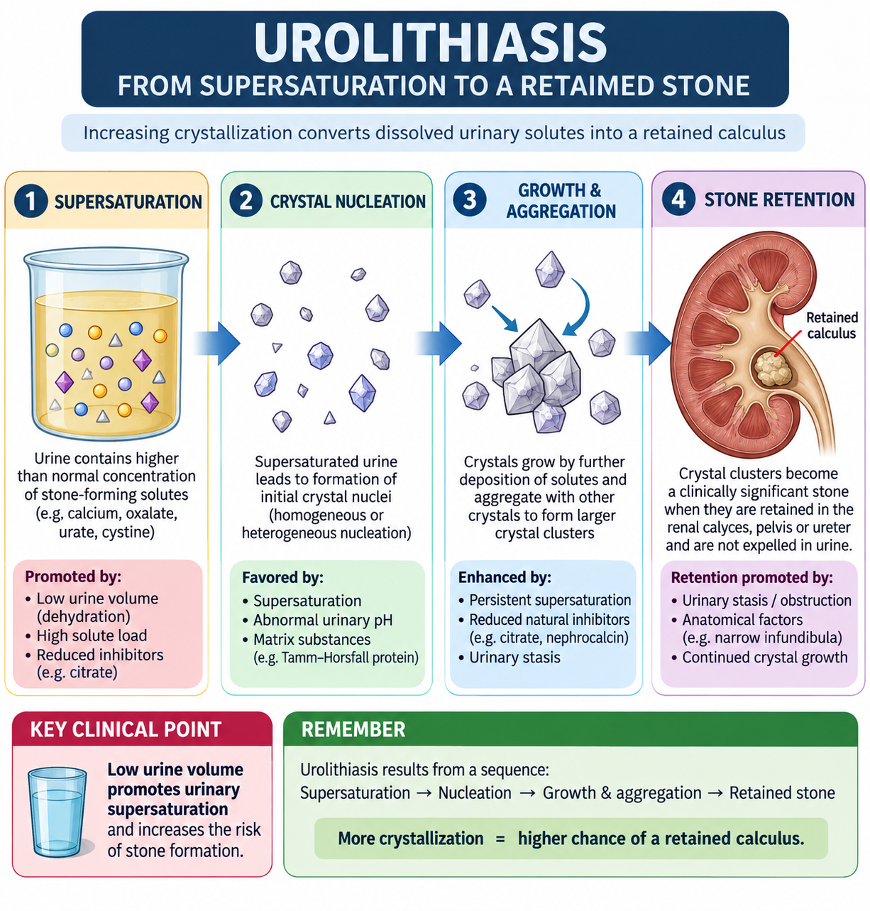
A urinary stone is a solid crystalline mass formed from substances normally present in urine. Stone formation is not simply the presence of calcium, uric acid or another solute. It occurs when the urinary environment favors crystallization and allows crystals to grow rather than remain dissolved and pass out in urine.
How a stone forms
The first important step is supersaturation. This means that urine contains more of a stone-forming substance than can remain freely dissolved. Supersaturation may result from low urine volume, increased urinary excretion of a solute or a urine pH that decreases the solubility of that substance.
During nucleation, the first small crystal forms. Additional material can then deposit on this crystal, producing crystal growth. Several crystals may join together by aggregation. A clinically important stone develops when these crystals are retained within the kidney or urinary tract rather than being washed away.
Urine also contains substances that normally reduce crystallization. Citrate is important because it binds calcium and decreases the amount of free calcium available for calcium-stone formation. Therefore, hypocitraturia can promote calcium stones.
Main types of urinary stones
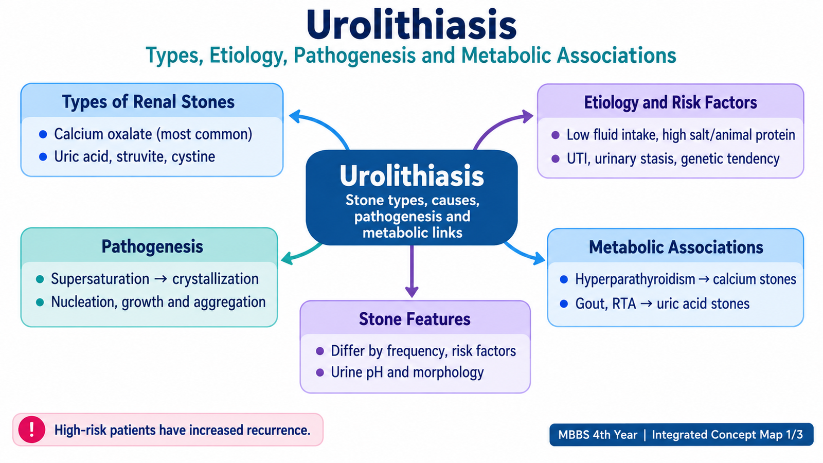
Urinary stones are classified mainly according to their chemical composition. The composition matters because it points toward the underlying metabolic or infectious process and influences imaging and treatment.
- Calcium stones — mainly calcium oxalate, often with calcium phosphate; these are the most common stones.
- Uric acid stones — form particularly when urine remains acidic.
- Struvite stones — composed mainly of magnesium ammonium phosphate and associated with urinary infection by urease-producing organisms.
- Cystine stones — uncommon stones caused by inherited cystinuria.
B. Major Stone Types — Predisposing Factors, Urine pH and Morphology
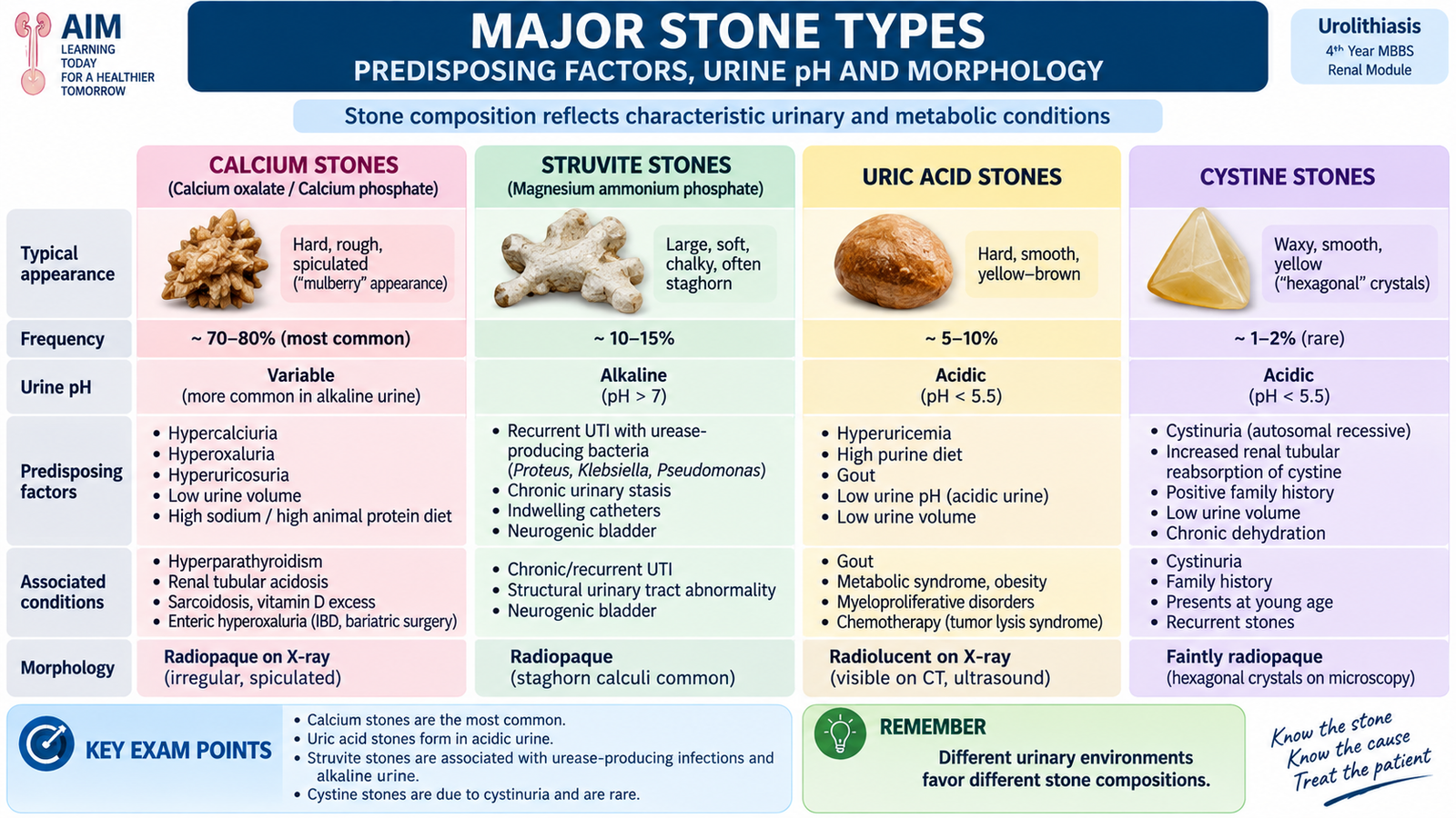
Different urinary stones form under different biochemical conditions. Recognizing the relationship between stone composition, metabolic disease, urine pH and morphology helps explain why a particular patient develops a particular type of stone.
Calcium stones
Calcium-containing stones are the commonest urinary stones. Calcium oxalate is the dominant component in many cases, although calcium phosphate may also be present. Increased urinary calcium, increased urinary oxalate, reduced urinary citrate and low urine volume can all favor formation.
Important metabolic associations include:
- Hypercalciuria, which may occur even when the serum calcium is normal.
- Hyperparathyroidism, where increased calcium availability can promote hypercalciuria.
- Hyperoxaluria, which increases urinary calcium oxalate supersaturation.
- Hypocitraturia, which removes an important inhibitor of calcium crystallization.
- Distal renal tubular acidosis, particularly in relation to calcium phosphate stones and alkaline urine.
Calcium oxalate stones are often hard and irregular or rough. Their calcium content makes them radiopaque, so they are usually visible on a plain X-ray KUB.
Struvite stones
Struvite stones develop in association with urinary infection by organisms that produce the enzyme urease. Urease splits urea and increases urinary ammonia, which makes the urine alkaline. This favors precipitation of magnesium ammonium phosphate.
These stones can become very large and may branch through the renal pelvis and calyces, producing a staghorn calculus. Their association with persistent infection is clinically important because treating only the stone without addressing infection can lead to recurrence.
Uric acid stones
Uric acid becomes less soluble as urine becomes more acidic. Persistent acidic urine therefore promotes uric acid crystallization. Hyperuricemia or increased uric acid production can add to this risk, but acidic urine is a particularly important factor.
Uric acid stones may be associated with disorders of increased urate production or turnover, including gout and some high-cell-turnover states. They are usually smooth and yellow to reddish-brown. Because they contain no calcium, they are radiolucent on plain X-ray KUB, although they are visible on CT.
Cystine stones
Cystine stones occur in cystinuria, an inherited disorder in which renal tubular reabsorption of cystine and certain other dibasic amino acids is impaired. Excess cystine enters the urine. Because cystine has poor solubility, particularly in acidic urine, crystals and stones can form.
The classic urinary finding is a hexagonal cystine crystal. Stones are often smooth and waxy. Cystinuria should be considered particularly when stone disease begins at a young age or repeatedly recurs.
Important comparison of stone types
| Stone | Relative frequency | Important predisposition | Urine environment | Typical clue |
|---|---|---|---|---|
| Calcium oxalate ± phosphate | Most common | Hypercalciuria, hyperoxaluria, hypocitraturia | Variable; calcium phosphate favored by alkaline urine | Radiopaque; often rough/irregular |
| Struvite | Less common | Urease-producing urinary infection | Alkaline | May form staghorn calculus |
| Uric acid | Less common | Persistently acidic urine; hyperuricemia/hyperuricosuria | Acidic | Radiolucent on plain KUB |
| Cystine | Rare | Inherited cystinuria | Poor solubility in acidic urine | Hexagonal urinary crystals |
C. Clinical Presentation, Diagnosis and Evaluation of the Stone Former
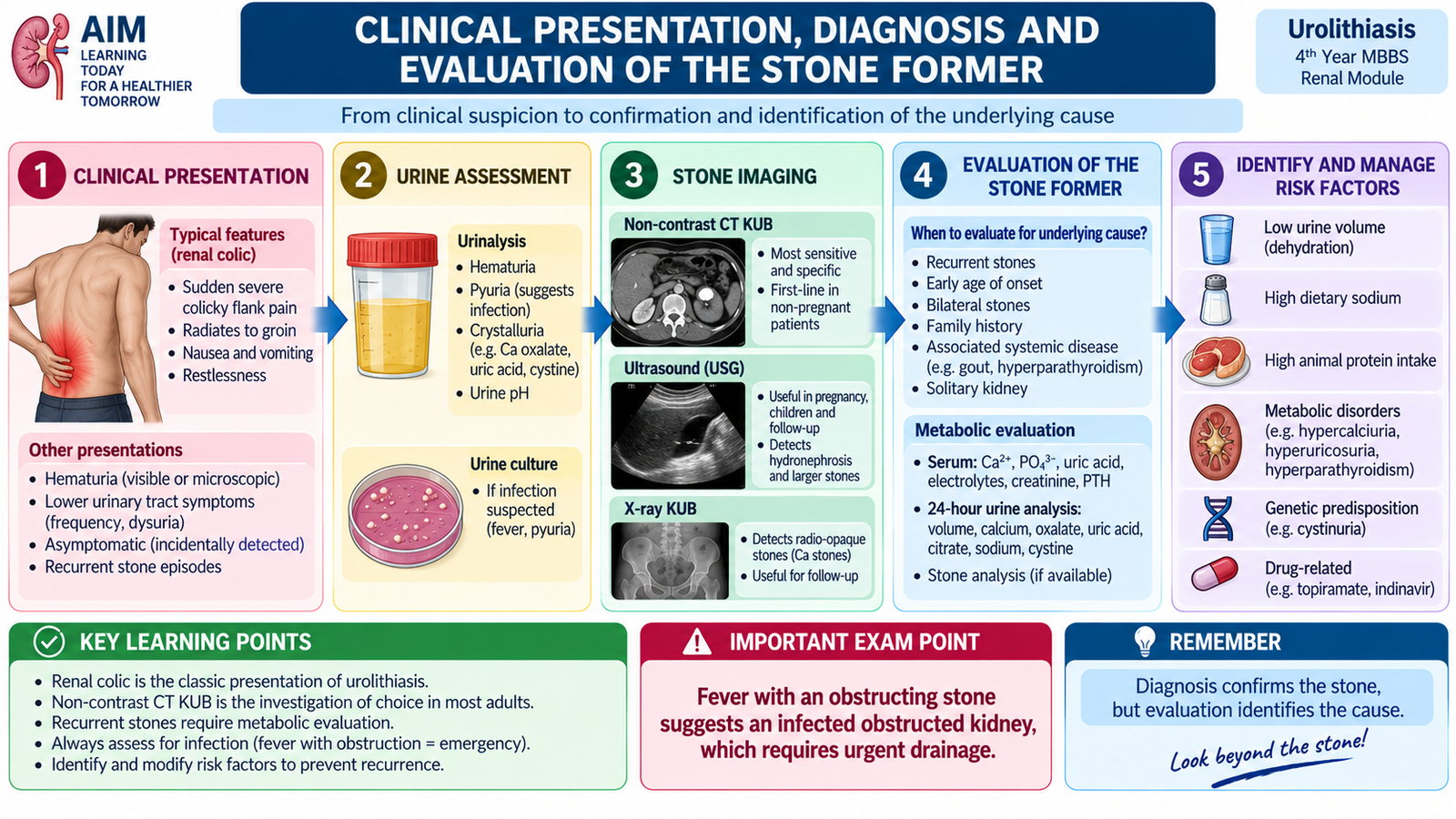
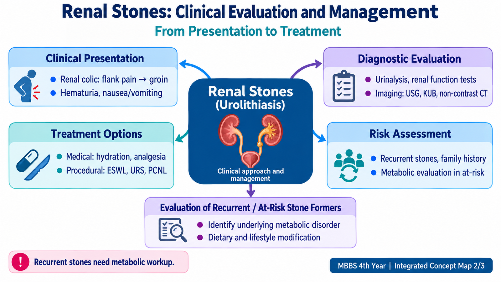
A stone may remain silent while it lies within the kidney. Symptoms commonly appear when it moves into the ureter and causes obstruction. Distension and vigorous ureteric contraction against the obstruction produce the characteristic pain of renal or ureteric colic.
Clinical presentation
The classical presentation is sudden severe flank pain. As a ureteric stone moves distally, pain may radiate toward the lower abdomen, groin, testis or labium. Unlike many patients with peritoneal inflammation, a patient with renal colic is often restless because changing position does not reliably relieve the pain.
- Severe colicky flank pain, often radiating toward the groin.
- Hematuria, microscopic or visible, due to mucosal irritation.
- Nausea and vomiting, commonly accompanying severe visceral pain.
- Urinary frequency or urgency when a stone approaches the lower ureter or vesicoureteric junction.
- Costovertebral-angle tenderness may be present.
Initial diagnostic assessment
Diagnosis combines the clinical pattern with urine testing, renal-function assessment and imaging. Investigations should not merely confirm a stone; they should also identify infection, impaired renal function and other features that change management.
- Urinalysis: looks for blood, urinary pH and features suggesting infection.
- Urine culture: important when infection is suspected.
- Renal function: serum creatinine helps identify impaired kidney function.
- Blood chemistry: selected measurements such as calcium, uric acid, electrolytes and bicarbonate help identify an underlying metabolic cause when appropriate.
- Imaging: confirms the presence, location and burden of stone disease and assesses obstruction.
Who needs more detailed metabolic evaluation?
A single uncomplicated stone does not always require the same depth of metabolic evaluation as recurrent or high-risk stone disease. More detailed evaluation becomes important when the probability of an underlying persistent abnormality is higher.
Examples of recurrent or higher-risk situations include:
- recurrent stone formation;
- multiple or bilateral stones;
- stone disease beginning at a young age;
- a strong family history;
- a solitary functioning kidney;
- unusual stone composition such as cystine or infection stones;
- features suggesting an associated metabolic disorder.
Evaluation of recurrent or at-risk stone formers
Stone analysis, whenever a passed or removed stone is available, can directly identify its composition. This is useful because prevention should target the process actually producing the stone.
A detailed metabolic evaluation can include appropriate serum studies together with timed urine assessment. Urine testing may assess volume and excretion of substances such as calcium, oxalate, uric acid, citrate, sodium and, when clinically suspected, cystine. The objective is to find a correctable reason for recurrent supersaturation.
D. Management of Urolithiasis — Medical and Procedural Principles
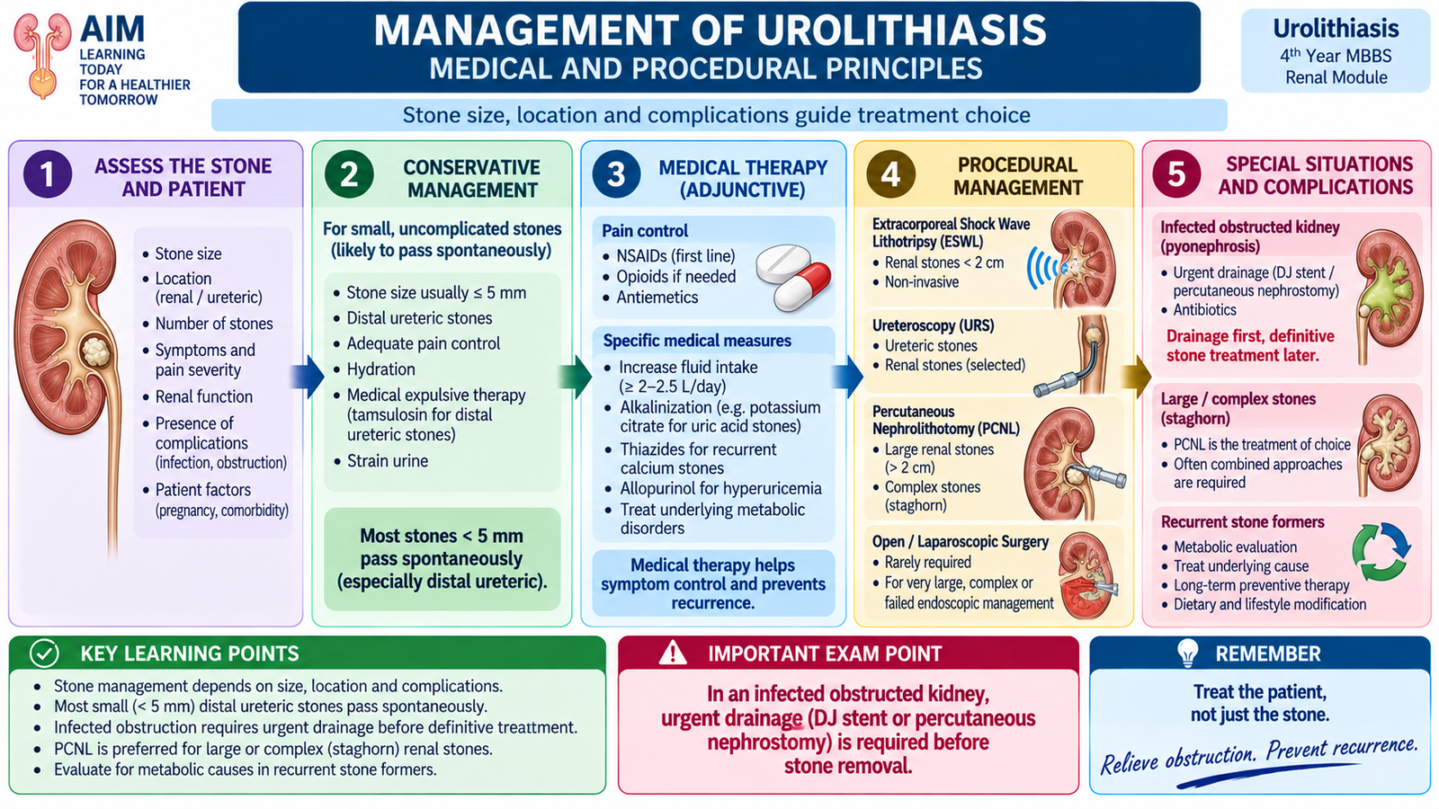
Management depends on the patient’s clinical condition, stone location and burden, presence of obstruction or infection, renal function and the likelihood of spontaneous passage. The aim is not only to relieve pain but also to preserve renal function, treat complications and reduce recurrence.
Initial medical management
In an uncomplicated patient, immediate treatment focuses on pain control, nausea or vomiting when present, assessment of hydration and confirmation that no dangerous obstruction or infection is being missed. Selected ureteric stones may pass spontaneously.
Non-steroidal anti-inflammatory drugs are useful for renal colic because reducing prostaglandin-mediated effects can decrease ureteric and renal pelvic pressure as well as pain. Other analgesia may be required when pain remains severe or NSAIDs are unsuitable.
An alpha-adrenergic blocker may be considered as medical expulsive therapy in appropriately selected ureteric stones. Relaxation of ureteric smooth muscle can facilitate passage in some patients. This is a selection-based treatment rather than a universal treatment for every stone.
Correcting the mechanism of recurrent stones
Long-term medical treatment should target the identified mechanism rather than treating all stone formers in the same way.
- Increasing urine volume reduces urinary supersaturation and is a central preventive principle.
- Urinary alkalinization increases the solubility of uric acid and cystine and can be used when appropriate.
- Thiazide-type therapy can reduce urinary calcium excretion in selected recurrent calcium-stone formers with hypercalciuria.
- Citrate therapy may be useful when hypocitraturia contributes to calcium stones and also provides urinary alkalinization.
- Urate-lowering therapy may be appropriate when excessive uric acid production or urinary uric acid is an important contributor.
- Infection control is essential in struvite disease because continuing urease-producing infection promotes further stone formation.
When procedural treatment is needed
Intervention is considered when a stone is unlikely to pass, symptoms remain uncontrolled, obstruction threatens renal function, infection coexists with obstruction, or conservative treatment has failed. The choice of procedure depends on stone site, size and anatomy as well as patient factors.
- Extracorporeal shock-wave lithotripsy (ESWL): externally generated shock waves fragment a suitable stone so that smaller pieces can pass through the urinary tract.
- Ureteroscopy: an endoscope is passed through the urinary tract to visualize and remove or fragment ureteric and selected renal stones.
- Percutaneous nephrolithotomy (PCNL): a percutaneous tract into the kidney permits removal of a large or complex renal stone.
E. Renal Trauma — Mechanism, Initial Resuscitation, Grading and Assessment
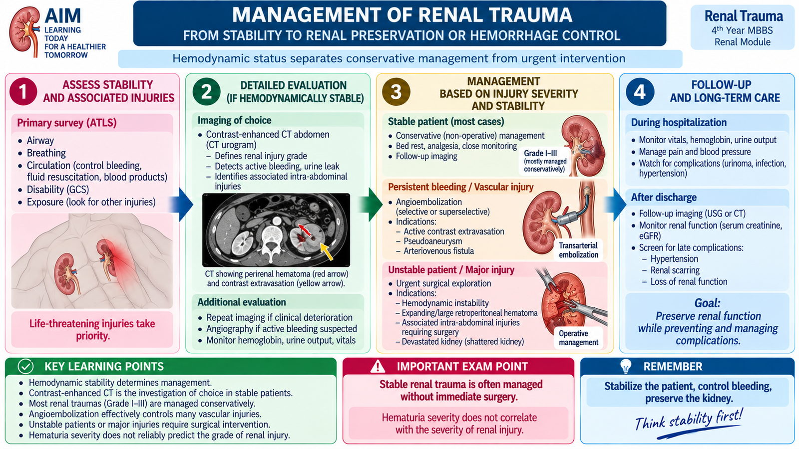
Renal trauma ranges from a minor renal contusion to major disruption of the kidney or its blood supply. The first decision is not the anatomical grade of injury. The first priority is whether the injured patient is physiologically stable. Life-threatening problems are treated before detailed renal assessment.
Mechanisms of injury
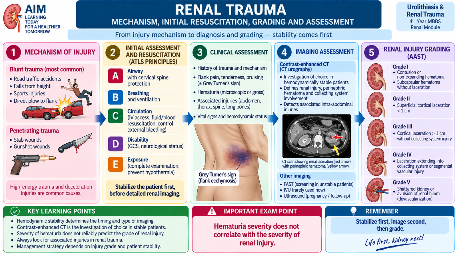
Renal injuries may result from blunt or penetrating trauma. Blunt trauma can compress the kidney against the spine or ribs or produce rapid deceleration that stretches renal vessels. Penetrating trauma may directly damage renal tissue, the collecting system or renal vessels.
- Blunt trauma: road-traffic injury, fall, crush injury or a direct blow to the flank.
- Rapid deceleration: can particularly threaten the renal vascular pedicle.
- Penetrating trauma: may cause a less predictable path through parenchyma and vessels.
Initial resuscitation
The injured patient is assessed using standard trauma priorities. Airway, breathing and circulation are addressed before focusing on the kidney. Hemodynamic instability raises concern for major bleeding and can change both imaging and management decisions.
Clinical assessment
A history of significant flank trauma, rapid deceleration or penetrating injury should raise suspicion. Examination may show flank tenderness, bruising, abdominal tenderness or signs of blood loss. Hematuria is common but its absence does not completely exclude important renal injury.
Urinalysis documents hematuria, while blood tests help assess hemoglobin, renal function and the physiological effect of injury. In a stable patient with a significant suspected renal injury, CT provides the major anatomical assessment.
Renal injury grading
The commonly used renal trauma grading system progresses from minor contusion or hematoma to deep laceration, collecting-system injury and major vascular destruction. The grade helps describe the anatomy and supports management planning, but it must always be interpreted together with hemodynamic stability.
| Grade | Core injury pattern |
|---|---|
| I | Renal contusion or subcapsular hematoma without laceration. |
| II | Perirenal hematoma confined around the kidney or superficial cortical laceration without urinary extravasation. |
| III | Deeper renal parenchymal laceration without collecting-system rupture. |
| IV | Laceration involving the collecting system with urinary extravasation and/or significant segmental vascular injury. |
| V | Shattered kidney or major hilar vascular injury causing severe devascularization. |
Radiological assessment
In a hemodynamically stable patient with significant suspected renal trauma, contrast-enhanced CT can demonstrate renal laceration, hematoma, active bleeding, devascularized areas and urinary extravasation. Delayed images may help demonstrate injury to the collecting system.
F. Management of Renal Trauma
Management of renal trauma is guided mainly by hemodynamic stability, ongoing bleeding and the anatomical injury. Modern renal-trauma care aims to preserve functioning renal tissue whenever this can be achieved safely. Therefore, the presence of a renal laceration alone does not automatically mean that open surgery is required.
Stable patient
Hemodynamically stable patients with minor injuries are usually managed non-operatively with observation and clinical monitoring. Selected stable patients with more significant injuries can also be managed without immediate surgery when bleeding is controlled and there is no other indication for exploration.
Monitoring may include:
- vital signs and clinical evidence of ongoing bleeding;
- hemoglobin when clinically required;
- renal function;
- urine output;
- follow-up imaging when the injury or clinical course makes it necessary.
Angioembolization
When CT demonstrates significant arterial bleeding in a patient who can be managed by an endovascular approach, selective angiographic embolization can control bleeding while preserving more renal tissue than nephrectomy. It is therefore an important option between simple observation and open surgery.
Urinary extravasation
Collecting-system injury may allow urine to leak outside the urinary tract. Some urinary extravasation resolves with observation. Persistent or complicated leakage may require urinary diversion or drainage, such as ureteric stenting or percutaneous drainage, according to the clinical situation.
When operative management becomes necessary
The strongest concern is uncontrolled hemorrhage with persistent hemodynamic instability. Major vascular disruption, a severely shattered kidney or associated abdominal injury may also require operative exploration. The surgical goal is control of bleeding and preservation of viable renal tissue whenever safely possible; nephrectomy is reserved for situations where the kidney cannot be salvaged or life-threatening bleeding cannot otherwise be controlled.
G. Diagnostic Imaging of the Urinary Tract
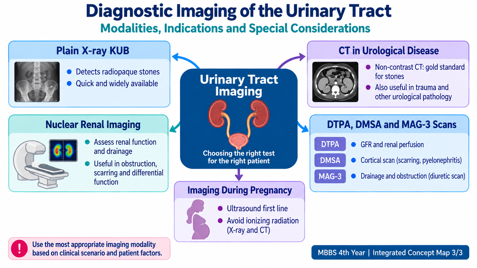
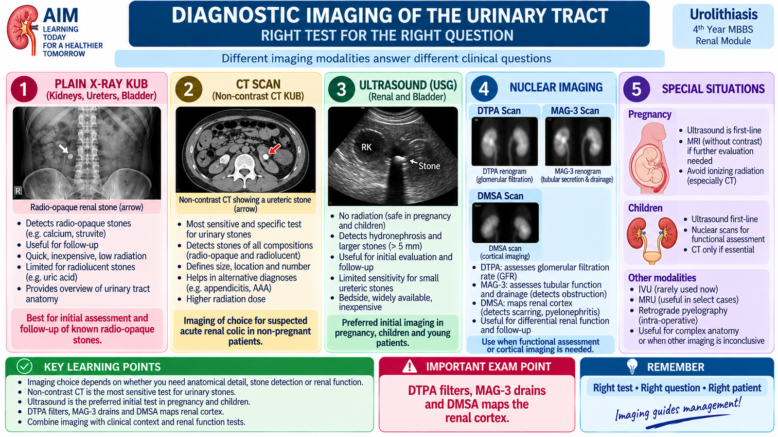
Different urinary investigations answer different questions. Plain X-ray mainly provides structural information about radiopaque material. CT gives detailed cross-sectional anatomy. Nuclear renal scans provide functional information such as cortical function, filtration or drainage. Choosing the correct investigation therefore requires knowing what each modality can and cannot demonstrate.
Plain X-ray KUB
A plain X-ray of the kidneys, ureters and bladder can demonstrate many calcium-containing stones because calcium attenuates X-rays and appears radiopaque. It can be useful for identifying or following a known radiopaque calculus.
Its major limitation is that not every urinary stone is clearly visible. Uric acid stones are radiolucent on plain X-ray, and bowel contents or overlying structures can also obscure urinary calculi. A plain film gives much less information than CT about obstruction, surrounding tissues and alternative diagnoses.
CT in urological disease
Non-contrast CT is highly useful for suspected urinary calculi because it can detect both radiopaque and radiolucent stones, identify their anatomical location and demonstrate secondary signs of obstruction. This explains why a uric acid stone that is invisible on plain KUB can still be detected on CT.
Contrast-enhanced CT is valuable when assessment of renal parenchyma, renal vessels, trauma or other urological pathology is needed. In renal trauma, it can demonstrate laceration, hematoma, vascular injury, active bleeding and urinary extravasation in an appropriately selected stable patient.
Nuclear renal imaging — the basic idea
Nuclear renal imaging uses a small amount of radiopharmaceutical and a gamma camera to follow its distribution and handling by the kidneys. Unlike an ordinary anatomical image, a renogram can provide information about how each kidney functions and how effectively tracer passes from the kidney into the collecting system.
DTPA scan
DTPA is handled predominantly by glomerular filtration. This makes it useful for assessing renal perfusion, relative renal function, glomerular filtration and urinary drainage. Dynamic images can generate a renogram showing tracer arrival, renal uptake and excretion.
MAG-3 scan
MAG-3 is predominantly handled by tubular secretion and is efficiently extracted by the kidneys. It provides good dynamic assessment of renal function and drainage and is particularly useful when evaluating whether dilatation of the collecting system represents functionally significant obstruction.
DMSA scan
DMSA localizes mainly in functioning renal cortex and therefore provides a cortical image rather than a drainage study. It is useful for assessing differential cortical function and identifying areas of cortical damage or scarring.
DTPA, MAG-3 and DMSA compared
| Scan | Main renal handling | Main information | Key concept |
|---|---|---|---|
| DTPA | Glomerular filtration | Function, filtration and drainage | Dynamic renogram |
| MAG-3 | Predominantly tubular secretion | Function and urinary drainage | Very useful for obstruction assessment |
| DMSA | Cortical localization | Cortical integrity and differential cortical function | Cortical defects/scarring |
Imaging the renal system during pregnancy
Pregnancy changes imaging selection because fetal exposure to ionizing radiation should be minimized. Ultrasonography is the preferred initial renal and urinary-tract investigation because it does not use ionizing radiation and can demonstrate hydronephrosis and many clinically important urinary abnormalities.
If ultrasound does not answer an important clinical question, MR urography without ionizing radiation may provide additional anatomical information. Plain X-ray KUB and CT are not routine first-line investigations during pregnancy because they use ionizing radiation. When a serious clinical problem remains unresolved and the information is essential for management, the need for further imaging is decided by balancing maternal benefit against fetal radiation risk rather than simply withholding a necessary investigation.
Integrated Mechanism Flow — From Stone Risk to Clinical Disease
⭐ AIM High-Yield Review
Urolithiasis, Renal Trauma & Diagnostic Imaging
Use these videos after completing the AIM learning material to reinforce the major clinical and imaging concepts.
1. Urolithiasis — Clinical Evaluation & Management
Reinforces presentation, diagnostic evaluation, obstruction and management principles of urinary stones.
2. Renal Trauma — Assessment, Grading & Management
Reviews renal trauma assessment, injury severity and major management principles.
3. Renal Imaging — Ultrasound, DTPA & MAG-3 Renal Scan
Reinforces functional renal imaging, including assessment of renal function, drainage and obstruction.