
Topic 8 — Renal Tumors: Renal Cell Carcinoma and Wilms Tumor
1. Topic Introduction
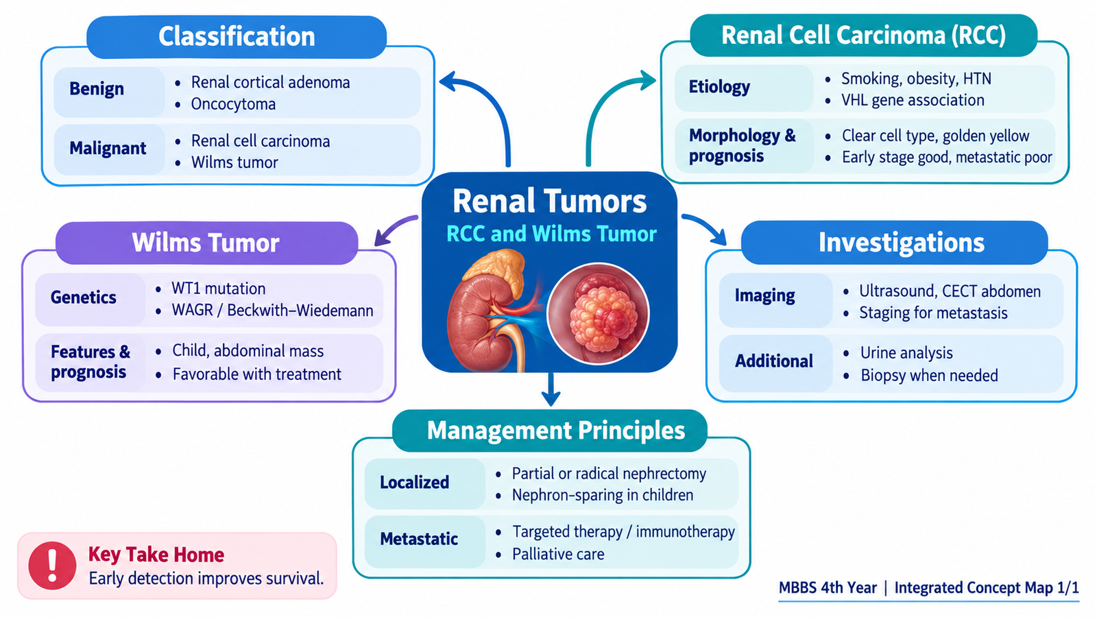
Renal tumors are abnormal proliferations arising from structures within the kidney. They range from benign lesions that usually remain localized to malignant tumors capable of invading blood vessels and spreading to distant organs. In adults, the most important malignant renal parenchymal tumor is renal cell carcinoma (RCC), whereas Wilms tumor is an important embryonal renal malignancy of childhood. Understanding these tumors requires linking their cellular origin and genetic abnormalities with their gross and microscopic appearance, clinical presentation, investigations and treatment. This chapter first organizes the major renal tumors, then explains RCC and Wilms tumor in detail, followed by the diagnostic approach, management principles and major prognostic features.
A. Classification and Core Orientation of Renal Tumors
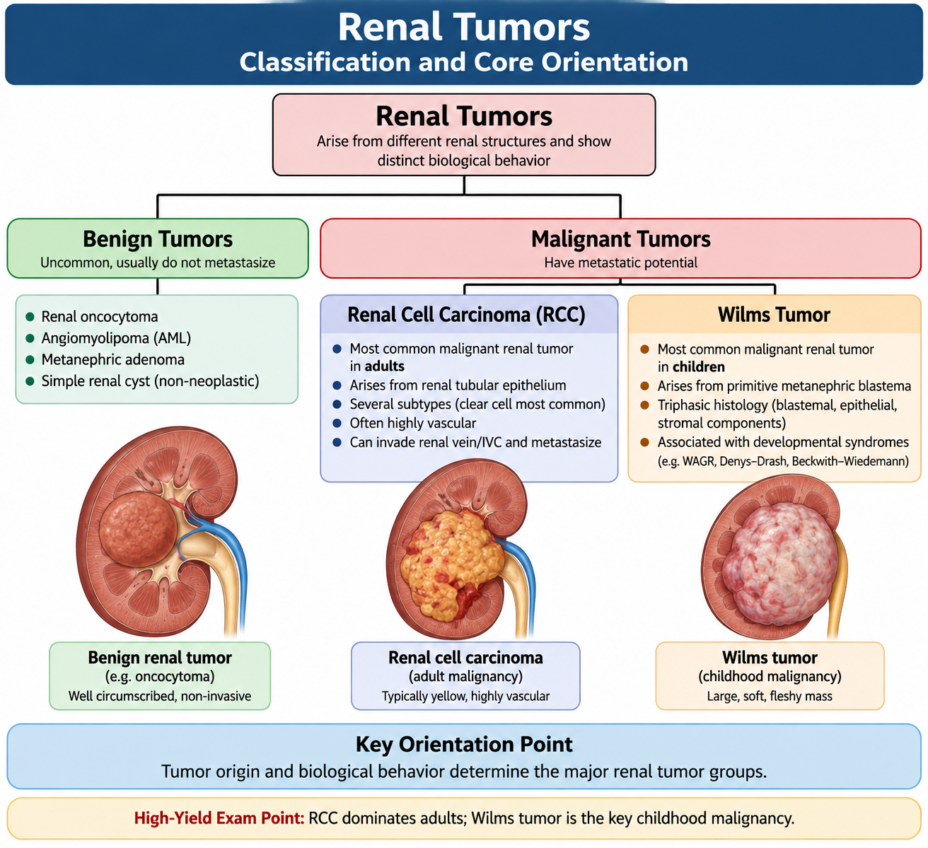
Renal tumors can arise from renal tubular epithelium, embryonal renal tissue or mesenchymal components of the kidney. The first useful distinction is between benign tumors, which generally remain localized, and malignant tumors, which may invade surrounding structures, enter blood vessels or metastasize. For this topic, the two malignant tumors that require the greatest attention are RCC in adults and Wilms tumor in children.
Important benign renal tumors
- Papillary adenoma: a small benign epithelial tumor arising from renal tubular epithelium.
- Renal oncocytoma: a benign epithelial neoplasm composed of cells rich in mitochondria. It may form a well-circumscribed renal mass and can resemble RCC on imaging.
- Angiomyolipoma: a benign mesenchymal tumor containing variable amounts of blood vessels, smooth muscle and adipose tissue. Recognition of macroscopic fat on imaging can suggest the diagnosis.
Important malignant renal tumors
- Renal cell carcinoma: malignant epithelial tumor arising from renal tubular cells. Major types include clear cell, papillary and chromophobe RCC.
- Wilms tumor (nephroblastoma): an embryonal malignant tumor that mainly occurs in young children.
- Other malignant renal tumors: uncommon malignant mesenchymal tumors may occur, but they are not the central focus of this topic.
| Tumor | Nature | Main Origin / Composition | Key Orientation |
|---|---|---|---|
| Papillary adenoma | Benign | Renal tubular epithelium | Usually small and incidental |
| Oncocytoma | Benign | Renal epithelial cells | May mimic RCC clinically or radiologically |
| Angiomyolipoma | Benign | Vessels, smooth muscle and fat | Macroscopic fat is an important clue |
| Renal cell carcinoma | Malignant | Renal tubular epithelium | Major adult malignant renal tumor |
| Wilms tumor | Malignant | Embryonal renal tissue | Important childhood renal malignancy |
B. Renal Cell Carcinoma: Etiology, Pathogenesis and Morphology
Renal cell carcinoma is a malignant epithelial tumor arising from renal tubular cells. Different RCC subtypes develop from different molecular abnormalities and have characteristic microscopic appearances. The most important subtype for understanding pathogenesis is clear cell RCC, in which loss of normal VHL tumor-suppressor activity promotes abnormal signaling that supports tumor growth and new blood-vessel formation.
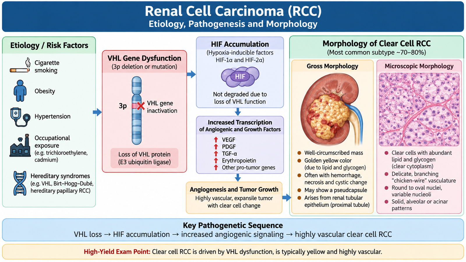
Etiology and important risk factors
Most RCCs are sporadic, but environmental, acquired and hereditary factors can increase risk. These factors do not produce a unique clinical form of RCC; instead, they increase the probability that renal epithelial cells will acquire the changes necessary for malignant growth.
- Cigarette smoking is an established modifiable risk factor.
- Obesity and hypertension are important associations.
- Patients with acquired cystic kidney disease, particularly in the setting of prolonged severe renal disease, have increased RCC risk.
- Von Hippel-Lindau disease predisposes to clear cell RCC because the VHL tumor-suppressor pathway is abnormal.
Clear cell RCC
Clear cell RCC is strongly associated with loss or inactivation of the VHL tumor-suppressor gene. Normal VHL protein helps regulate hypoxia-inducible factors. When VHL function is lost, hypoxia-inducible factors accumulate even when oxygen is available. This increases expression of growth-promoting and angiogenic factors, including VEGF. The resulting tumor is therefore characteristically highly vascular.
Gross morphology
Clear cell RCC commonly forms a solitary cortical renal mass. The cut surface is often yellow to orange because tumor cells contain lipid. Areas of hemorrhage, cystic change and necrosis may produce a variegated appearance. As the tumor enlarges, it may extend into perinephric tissue or invade the renal vein.
Microscopic morphology
The tumor is typically composed of nests or sheets of cells with clear cytoplasm separated by a delicate network of blood vessels. The cytoplasm appears clear because intracellular lipid and glycogen are removed during routine tissue processing.
Papillary RCC
Papillary RCC is another important RCC subtype and is associated with abnormalities involving growth-signaling pathways, particularly the MET pathway in hereditary forms. Microscopically, the tumor forms papillary or tubular structures. Foamy macrophages may be present within papillary cores, and calcified psammoma bodies can occur.
Chromophobe RCC
Chromophobe RCC arises from cells related to the collecting duct intercalated cell lineage. The cells are usually pale or eosinophilic, have prominent cell membranes and may show a characteristic perinuclear halo. When localized, this subtype often behaves less aggressively than clear cell RCC.
C. RCC: Clinical Presentation, Spread and Prognosis
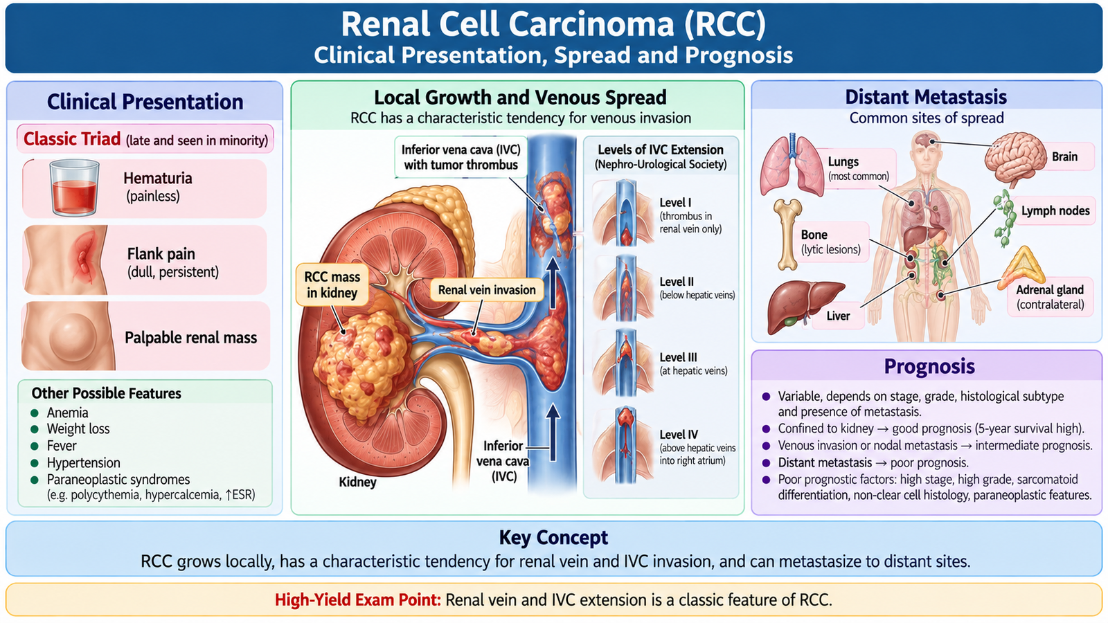
RCC may remain clinically silent while confined to the kidney, so many tumors are detected incidentally during abdominal imaging. Symptoms appear when the tumor causes bleeding, stretches or invades nearby structures, produces systemic mediators or spreads beyond the kidney. Understanding the pattern of presentation therefore requires linking the location and biological behavior of the tumor with its clinical effects.
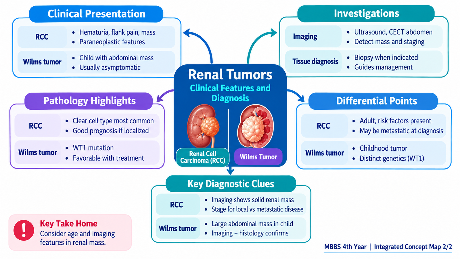
Clinical presentation
- Hematuria: may occur when the tumor communicates with or invades the collecting system.
- Flank pain: can result from enlargement of the renal mass, local tissue involvement or hemorrhage.
- Palpable renal or abdominal mass: usually indicates a larger tumor.
- Constitutional features: fatigue, loss of appetite, weight loss or fever may occur in more advanced disease.
Paraneoplastic manifestations
RCC can produce hormones or hormone-like substances. These effects may occasionally bring the patient to medical attention before the renal mass itself produces major local symptoms.
- Polycythemia may occur because some tumors produce erythropoietin.
- Hypercalcemia may occur through tumor-related humoral mechanisms.
- Hypertension may be associated with increased renin activity or underlying renal disease.
- Other endocrine-like manifestations can occur but are less central to undergraduate recognition.
Local invasion and metastatic spread
A characteristic biological feature of RCC is its tendency to invade veins. The tumor may extend from intrarenal veins into the renal vein and then into the inferior vena cava. This does not necessarily mean distant metastasis is already present, but it is an important feature of local tumor extension and influences surgical planning.
Hematogenous spread is common in advanced disease. Important metastatic sites include the lungs, bones and liver. Regional lymph nodes may also be involved.
Clinical link: Venous obstruction may produce new varicocele or lower-body venous congestion. Persistent or unusual venous findings in a patient with a renal mass should therefore increase concern about vascular extension.
Prognosis
The most important determinant of RCC prognosis is the anatomical extent of disease. A tumor confined to the kidney has a substantially better outlook than one that has invaded surrounding tissues, major veins, lymph nodes or distant organs. Histological grade and RCC subtype also influence biological behavior, but stage remains central to clinical assessment.
D. Investigation and Diagnosis of Renal Tumors
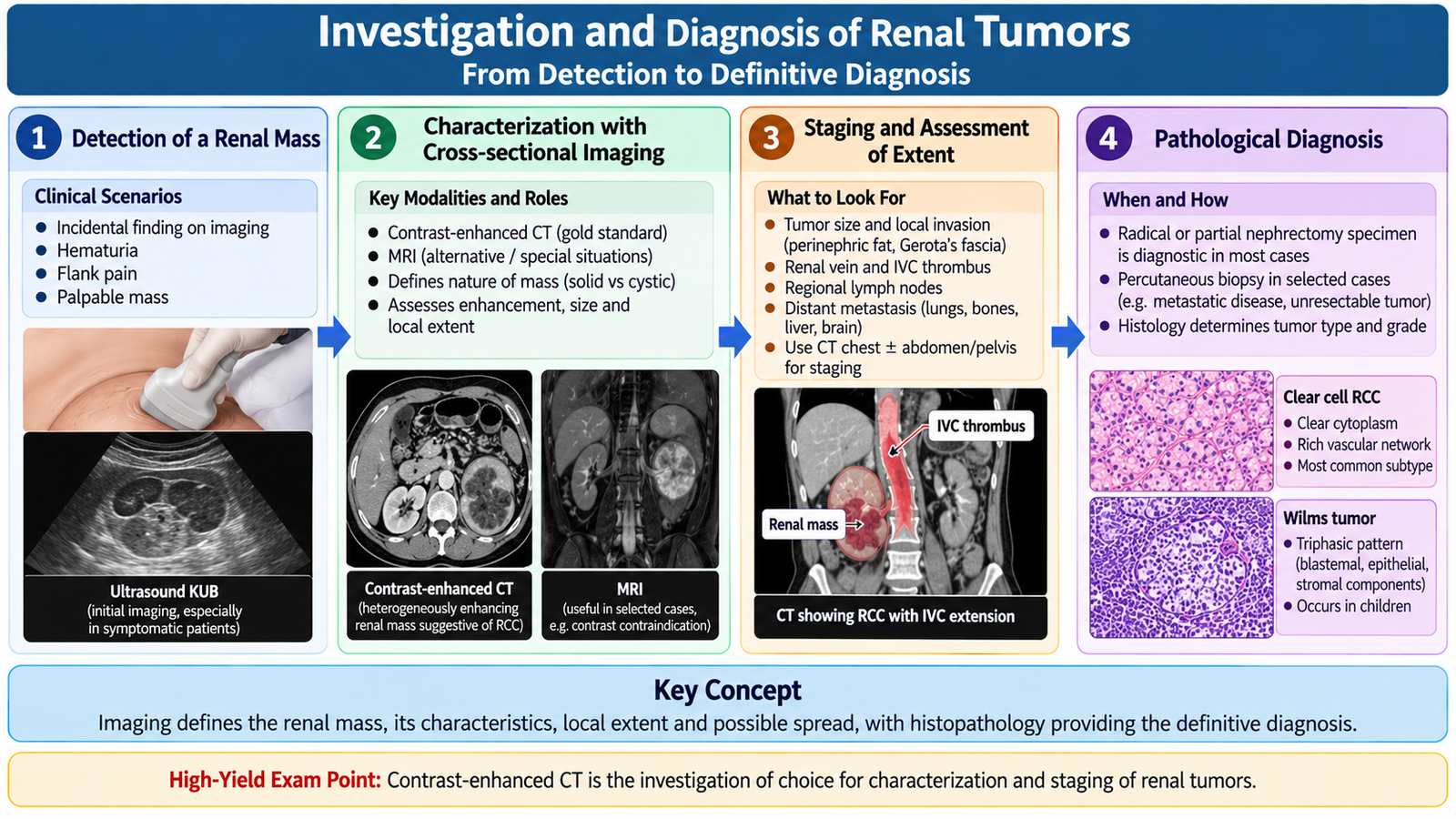
The diagnostic approach to a renal tumor should first confirm that a renal lesion is present, determine whether it is solid or cystic, assess its local extent and look for evidence of spread. Imaging is therefore central. Laboratory tests complement imaging by assessing hematuria, renal function and possible systemic effects of the tumor.
Initial laboratory assessment
- Urinalysis: detects microscopic or gross hematuria.
- Complete blood count: may show anemia from chronic disease or blood loss; occasionally polycythemia may be present.
- Renal function tests: establish baseline kidney function before treatment and help assess the opposite kidney.
- Serum calcium: may identify tumor-associated hypercalcemia when clinically suspected.
- Liver-related biochemical tests: may contribute to assessment when systemic or metastatic disease is suspected.
Ultrasonography
Ultrasound is useful for identifying a renal mass and determining whether a lesion is predominantly solid or cystic. It is particularly useful as an initial investigation and is also valuable in children because it avoids ionizing radiation.
Contrast-enhanced CT
Cross-sectional CT imaging is central to the evaluation of a suspected RCC. It demonstrates the renal mass, enhancement pattern, relation to surrounding structures, involvement of the opposite kidney, regional lymph nodes and possible extension into the renal vein or inferior vena cava. CT of appropriate additional regions can also help assess metastatic disease.
MRI
MRI provides excellent soft-tissue and vascular detail. It is particularly useful when the extent of venous tumor involvement needs clarification or when CT contrast cannot be used appropriately.
Assessment for distant spread
The lungs are an important site of RCC metastasis, so chest imaging is part of staging when malignancy is suspected or confirmed. Additional investigations are directed by symptoms and clinical findings rather than performed simply as an exhaustive test list.
Role of tissue diagnosis
Histopathology establishes the definitive tumor type after tissue has been obtained. However, a needle biopsy is not automatically required before surgery for every radiologically typical, resectable renal mass. Percutaneous biopsy is particularly useful when the result is expected to change management, such as before selected ablative or systemic treatments, or when imaging leaves significant diagnostic uncertainty.
Investigation of suspected Wilms tumor
In a child with a renal or abdominal mass, ultrasound helps confirm that the lesion arises from the kidney. CT or MRI then defines the renal mass, its relation to vessels and surrounding structures, the condition of the opposite kidney and possible regional spread. Chest imaging is important because the lungs are a major site of distant spread. Definitive pathological assessment identifies the characteristic histology and determines whether adverse features such as anaplasia are present.
E. RCC Management: Localized and Metastatic Disease
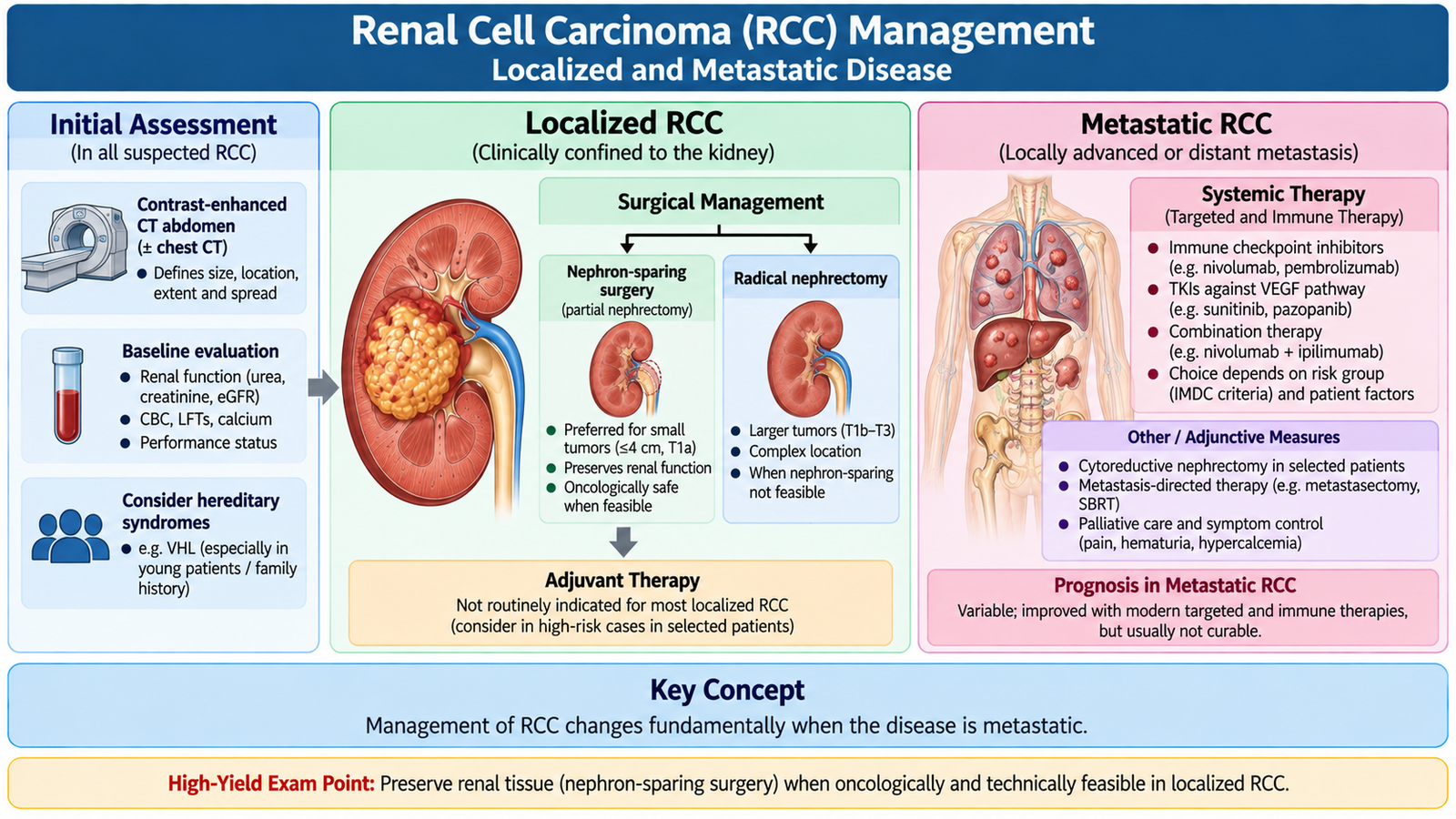
The management of RCC depends mainly on whether the tumor is confined to the kidney and surgically removable or has developed metastatic disease. Surgery is the principal curative treatment for localized RCC. In metastatic RCC, treatment becomes primarily systemic, although surgery or local treatment may still have a role in carefully selected patients.
Localized RCC
The main surgical objective is to remove the malignant tumor completely while preserving as much functioning renal tissue as is safely possible.
Partial nephrectomy
Partial nephrectomy removes the tumor with a margin of surrounding renal tissue while preserving the remainder of the kidney. It is particularly valuable when the tumor is localized and technically suitable for nephron-sparing surgery. Preserving renal tissue becomes especially important when future renal function is a concern.
Radical nephrectomy
Radical nephrectomy involves removal of the affected kidney together with appropriate surrounding tissues according to the extent of disease. It is considered when the tumor cannot be safely or adequately managed by nephron-sparing surgery because of its size, position or local extent.
Selected alternatives
Some small renal masses in carefully selected patients may be managed by active surveillance or local ablative techniques. These approaches are chosen according to tumor characteristics, patient fitness, kidney function and expected benefit from intervention rather than being routine alternatives for every patient.
Metastatic RCC
Once distant metastases are present, surgery alone cannot eradicate all disease. Modern management therefore relies mainly on systemic therapy. Two major therapeutic approaches are immune-based treatment and drugs that inhibit signaling pathways involved in tumor growth and angiogenesis.
- Immune checkpoint inhibition enhances antitumor immune activity.
- VEGF-pathway targeted therapy, including tyrosine kinase inhibition, reduces signaling that supports tumor angiogenesis and growth.
- These systemic approaches may be used in combinations selected by the oncology team according to the patient’s disease and overall clinical condition.
Removal of the primary kidney tumor in metastatic disease is not automatically appropriate for every patient. Cytoreductive nephrectomy may be considered in selected patients when the overall disease burden, symptoms, performance status and planned systemic treatment make surgery beneficial. Selected metastatic deposits may also receive local treatment, while radiotherapy can be useful for palliation of troublesome metastatic symptoms such as bone pain.
Conventional cytotoxic chemotherapy has relatively limited effectiveness against typical RCC compared with the systemic targeted and immune therapies used for advanced disease.
F. Wilms Tumor: Genetics, Clinicopathological Features, Morphology and Prognosis
Wilms tumor, or nephroblastoma, is a malignant embryonal tumor of the kidney that primarily affects young children. It develops from immature renal precursor tissue and therefore resembles structures seen during normal kidney development. The disease is particularly important because characteristic developmental genetic abnormalities are linked with its pathogenesis, while its microscopic appearance often reproduces different stages of renal differentiation.
Genetic basis
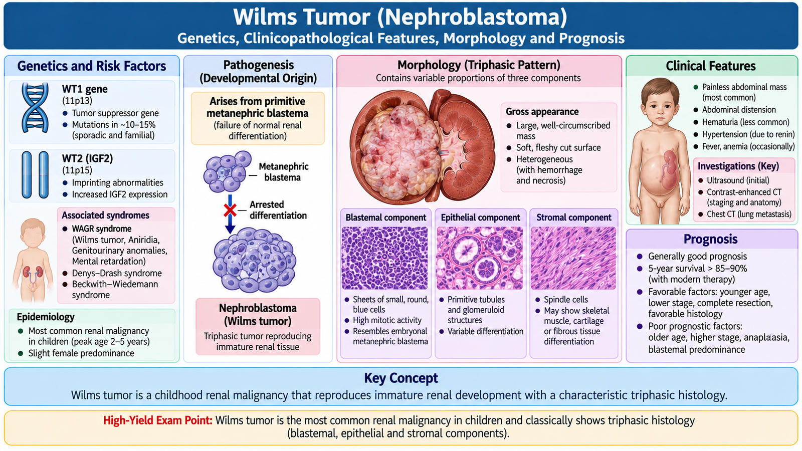
Wilms tumor can arise sporadically or in association with inherited developmental abnormalities. Important genetic regions include WT1 on chromosome 11p13 and abnormalities involving the 11p15 region, where disturbed regulation of growth-related genes can promote persistent proliferation of embryonal renal tissue.
Important clinical associations help demonstrate the connection between abnormal renal development and tumor formation:
- WAGR syndrome: associated with WT1-region abnormalities and characterized by Wilms tumor susceptibility together with developmental abnormalities including aniridia and genitourinary abnormalities.
- Denys-Drash syndrome: associated with WT1 abnormalities and increased risk of Wilms tumor in the setting of severe renal and genitourinary developmental disease.
- Beckwith-Wiedemann syndrome: an overgrowth syndrome associated with abnormalities involving chromosome 11p15 and increased risk of embryonal tumors, including Wilms tumor.
Clinical features
The most typical presentation is a large abdominal or flank mass in a young child. Because the tumor may become large before producing major symptoms, the mass may first be noticed by a parent or during examination.
- Abdominal or flank mass is the major clinical clue.
- Abdominal discomfort or pain may occur.
- Hematuria may occur when the tumor involves the collecting system.
- Hypertension may occur because the renal tumor can alter renal vascular and renin-related mechanisms.
- Some children develop fever or other constitutional symptoms.
Gross morphology
Wilms tumor typically forms a large, well-demarcated renal mass. The cut surface is usually soft and tan-gray and may contain areas of hemorrhage, necrosis or cystic degeneration. A large tumor may replace much of the normal kidney and compress the remaining renal tissue.
Microscopic morphology: the classic triphasic pattern
The classic histological pattern contains three components that imitate stages of developing kidney tissue:
- Blastemal component: sheets of small, primitive blue cells representing immature renal precursor tissue.
- Epithelial component: primitive tubules or glomerulus-like structures showing attempted renal differentiation.
- Stromal component: immature mesenchymal tissue that may show varying degrees of differentiation.
Not every tumor contains the three elements in equal proportions, but recognition of the triphasic pattern is highly characteristic.
Anaplasia and prognosis
Anaplasia refers to marked nuclear enlargement, hyperchromasia and abnormal mitotic activity. Its presence, particularly when widespread, is an important adverse pathological feature because it is associated with more resistant and aggressive tumor behavior.
Overall prognosis depends mainly on the extent of disease and histological features. Children with localized tumors and favorable histology generally have a good outlook, while advanced disease and unfavorable histology such as diffuse anaplasia worsen prognosis.
Management principles
Wilms tumor is treated by a multimodal approach. Surgery and chemotherapy are central components, while radiotherapy is used selectively according to disease extent and pathological risk. The exact sequence of therapy varies between established pediatric oncology protocols, so the undergraduate principle is to recognize that treatment requires coordinated pediatric surgical and oncology care rather than nephrectomy alone.
When both kidneys are involved, preservation of functioning renal tissue becomes especially important and treatment planning aims to achieve tumor control while retaining as much renal function as possible.
Integrated Mechanism Flow — Clear Cell RCC from Molecular Change to Clinical Disease
Loss of normal tumor-suppressor control
Growth and hypoxia pathways remain activated
Increased VEGF supports vascular tumor growth
Clear cells form a highly vascular renal tumor
Local tissues and renal vein may become involved
Metastatic disease requires systemic treatment
Important Comparison — RCC versus Wilms Tumor
| Feature | Renal Cell Carcinoma | Wilms Tumor |
|---|---|---|
| Typical setting | Adult renal malignancy | Young-child renal malignancy |
| Cellular origin | Renal tubular epithelium | Embryonal renal precursor tissue |
| Important genetics | VHL pathway important in clear cell RCC | WT1 and 11p15 abnormalities are important |
| Characteristic morphology | Clear cells and rich vascular network in clear cell type | Blastemal, epithelial and stromal components |
| Common clinical clue | Hematuria or incidentally detected renal mass | Large abdominal mass in a child |
| Characteristic spread issue | Renal vein and IVC invasion | Local spread and distant metastasis, especially to lungs |
| Core treatment principle | Surgery for localized disease; systemic targeted/immune therapy for metastatic disease | Multimodal pediatric treatment using surgery and chemotherapy, with selective radiotherapy |
| Major prognostic principle | Anatomical stage is central | Stage and histology, especially anaplasia |
⭐ AIM High-Yield Review
“`
“`
🎥 Video Learning — Renal Cell Carcinoma and Wilms Tumor
Review the key pathological concepts of renal cell carcinoma and Wilms tumor, including their important morphology and distinguishing features.
If the video does not load, open it on YouTube .