
Respiratory Structure, Function and Clinical Assessment
Topic Introduction
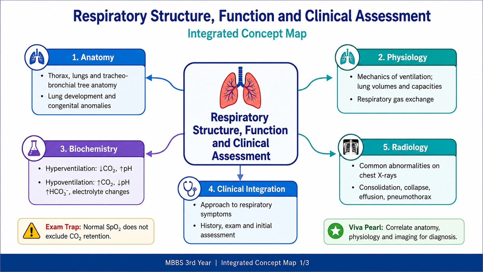
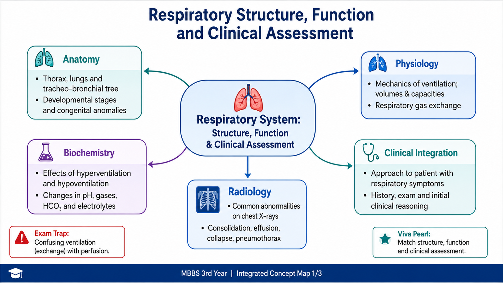
The respiratory system provides a pathway for air to reach the lungs and allows oxygen and carbon dioxide to move between the atmosphere and blood. Understanding respiratory disease therefore requires a clear connection between structure, ventilation, gas exchange and clinical assessment. In third year, the basic sciences are revisited mainly to support understanding of clinical and pathological respiratory problems rather than to repeat complete earlier-year chapters. This topic therefore focuses on the clinically relevant anatomy of the thorax and airways, essential lung development, mechanics of breathing, gas exchange, effects of abnormal ventilation on blood gases and acid–base balance, assessment of respiratory symptoms and basic interpretation of chest radiographs.
A. Clinical Anatomy of the Thorax, Lungs and Airways
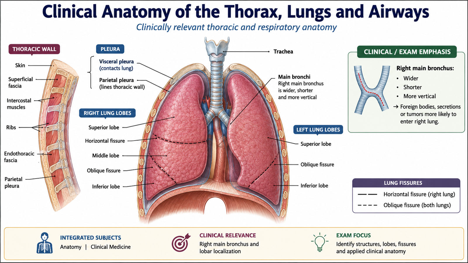
The thorax contains the lungs and mediastinal structures within a protective but mobile cage formed by the ribs, sternum and thoracic vertebrae. Its structure allows the chest to expand during inspiration and become smaller during expiration. For third-year learning, the most important anatomical relationships are those that help explain respiratory examination, localization of disease and common clinical findings.
Thoracic Wall and Pleura
The thoracic wall is formed mainly by the ribs, intercostal muscles, sternum and thoracic vertebrae. The intercostal muscles participate in respiratory movements, while the diaphragm forms the major muscular floor of the thoracic cavity. Each lung is covered directly by visceral pleura, while the inner surface of the thoracic wall is lined by parietal pleura. The potential space between them is the pleural cavity. A thin layer of pleural fluid permits smooth movement while the lungs expand and recoil.
Lungs and Lobes
The right lung is divided into upper, middle and lower lobes by horizontal and oblique fissures. The left lung has upper and lower lobes separated by an oblique fissure. The smaller left lung accommodates the heart and therefore has a cardiac notch.
Tracheobronchial Tree
Air passes through a progressively branching conducting system before reaching the gas-exchanging portion of the lung.
The right main bronchus is shorter, wider and more vertical than the left. This anatomical arrangement makes aspirated material more likely to enter the right bronchial tree.
Clinically Relevant Surface Anatomy
Surface markings help relate internal structures to findings during inspection, percussion and auscultation. The lung apices extend slightly above the clavicles, while the inferior lung borders move downward during inspiration. The lower lobes occupy a large part of the posterior chest, making examination of the back particularly important when lower-lobe disease is suspected. Knowledge of lobes and fissures also helps correlate abnormal breath sounds, percussion findings and radiological abnormalities with the likely anatomical site of disease.
B. Lung Development and Major Congenital Correlations
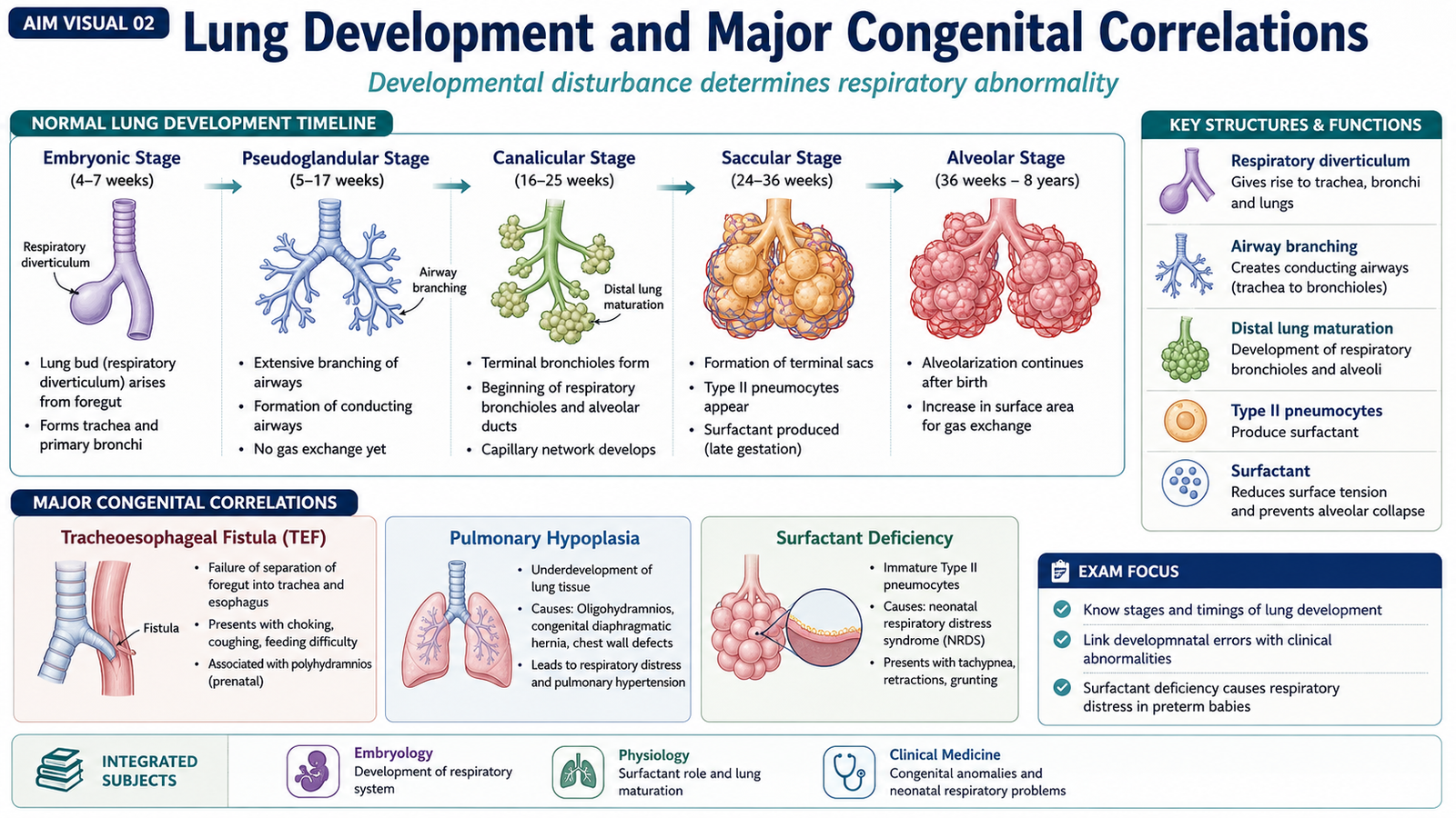
The respiratory system begins as an outgrowth from the primitive foregut and progressively develops into the conducting airways and gas-exchanging portions of the lungs. At third-year level, the important principle is not detailed embryological memorization but understanding that disruption of normal development can produce clinically important congenital abnormalities. The early respiratory diverticulum develops into the trachea and bronchial tree. Repeated branching produces progressively smaller airways. The distal portions of the developing lungs then become increasingly suitable for gas exchange as vascularization increases and the alveolar region matures. Lung maturation occurs through recognizable developmental stages. The clinically important progression is from formation of the conducting passages toward development of increasingly thin, vascular gas-exchanging structures. In later fetal life, maturation of type II pneumocytes and increasing production of surfactant become particularly important.
Surfactant lowers surface tension within the alveoli and therefore reduces their tendency to collapse. Insufficient pulmonary maturity in premature infants can consequently impair effective lung expansion.
Important Congenital Correlations
- Tracheoesophageal fistula: abnormal communication between the airway and esophagus due to abnormal separation during development.
- Pulmonary hypoplasia: incomplete development of lung tissue, resulting in reduced functional lung volume.
- Congenital pulmonary airway abnormalities: abnormal formation of portions of the developing lower respiratory tract.
The central clinical relationship is that the timing and nature of disturbed development determine which respiratory structure is abnormal and how severely breathing may be affected after birth.
 .
.C. Mechanics of Ventilation and Lung Volumes
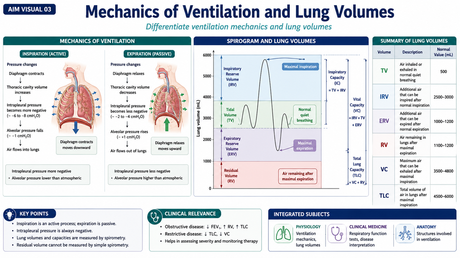
Ventilation is the movement of air into and out of the lungs. Air moves because respiratory muscles change the volume of the thoracic cavity and therefore create pressure differences between the atmosphere and the alveoli.
Inspiration
During quiet inspiration, the diaphragm contracts and moves downward. The external intercostal muscles assist expansion of the thoracic cage. As thoracic volume increases, intrapulmonary pressure falls below atmospheric pressure and air moves into the lungs.
Expiration
Quiet expiration is mainly passive. Relaxation of the inspiratory muscles and elastic recoil of the lungs decrease thoracic volume. Alveolar pressure rises above atmospheric pressure and air moves out of the lungs.
Major Lung Volumes
- Tidal volume: air inspired or expired during normal quiet breathing.
- Inspiratory reserve volume: additional air that can be inspired after a normal inspiration.
- Expiratory reserve volume: additional air that can be expired after a normal expiration.
- Residual volume: air remaining in the lungs after maximal expiration.
Lung capacities are combinations of two or more lung volumes. Clinically important examples include vital capacity and total lung capacity. These measurements help identify abnormal patterns of ventilation. Restrictive disorders generally reduce the amount of air that the lungs can contain, whereas obstructive disorders primarily interfere with effective airflow, particularly during expiration.
D. Respiratory Gas Exchange
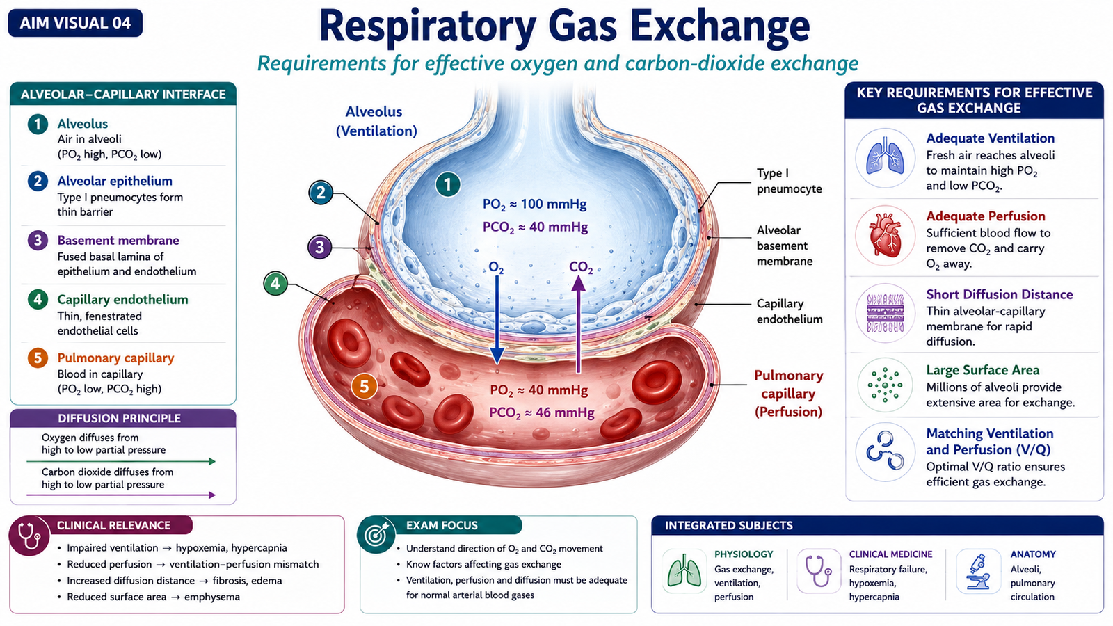
Gas exchange occurs mainly across the thin alveolar-capillary membrane. The purpose is to transfer oxygen from inspired air into pulmonary capillary blood and remove carbon dioxide from the blood so that it can be exhaled. Oxygen moves from the alveoli into pulmonary blood because its partial pressure is higher in alveolar air than in deoxygenated blood reaching the lungs. Carbon dioxide moves in the opposite direction, from pulmonary capillary blood into the alveoli.
Efficient gas exchange requires several processes to work together:
- Adequate ventilation of the alveoli.
- Adequate pulmonary blood flow.
- An intact and sufficiently thin alveolar-capillary membrane.
- Reasonable matching between ventilation and perfusion.
If an alveolus receives blood but insufficient ventilation, oxygen transfer becomes impaired. Similarly, diseases that fill alveoli, reduce pulmonary blood flow or damage the alveolar-capillary interface can interfere with normal gas exchange. This relationship explains why respiratory diseases may produce hypoxemia. Carbon dioxide abnormalities become particularly important when effective alveolar ventilation is significantly reduced or increased.
E. Ventilation, Blood Gases and Acid–Base Changes
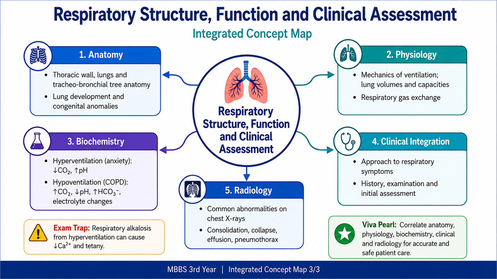
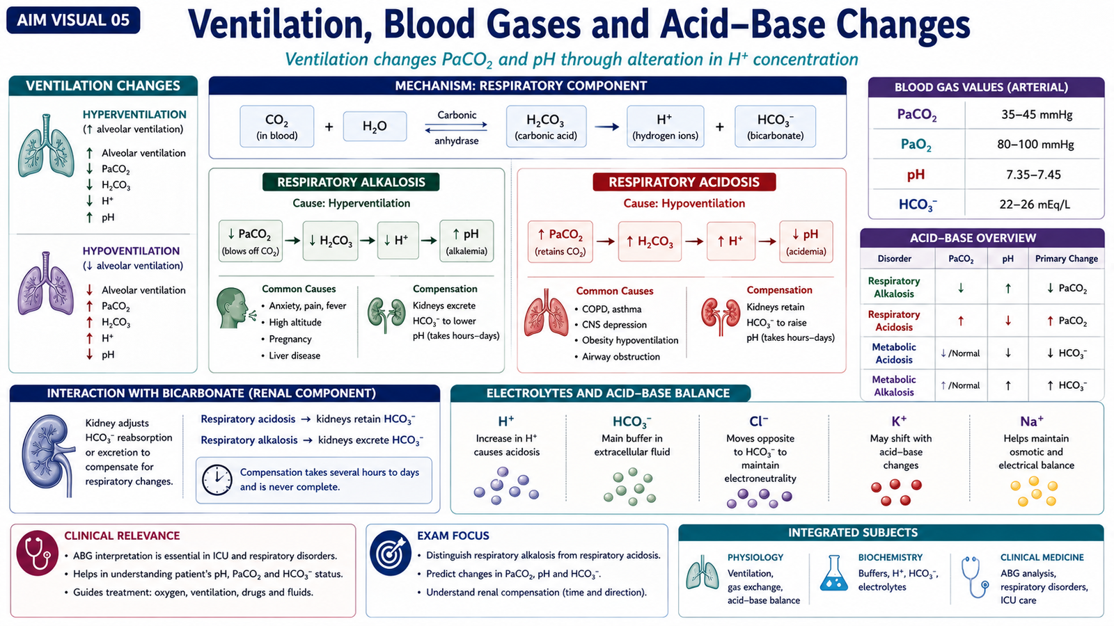
Ventilation directly influences the amount of carbon dioxide in arterial blood. Because carbon dioxide participates in the bicarbonate buffer system, excessive removal or retention of carbon dioxide produces predictable changes in blood pH. This connection is clinically important when interpreting respiratory disorders and arterial blood gases.
Hyperventilation
In hyperventilation, alveolar ventilation exceeds the amount required for carbon-dioxide elimination. Anxiety is a common clinical example. Excessive carbon dioxide is removed from the body, causing arterial carbon dioxide pressure to fall.
If respiratory alkalosis persists, renal compensation gradually reduces bicarbonate retention, so plasma bicarbonate falls.
Hypoventilation
In hypoventilation, alveolar ventilation is insufficient to remove the carbon dioxide produced by metabolism. This may occur when effective ventilation is seriously impaired, including in severe respiratory disease.
When respiratory acidosis persists, the kidneys increase bicarbonate retention, helping to reduce the fall in pH.
| Ventilatory Change | PaCO₂ | pH | Persistent Compensation |
|---|---|---|---|
| Hyperventilation | Decreases | Increases | Bicarbonate decreases |
| Hypoventilation | Increases | Decreases | Bicarbonate increases |
Acid–base disturbances can also influence electrolyte distribution, particularly potassium. The direction and degree of electrolyte change depend on the severity, duration and underlying cause, so the main third-year concept is to recognize that ventilatory disturbances affect not only PaCO₂ and pH but also bicarbonate and electrolyte balance.
F. Clinical Approach to Respiratory Symptoms
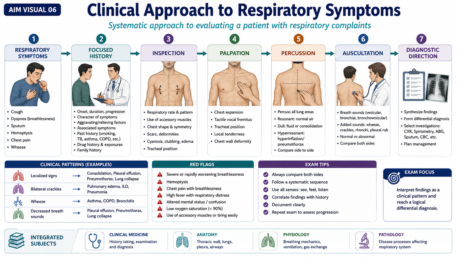
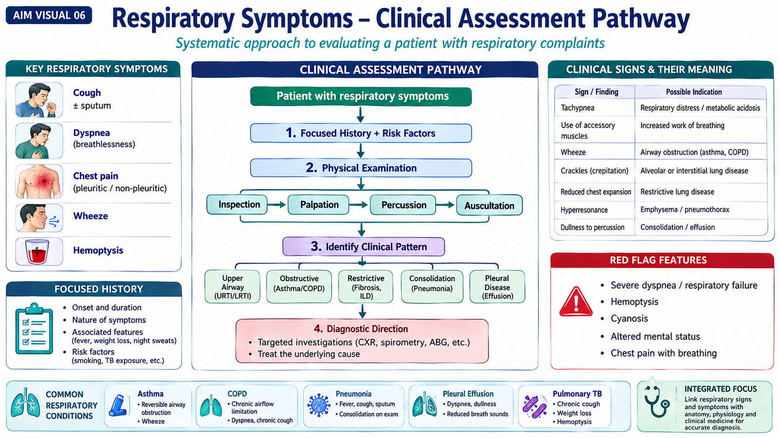
Respiratory assessment begins by identifying the patient’s main symptom and determining whether the problem is primarily affecting the airways, lung parenchyma, pleura or respiratory function. History and examination should therefore be performed systematically and interpreted together.
Important Respiratory Symptoms
- Cough.
- Sputum production.
- Shortness of breath.
- Wheeze.
- Chest pain.
- Hemoptysis.
- Fever associated with respiratory symptoms.
The history should determine the onset, duration, progression, severity and associated features of the symptoms. Relevant previous respiratory disease and exposures such as cigarette smoking, occupational exposures or infectious contact should be considered when appropriate.
Respiratory Examination
Examination follows the familiar sequence of inspection, palpation, percussion and auscultation. Each stage provides different information about respiratory structure and function.
- Inspection: assesses breathing pattern, respiratory effort, symmetry of chest movement and visible signs of distress.
- Palpation: helps assess chest expansion and other physical signs.
- Percussion: helps distinguish normally aerated lung from areas containing increased fluid or tissue, or excessive air.
- Auscultation: identifies breath sounds and added sounds such as wheeze and crackles.
Clinical signs should be connected with their underlying mechanism. Wheeze commonly results from airflow through narrowed airways, whereas crackles may occur when previously closed or fluid-affected small airways and alveolar regions open during breathing.
Severe respiratory distress, marked hypoxemia, altered consciousness or other evidence of physiological instability should be recognized as warning features requiring urgent assessment.
G. Basic Chest Radiology and Common Abnormalities
Chest radiography provides a basic structural view of the lungs, pleural spaces, central airways, heart, diaphragms and thoracic cage. A useful interpretation depends on examining the image systematically instead of focusing immediately on a single abnormal shadow.
Systematic Review
After considering whether the image is technically adequate, important regions should be reviewed in an orderly manner:
- Trachea and central airways.
- Lung fields.
- Hilar regions.
- Pleural spaces.
- Diaphragms and costophrenic angles.
- Cardiac silhouette.
- Visible bones and soft tissues.
Many common abnormalities can be understood by asking whether the affected region has become more opaque, more lucent, reduced in volume or altered in contour.
Consolidation
When air within alveoli is replaced by inflammatory material or fluid, the affected lung becomes more radiopaque. Consolidation therefore appears as an area of increased pulmonary opacity.
Pleural Effusion
Fluid accumulating in the pleural cavity produces increased opacity in the dependent portion of the chest and commonly causes blunting of the costophrenic angle.
Pneumothorax
Air in the pleural cavity separates the lung from the chest wall. The affected peripheral region becomes abnormally lucent and normal lung markings are absent beyond the visible pleural line.
Atelectasis
Atelectasis means collapse or incomplete expansion of lung tissue. It commonly produces increased opacity together with evidence of volume loss, which helps distinguish it from some other causes of pulmonary opacity.
Hyperinflation
Excessive air trapping increases lung volume. The lungs may appear unusually expanded and lucent, and the diaphragms may appear flattened.
| Abnormality | Main Structural Change | Basic X-Ray Clue |
|---|---|---|
| Consolidation | Alveolar air replaced by inflammatory material/fluid | Increased pulmonary opacity |
| Pleural effusion | Fluid in pleural space | Dependent opacity and blunted costophrenic angle |
| Pneumothorax | Air in pleural space | Increased lucency with absent peripheral lung markings |
| Atelectasis | Loss of lung expansion | Opacity with volume loss |
| Hyperinflation | Excessive air trapping | Expanded lucent lungs with flattened diaphragms |
Chest X-ray findings should always be interpreted together with the patient’s symptoms and physical examination rather than being treated as an isolated diagnosis.
Integrated Mechanism Flow
⭐ AIM High-Yield Review
- The right main bronchus is shorter, wider and more vertical than the left, making right-sided aspiration more likely.
- The right lung has three lobes; the left lung has two.
- The lower lobes occupy a large posterior area, so posterior chest examination is clinically important.
- Surfactant reduces alveolar surface tension and helps prevent alveolar collapse.
- Inspiration occurs when thoracic expansion lowers alveolar pressure and draws air into the lungs.
- Quiet expiration is mainly produced by relaxation and elastic recoil.
- Effective gas exchange requires adequate ventilation, perfusion and an intact alveolar-capillary interface.
- Hyperventilation lowers PaCO₂ and produces respiratory alkalosis.
- Hypoventilation raises PaCO₂ and produces respiratory acidosis.
- Persistent respiratory alkalosis causes compensatory reduction in bicarbonate, while persistent respiratory acidosis causes compensatory bicarbonate retention.
- Respiratory examination follows inspection, palpation, percussion and auscultation.
- Wheeze commonly reflects narrowed airways.
- Consolidation produces increased lung opacity, while pneumothorax produces increased peripheral lucency with absent lung markings.
- Pleural effusion commonly causes dependent opacity with blunting of the costophrenic angle.
- Atelectasis is distinguished by loss of lung volume, while hyperinflation reflects excessive air trapping.