
📌 AIM Study Tip
This chapter follows the KMU learning outcomes for laryngeal anatomy, laryngitis and laryngeal tumours. First understand how the structure of the larynx relates to voice, airway protection and clinical symptoms; then revise the high-yield points at the end.
Topic 5 — Laryngeal Anatomy, Inflammation and Tumours
Respiration Module • Clinical Anatomy of the Larynx • Acute and Chronic Laryngitis • Laryngeal Tumours
Topic Introduction
The larynx is a specialized part of the upper airway that connects the pharynx with the trachea. It performs three closely related functions: maintaining an open airway, protecting the lower respiratory tract during swallowing and producing the voice. Because these functions depend on the position and movement of the laryngeal cartilages and vocal folds, inflammation or tumour formation may produce characteristic symptoms such as hoarseness, throat discomfort, cough, difficulty swallowing or airway obstruction. In this chapter, you will first understand the clinically important anatomy of the larynx. You will then learn the causes, clinical features and management principles of acute and chronic laryngitis, followed by the major risk factors, morphology, clinical presentation and staging principles of laryngeal tumours.
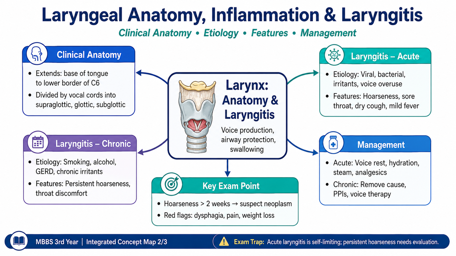
A. Clinical Anatomy of the Larynx
The larynx lies in the anterior part of the neck between the pharynx above and the trachea below. Its framework is formed by cartilages connected by joints, membranes and muscles. This framework allows the laryngeal inlet and vocal folds to change position during breathing, swallowing and phonation. Understanding these relationships is clinically important because disease at different laryngeal sites produces different symptoms and may spread through predictable anatomical pathways.
Laryngeal Framework
The principal laryngeal cartilages form a supporting skeleton around the airway.
- Thyroid cartilage: the largest cartilage and the main anterior and lateral framework of the larynx.
- Cricoid cartilage: lies below the thyroid cartilage and forms a complete ring around the airway.
- Epiglottis: a leaf-shaped elastic cartilage that helps protect the laryngeal inlet during swallowing.
- Arytenoid cartilages: paired cartilages located on the posterior part of the cricoid; movements of the arytenoids change the position and tension of the vocal folds.
Regions of the Larynx
Clinically, the larynx is divided into three important regions. This division is particularly useful when describing laryngeal tumours.
- Supraglottis: the region above the true vocal folds, including the epiglottis and related structures.
- Glottis: principally the true vocal folds and the space between them, called the rima glottidis.
- Subglottis: extends below the vocal folds toward the lower border of the cricoid cartilage.
True and False Vocal Folds
The true vocal folds contain the vocal ligaments and are the main structures involved in phonation. Air passing between the approximated vocal folds causes them to vibrate and produce sound. The false vocal folds, or vestibular folds, lie above them and mainly contribute to protection rather than normal voice production.
Intrinsic Muscles and Vocal Fold Movement
Intrinsic laryngeal muscles move the vocal folds by acting on the laryngeal cartilages. Their coordinated action opens the glottis during inspiration, closes it during airway protection and adjusts vocal-fold tension for phonation.
The posterior cricoarytenoid is particularly important because it abducts the vocal folds and therefore opens the rima glottidis. Most other intrinsic muscles primarily adduct or adjust the tension of the folds.
Innervation
Motor and sensory innervation of the larynx is supplied mainly through branches of the vagus nerve.
- The recurrent laryngeal nerve supplies nearly all intrinsic muscles of the larynx.
- The external branch of the superior laryngeal nerve supplies the cricothyroid muscle.
- Sensory supply above and below the vocal folds is also carried through branches of the vagus nerve.
Clinical link: Injury to a recurrent laryngeal nerve may impair vocal-fold movement and cause hoarseness. Bilateral severe impairment may compromise the airway because the vocal folds cannot abduct normally.
🖼 AIM VISUAL 01 — Clinical Anatomy of the Larynx

B. Acute Laryngitis
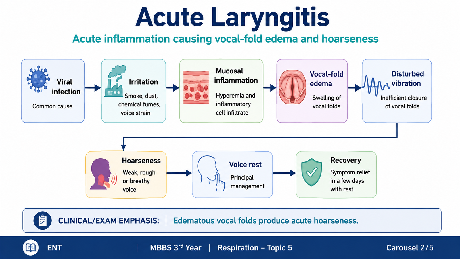
Acute laryngitis is a short-duration inflammation of the laryngeal mucosa, particularly involving the vocal folds. Inflammation causes mucosal congestion and edema. Because normal phonation requires smooth and regular vibration of the vocal folds, even mild swelling can alter the voice and produce hoarseness.
Etiology
Acute laryngitis most often occurs as part of an acute upper respiratory illness, although irritation or excessive mechanical use of the voice may also contribute.
- Viral upper respiratory tract infections
- Secondary bacterial infection in some cases
- Excessive voice use or vocal strain
- Exposure to smoke or other inhaled irritants
How the Symptoms Develop
Inflammation increases blood flow and vascular permeability in the laryngeal mucosa. Fluid enters the tissues and produces edema. When the true vocal folds become swollen, their normal vibration is disturbed.
Clinical Features
- Hoarseness or alteration in voice is the characteristic feature.
- Throat discomfort, dryness or soreness may occur.
- Dry cough or frequent throat clearing may be present.
- Symptoms of an accompanying upper respiratory infection may occur.
- Temporary loss of voice may occur when inflammation is marked.
Management Principles
Management is mainly directed toward reducing irritation of the inflamed vocal folds and treating the underlying cause when appropriate.
- Voice rest: limits repeated mechanical trauma to swollen vocal folds.
- Adequate hydration: helps maintain moist mucosal surfaces.
- Avoidance of smoke and other irritants.
- Symptomatic treatment may be used for associated discomfort.
- Antibacterial therapy is not routinely required for uncomplicated viral laryngitis.
🖼 AIM VISUAL 02 — Acute Laryngitis
C. Chronic Laryngitis
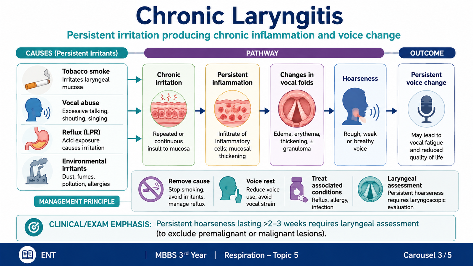
Chronic laryngitis is persistent inflammation of the laryngeal mucosa caused by continuing or repeated irritation. Unlike acute laryngitis, in which edema develops rapidly and usually resolves, chronic exposure can produce sustained mucosal inflammation and epithelial changes. Persistent hoarseness is therefore an important clinical feature and should not automatically be attributed to a simple infection.
Etiology
- Long-term tobacco-smoke exposure
- Repeated exposure to environmental or occupational irritants
- Persistent or repeated vocal abuse
- Repeated episodes of laryngeal inflammation
- Gastroesophageal or laryngopharyngeal reflux may contribute to chronic irritation
Clinical Features
Repeated irritation causes persistent swelling and altered vibration of the vocal folds. Patients commonly complain of:
- Persistent or recurrent hoarseness
- Voice fatigue
- Throat discomfort or irritation
- Chronic cough or repeated throat clearing
- A sensation of dryness or something being present in the throat
Management Principles
Effective treatment depends on identifying and removing the factor that is continuously irritating the larynx.
- Stop smoking and avoid inhaled irritants.
- Reduce excessive or traumatic voice use.
- Maintain hydration and appropriate vocal hygiene.
- Address reflux when it is contributing to laryngeal irritation.
- Persistent unexplained hoarseness requires assessment of the larynx to exclude an underlying structural lesion or tumour.
Persistent hoarseness, particularly in a patient with important risk factors such as smoking, should not simply be labelled chronic laryngitis without evaluating the larynx.
🖼 AIM VISUAL 03 — Chronic Laryngitis
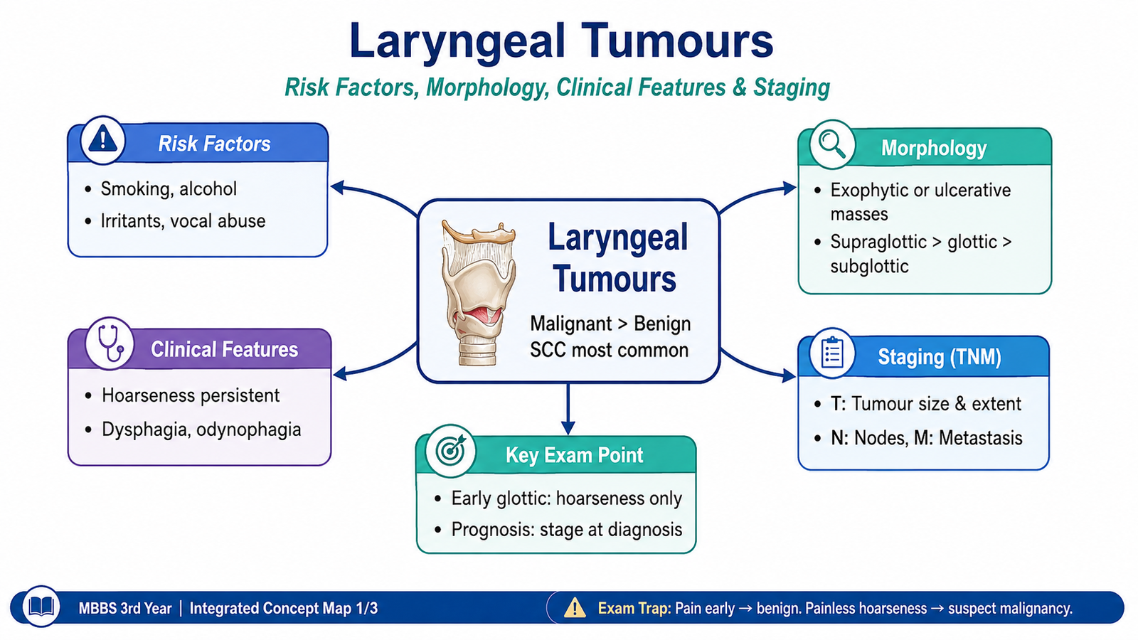
D. Laryngeal Tumours — Risk Factors and Morphology
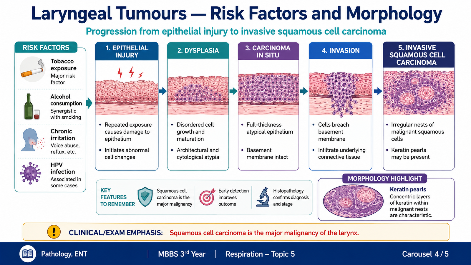
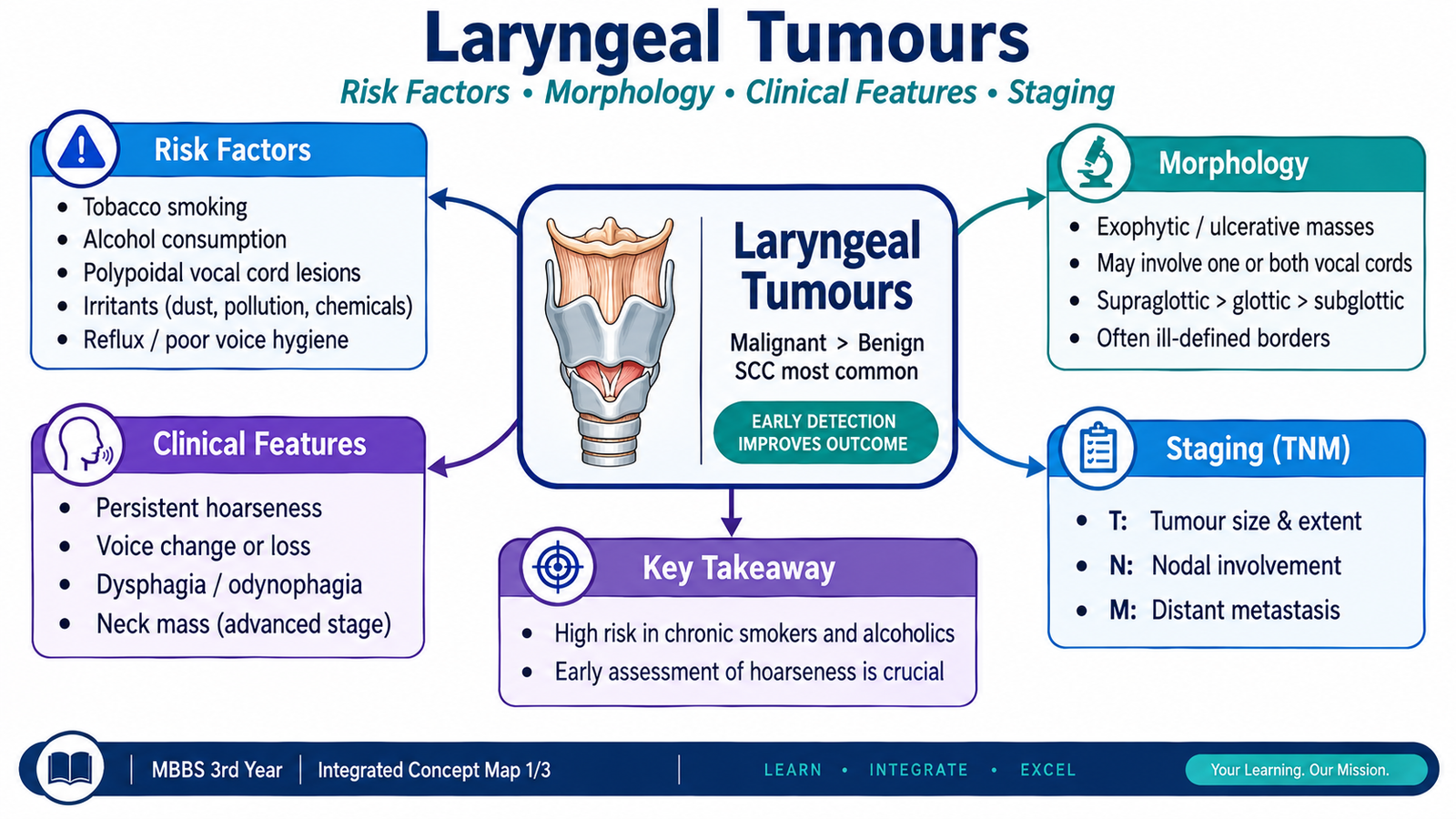
Laryngeal tumours may be benign or malignant, but the major malignant tumour of the larynx is squamous cell carcinoma. This is understandable because most of the laryngeal mucosal surface is lined by squamous-type epithelium or may undergo squamous metaplasia when exposed repeatedly to irritants. Repeated carcinogenic injury can lead to epithelial dysplasia, carcinoma in situ and eventually invasive carcinoma.
Major Risk Factors
The most important risk factor is prolonged exposure of the laryngeal epithelium to carcinogenic substances.
- Tobacco smoking is the dominant risk factor.
- Alcohol use can increase risk, especially when combined with tobacco exposure.
- Persistent exposure to some inhaled occupational or environmental irritants may contribute.
- Increasing age is associated with accumulated carcinogenic exposure.
Development of Squamous Cell Carcinoma
Chronic carcinogenic exposure repeatedly damages the epithelial cells. Over time, genetically altered cell clones may develop progressively abnormal maturation and architecture. The epithelial changes may progress through dysplasia before malignant cells cross the basement membrane and invade the underlying tissue.
Morphology
Gross Morphology
The tumour may form an irregular, thickened, ulcerative or exophytic mucosal lesion. As it enlarges, it may infiltrate the underlying laryngeal tissues and interfere with the mobility of the vocal folds.
Microscopic Morphology
Typical squamous cell carcinoma shows invasive nests, cords or sheets of malignant squamous cells. The cells show nuclear atypia and abnormal maturation. Well-differentiated tumours may demonstrate keratinization and formation of keratin pearls.
Invasion is the key feature that separates invasive carcinoma from carcinoma in situ. Once malignant cells penetrate the basement membrane, they can infiltrate local tissues and gain access to lymphatic channels.
🖼 AIM VISUAL 04 — Laryngeal Squamous Cell Carcinoma
E. Clinical Features and Staging of Laryngeal Carcinoma
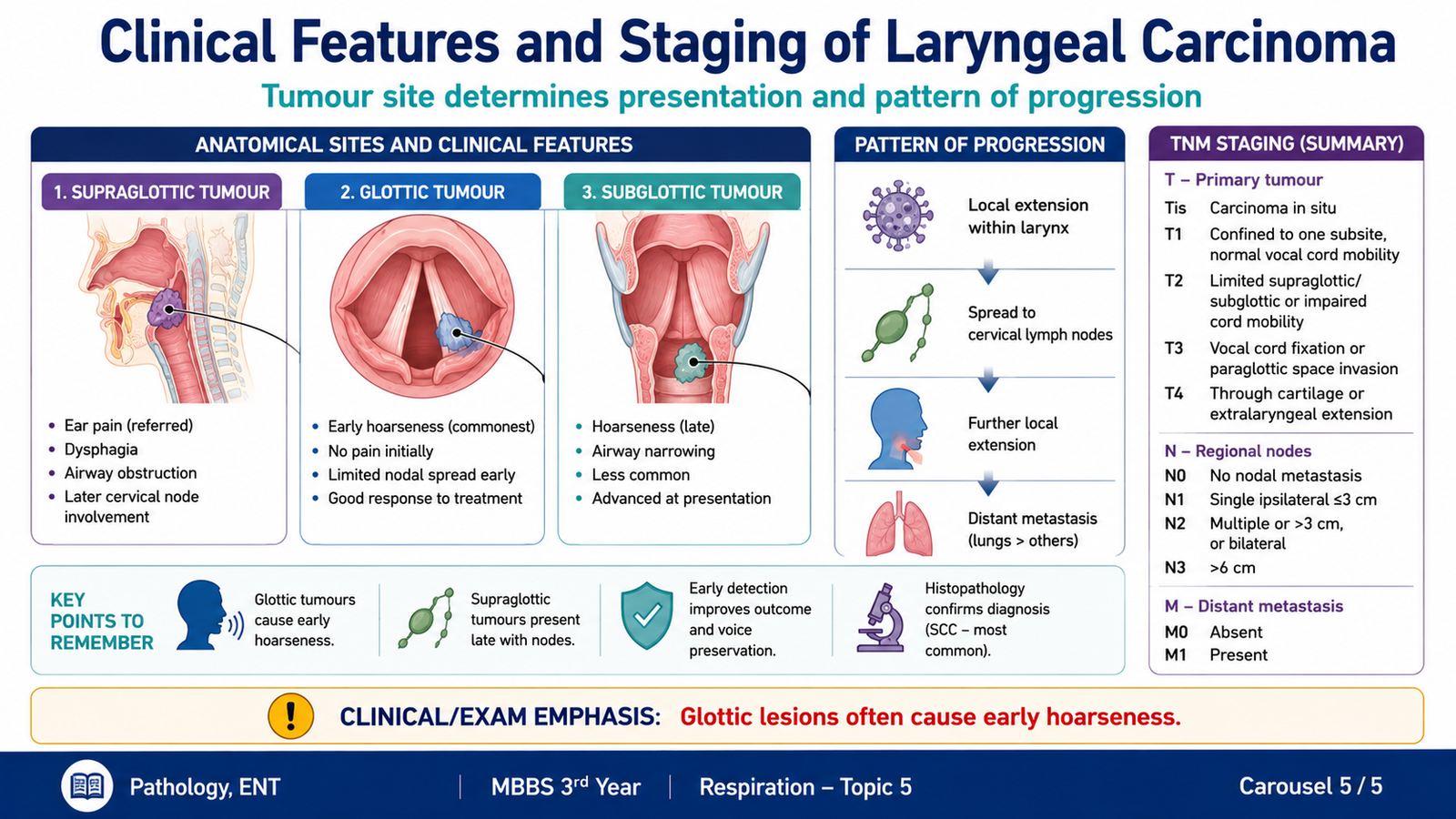
The clinical presentation of laryngeal carcinoma is strongly influenced by the anatomical site of the tumour. A small lesion involving a true vocal fold can interfere with vibration at an early stage and therefore produce hoarseness while the tumour is still relatively limited. Tumours arising away from the vocal folds may remain less obvious until they become larger or spread to nearby structures.
Clinical Features
- Persistent hoarseness: especially important in glottic disease because the vocal folds are directly involved in phonation.
- Throat discomfort or persistent throat symptoms.
- Dysphagia or odynophagia: may occur when more superior structures are involved.
- Persistent cough.
- Airway difficulty or stridor: may appear when a tumour significantly narrows the laryngeal airway.
- Neck lymph-node enlargement: may indicate regional lymphatic spread.
Importance of Tumour Site
Glottic tumours often produce hoarseness relatively early because even a small lesion disturbs normal vocal-fold vibration. This early symptom can lead to earlier recognition.
Supraglottic tumours may initially cause less obvious voice change because the true vocal folds are not involved immediately. They may therefore present with throat symptoms, swallowing difficulty or regional lymph-node involvement.
Subglottic tumours are less common and may remain clinically silent until they narrow the airway or extend into adjacent regions.
Principles of Staging
Staging describes the anatomical extent of a malignant tumour and helps communicate how far the disease has progressed. Laryngeal carcinoma is assessed using the TNM system.
- T — Primary tumour: considers the site and local extent of the primary tumour, including involvement of adjacent laryngeal regions and impairment or fixation of vocal-fold movement.
- N — Regional lymph nodes: records whether metastatic tumour has spread to cervical lymph nodes and the extent of that involvement.
- M — Distant metastasis: records whether distant metastatic spread is present.
The detailed T definitions differ for supraglottic, glottic and subglottic cancers because each region has different anatomical boundaries. For undergraduate learning, the essential principle is that increasing local extension, impaired laryngeal function, nodal spread and distant metastasis represent progressively more advanced disease.
A persistent change in voice in a patient with a strong smoking history should raise concern for a laryngeal lesion, particularly when symptoms do not resolve.
🖼 AIM VISUAL 05 — Clinical Presentation and Staging
Integrated Mechanism Flow
↓
Repeated epithelial injury and accumulation of abnormal cellular changes
↓
Squamous dysplasia and carcinoma in situ
↓
Basement-membrane penetration and invasive squamous cell carcinoma
↓
Local interference with vocal-fold movement or laryngeal function
↓
Hoarseness, swallowing symptoms or airway symptoms depending on tumour site
↓
Further local invasion and possible regional lymph-node or distant spread
Important Comparison — Acute vs Chronic Laryngitis
| Feature | Acute Laryngitis | Chronic Laryngitis |
|---|---|---|
| Duration | Short duration | Persistent or recurrent |
| Typical cause | Usually acute respiratory infection | Continuing irritation such as smoking or vocal abuse |
| Main tissue change | Acute congestion and edema | Persistent mucosal inflammation |
| Voice change | Temporary hoarseness | Persistent or recurrent hoarseness |
| Basic management | Voice rest, hydration, avoid irritants | Remove chronic cause and assess persistent symptoms |
⭐ AIM High-Yield Review
- The larynx maintains the airway, protects the lower respiratory tract during swallowing and produces the voice.
- The clinically important laryngeal regions are the supraglottis, glottis and subglottis.
- The true vocal folds are the principal structures responsible for phonation.
- The posterior cricoarytenoid abducts the vocal folds and opens the rima glottidis.
- The recurrent laryngeal nerve supplies nearly all intrinsic laryngeal muscles; injury may cause hoarseness.
- Acute laryngitis causes vocal-fold edema, which disturbs vibration and produces hoarseness.
- Chronic laryngitis results from persistent irritation such as smoking, repeated vocal strain or other chronic irritants.
- Persistent unexplained hoarseness requires assessment rather than repeated treatment as simple laryngitis.
- ⭐ The major malignant laryngeal tumour is squamous cell carcinoma.
- ⭐ Tobacco smoking is the major risk factor for laryngeal squamous cell carcinoma.
- Squamous carcinogenesis may progress from dysplasia to carcinoma in situ and then invasive carcinoma.
- Well-differentiated squamous cell carcinoma may show keratinization and keratin pearls.
- ⭐ Glottic tumours commonly produce hoarseness early because they directly disturb vocal-fold vibration.
- Supraglottic tumours may present later and can be associated with cervical lymph-node involvement.
- Laryngeal carcinoma staging follows the principles of T = primary tumour, N = regional nodes and M = distant metastasis.
Laryngeal Cancer — Anatomy, Clinical Understanding & Staging
Watch this short video after completing the learning material. Focus particularly on the anatomical regions of the larynx and the principles used to stage laryngeal carcinoma.
If the embedded player does not open,
open the video on YouTube
.