
This chapter follows the supplied KMU learning outcomes in a logical sequence. First understand how each tumour develops and how its morphology explains its clinical behaviour; then use the high-yield review for rapid revision.
Topic 13 — Lung and Laryngeal Neoplasms
Respiration | Pathology + ENT
A structured explanation of lung carcinoma, bronchial carcinoids, malignant mesothelioma and neoplastic laryngeal lesions, with emphasis on etiology, pathogenesis, morphology, clinical course and essential management principles.
Topic Introduction
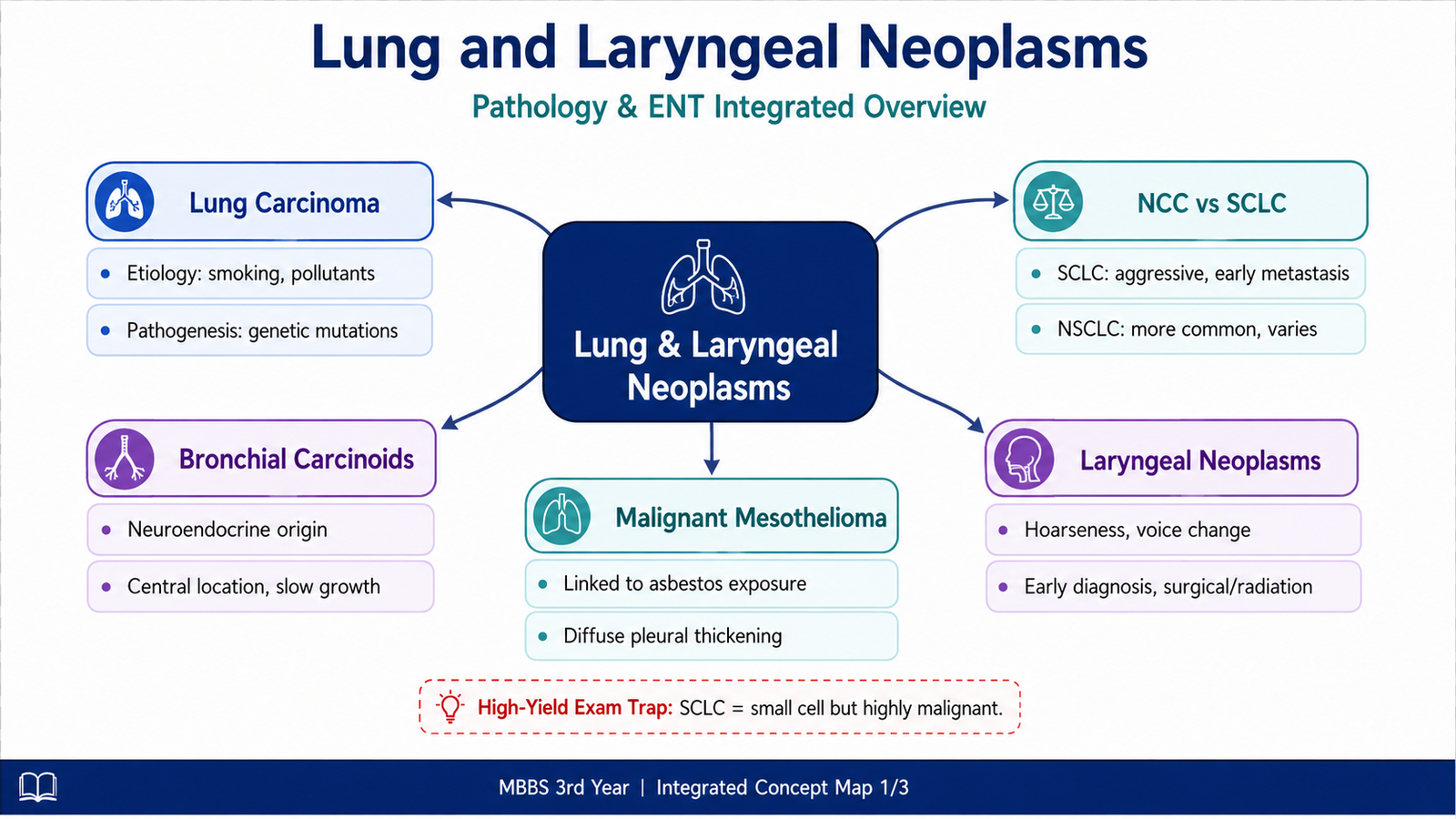
Neoplasms of the lung and larynx range from relatively slow-growing neuroendocrine tumours to highly aggressive carcinomas. In the lung, the major malignant epithelial tumours are grouped broadly into small-cell lung carcinoma and non-small-cell lung carcinoma. Their causes, microscopic appearance and biological behaviour are different, and these differences affect clinical presentation and treatment approach. Bronchial carcinoids form a separate group of neuroendocrine tumours, while malignant mesothelioma arises from mesothelial surfaces, particularly the pleura. In the larynx, squamous cell carcinoma is the major malignant neoplastic lesion. Understanding these tumours becomes easier when the student connects risk factors with molecular injury, morphology, local spread and clinical manifestations.
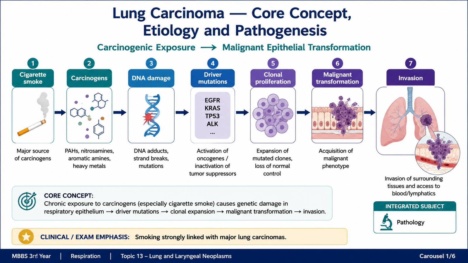
A. Lung Carcinoma — Core Concept, Etiology and Pathogenesis
Lung carcinoma is a malignant epithelial tumour arising from the respiratory epithelium. It is clinically important because it often remains silent until it has become locally advanced or metastatic. The major pathological groups are small-cell lung carcinoma (SCLC) and non-small-cell lung carcinoma (NSCLC). NSCLC includes adenocarcinoma, squamous cell carcinoma and large-cell carcinoma.
Etiology and Risk Factors
Cigarette smoking is the most important risk factor for lung carcinoma, especially for squamous cell carcinoma and small-cell carcinoma. Tobacco smoke contains multiple carcinogens that repeatedly injure bronchial epithelial cells. With continued exposure, genetic abnormalities accumulate and may eventually transform normal epithelial cells into malignant cells.
- Cigarette smoking: strongest established environmental association.
- Passive smoke exposure: increases exposure to tobacco carcinogens.
- Occupational carcinogens: asbestos and certain industrial agents may increase risk, especially in combination with smoking.
- Air pollution: contributes to risk through chronic exposure to inhaled carcinogens.
- Pre-existing pulmonary damage: areas of scarring may occasionally be associated with peripheral carcinomas.
Adenocarcinoma is particularly important because it is the most common lung carcinoma in people who have never smoked, although smoking can also contribute to its development.
Pathogenesis
Lung cancer develops through the progressive accumulation of genetic alterations that disturb normal control of cell proliferation, survival and DNA repair. Tobacco-related carcinogens produce repeated DNA damage. Cells that acquire growth-promoting mutations or lose tumour-suppressor mechanisms gain a survival advantage, expand clonally and eventually develop malignant behaviour.
Different lung carcinoma subtypes tend to show different molecular abnormalities. For undergraduate understanding, the important principle is that these molecular alterations are not random facts: they help explain why some tumours behave differently and why selected NSCLCs can be treated according to specific molecular targets.
B. Major Lung Carcinomas — Morphology and Clinical Course
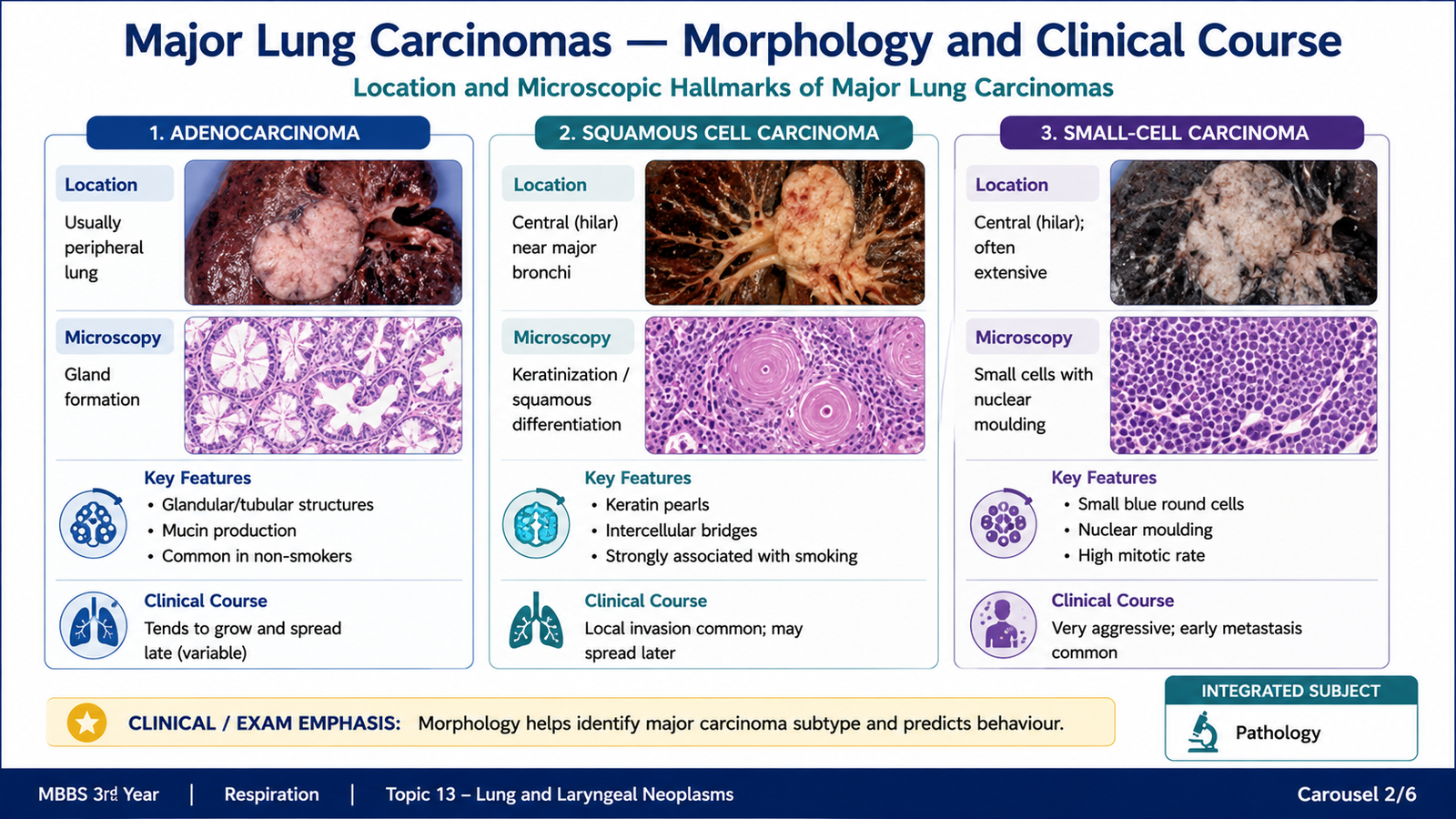
The morphology of lung carcinoma is important because tumour location and cell type help determine how the disease presents. Some tumours arise centrally near major bronchi, where they may cause obstruction early. Others arise more peripherally and may remain clinically silent until they invade adjacent structures or metastasize.
Adenocarcinoma
Adenocarcinoma is a malignant epithelial tumour showing glandular differentiation, mucin production or both. It commonly arises in the peripheral lung and is the most frequent type of primary lung carcinoma overall. It is also the major type encountered in never-smokers. Gross morphology: It is usually a peripheral, firm, grey-white lesion. Some tumours arise near areas of previous fibrosis or scarring and may cause pleural puckering. Microscopic morphology: Tumour cells may form glands, papillary structures or other adenocarcinoma patterns. Intracellular or extracellular mucin may be present. The degree of differentiation varies.
Squamous Cell Carcinoma
Squamous cell carcinoma is strongly related to cigarette smoking and commonly arises centrally from major bronchi. Chronic exposure to tobacco smoke may first produce squamous metaplasia and dysplasia in bronchial epithelium. With further genetic injury, carcinoma can develop. Gross morphology: These tumours are often central and may project into or obstruct a bronchus. Necrosis can produce cavitation. Microscopic morphology: Well-differentiated tumours show keratin formation and intercellular bridges, which reflect squamous differentiation.
Small-Cell Lung Carcinoma
Small-cell carcinoma is a highly malignant neuroendocrine carcinoma that is very strongly associated with cigarette smoking. It usually arises centrally and tends to spread early. Because dissemination often occurs before diagnosis, its clinical behaviour differs greatly from that of most NSCLCs. Microscopic morphology: The tumour consists of relatively small cells with scant cytoplasm, finely granular nuclear chromatin and poorly defined cell borders. Numerous mitotic figures and areas of necrosis are common. Nuclear moulding may be seen because adjacent nuclei press against one another.
Large-Cell Carcinoma
Large-cell carcinoma is an undifferentiated malignant epithelial tumour lacking the characteristic glandular differentiation of adenocarcinoma and the squamous differentiation of squamous cell carcinoma. The diagnosis depends on pathological assessment of poorly differentiated tumour cells.
Clinical Course of Lung Carcinoma
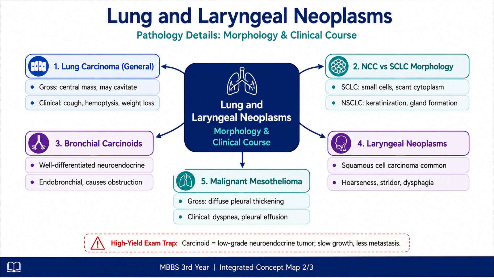
Clinical manifestations depend on tumour location, airway obstruction, invasion of neighbouring structures and metastasis. A centrally placed tumour may obstruct a bronchus and produce cough, recurrent infection or collapse of distal lung. Tumour erosion into vessels or airways may produce hemoptysis.
- Persistent or changing cough
- Hemoptysis
- Chest pain
- Dyspnea
- Recurrent or non-resolving pneumonia
- Weight loss and constitutional symptoms in advanced disease
Local extension may involve the pleura, chest wall, mediastinum or neighbouring nerves. Metastatic spread may occur through lymphatic and hematogenous routes. The brain, liver, bones and adrenal glands are important distant sites. Some lung carcinomas also produce hormones or hormone-like substances. These paraneoplastic syndromes may occasionally be an important clue to an underlying tumour. Small-cell carcinoma is particularly associated with ectopic hormone production, while squamous cell carcinoma may produce a parathyroid hormone-related protein effect leading to hypercalcemia.
C. Small-Cell versus Non-Small-Cell Lung Carcinoma
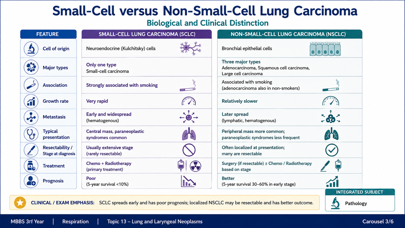
The distinction between SCLC and NSCLC is one of the most important practical classifications of lung cancer. The reason is not simply histological appearance. These two groups differ substantially in biological behaviour, pattern of spread and broad treatment approach. Small-cell carcinoma is a high-grade neuroendocrine carcinoma. It grows rapidly, metastasizes early and is usually disseminated at presentation. Therefore, treatment is mainly systemic rather than based on surgical removal of a localized lesion. Non-small-cell carcinoma includes adenocarcinoma, squamous cell carcinoma and large-cell carcinoma. These tumours generally spread less explosively than SCLC, and a localized resectable NSCLC may be treated surgically. Molecular testing may also identify therapeutically relevant abnormalities in selected NSCLCs.
| Feature | Small-Cell Lung Carcinoma | Non-Small-Cell Lung Carcinoma |
|---|---|---|
| Main types | Small-cell neuroendocrine carcinoma | Adenocarcinoma, squamous cell carcinoma, large-cell carcinoma |
| Smoking association | Very strong | Variable; strong in squamous carcinoma |
| Typical location | Usually central | Depends on subtype; adenocarcinoma commonly peripheral |
| Growth and spread | Rapid growth and early metastasis | Generally slower than SCLC |
| Histological clue | Small cells, scant cytoplasm, nuclear moulding, many mitoses | Subtype-specific glandular, squamous or poorly differentiated morphology |
| Broad treatment implication | Primarily systemic therapy because early dissemination is common | Surgical treatment may be possible when localized and resectable |
D. Bronchial Carcinoid Tumours
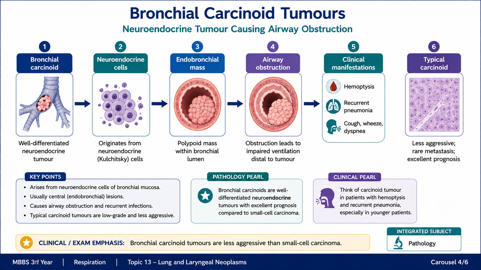
Bronchial carcinoids are neuroendocrine tumours of the lung. They are generally less aggressive than small-cell carcinoma and occur in a broader age range, including younger patients. Their biological behaviour is related to the degree of differentiation and proliferative activity. These tumours commonly arise in the central airways and may grow as polypoid masses projecting into the bronchial lumen. Because of this location, symptoms are often produced by mechanical obstruction rather than extensive metastatic disease.
Morphology
Gross morphology: A bronchial carcinoid is often a well-circumscribed, highly vascular, endobronchial mass. Its position in a bronchus may partially or completely obstruct airflow. Microscopic morphology: Typical carcinoids are composed of relatively uniform cells arranged in nests, trabeculae or organoid patterns. The cells have round-to-oval nuclei with finely granular neuroendocrine chromatin. Mitotic activity is low in typical carcinoids, and extensive necrosis is absent. Atypical carcinoids show greater proliferative activity and more aggressive behaviour than typical carcinoids, but they remain biologically distinct from small-cell carcinoma.
Clinical Features
Endobronchial growth explains many clinical manifestations. Obstruction prevents normal ventilation and drainage of distal airways, which can lead to recurrent localized infection or collapse.
- Persistent cough
- Hemoptysis because the tumour is vascular
- Wheezing due to partial bronchial obstruction
- Recurrent pneumonia distal to the obstructed bronchus
Some carcinoid tumours produce bioactive substances, but clinically obvious carcinoid syndrome is uncommon in primary bronchial carcinoids unless sufficient secreted products reach the systemic circulation.
E. Malignant Mesothelioma
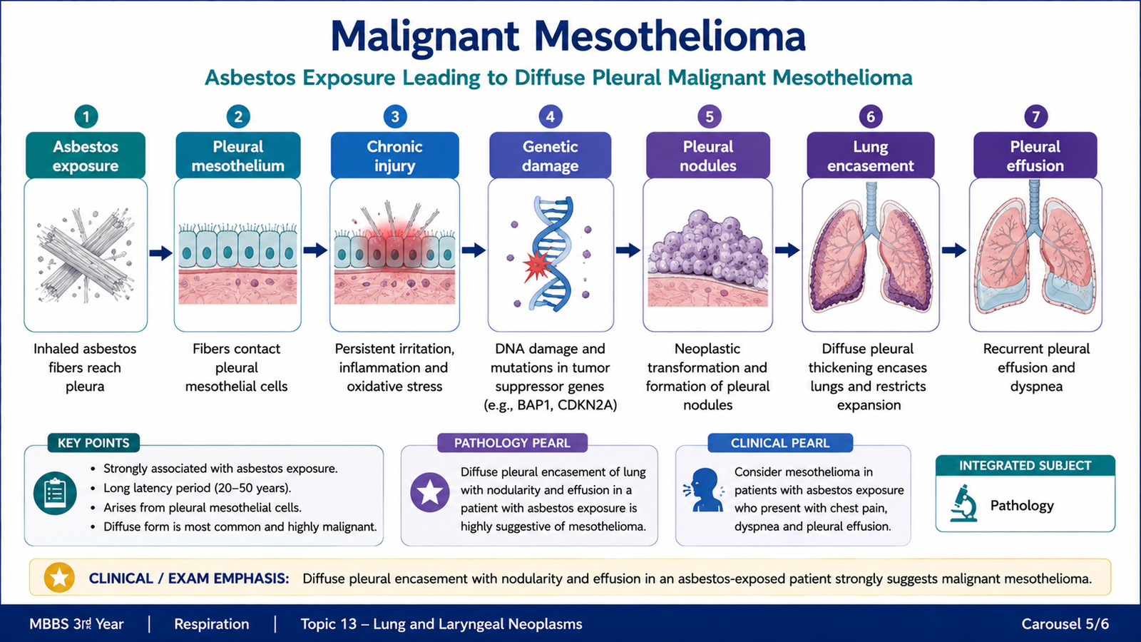
Malignant mesothelioma is an aggressive malignant tumour arising from mesothelial cells. In the thorax, it most commonly involves the pleura. Its most important etiological association is previous exposure to asbestos, usually after a long latent period.
Etiology and Pathogenesis
Inhaled asbestos fibres reach the distal lung and pleural surfaces. Persistent fibres produce chronic tissue injury, generation of reactive oxygen species and repeated mesothelial cell damage. Over time, accumulated genetic alterations may lead to malignant transformation.
An important examination distinction is that smoking greatly increases the risk of bronchogenic carcinoma in asbestos-exposed individuals, but smoking itself is not considered the major cause of malignant mesothelioma.
Gross Morphology
The tumour usually begins as multiple pleural nodules. These progressively enlarge and merge to form a diffuse thick tumour that can encase the lung like a rind. The pleural space may contain an associated effusion.
Microscopic Morphology
Malignant mesothelioma can show several patterns. The major morphological patterns are epithelial, sarcomatoid and biphasic. The epithelial pattern may form tubules or papillary structures and can resemble metastatic adenocarcinoma, making pathological distinction important.
Clinical Course
Patients may present with progressive chest pain, dyspnea and recurrent pleural effusion. Symptoms occur because the tumour diffusely thickens the pleura, restricts lung expansion and invades neighbouring thoracic structures. Malignant mesothelioma is usually locally aggressive and has a poor overall clinical course.
F. Neoplastic Laryngeal Lesions — Clinical Features and Management
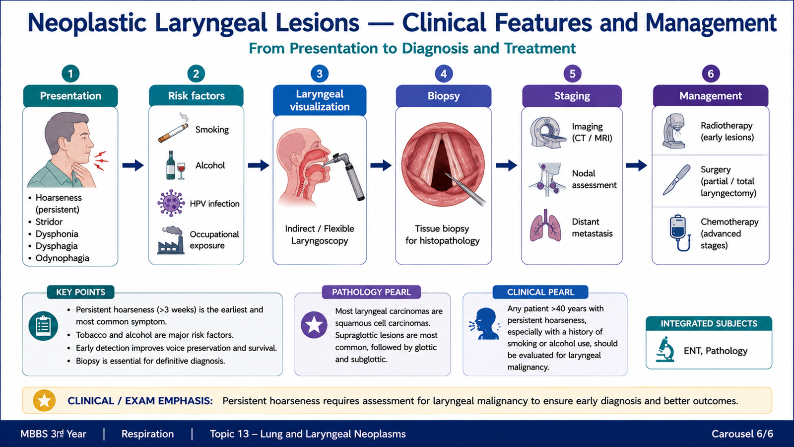
The most important malignant neoplasm of the larynx is squamous cell carcinoma. It develops from the laryngeal squamous epithelium, often after prolonged exposure to carcinogens. Cigarette smoking is the major risk factor, and alcohol exposure can further increase the risk. The clinical behaviour of a laryngeal tumour depends greatly on its anatomical site. A relatively small lesion involving the vocal cord can cause hoarseness early because even a minor disturbance of vocal-cord vibration alters the voice. In contrast, tumours outside the true vocal cord may enlarge considerably before producing obvious symptoms.
Clinical Features
- Persistent hoarseness: especially important in glottic lesions.
- Throat discomfort or persistent throat symptoms: may accompany local tumour growth.
- Dysphagia or odynophagia: may occur when a lesion interferes with swallowing or invades adjacent structures.
- Referred ear pain: may occur through shared sensory nerve pathways.
- Neck mass: may indicate regional lymph-node involvement.
- Stridor or respiratory difficulty: suggests significant airway narrowing and is an important red flag.
Clinical Assessment
Persistent unexplained hoarseness or other suspicious laryngeal symptoms require direct examination of the larynx. Visualization identifies the lesion and allows assessment of its site, extent and effect on vocal-cord movement. A suspicious lesion requires tissue diagnosis by biopsy because the definitive diagnosis of malignancy depends on histopathological examination.
Basic Management Principles
Management is determined mainly by tumour location, extent, functional considerations and the patient’s general condition. The major goals are tumour control, preservation of the airway and, where possible, preservation of speech and swallowing function.
- Localized lesions: may be treated using radiotherapy or appropriately selected surgical treatment.
- More extensive disease: may require larger surgical procedures, radiotherapy, systemic therapy or combined treatment.
- Airway compromise: requires urgent assessment and airway management.
- Advanced or complex disease: requires multidisciplinary specialist management.
The important undergraduate principle is that management should not be memorized as a single operation for every patient. The treatment approach changes according to the site and extent of the tumour while balancing cancer control with preservation of laryngeal function.
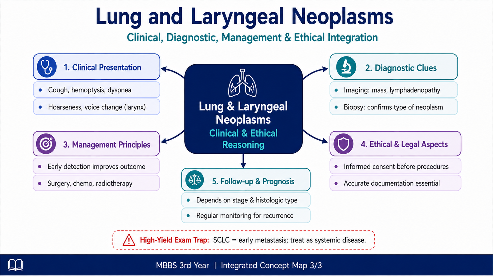
Integrated Mechanism Flow
Important Morphological Comparison
| Tumour | Typical Site | Key Morphology | High-Yield Behaviour |
|---|---|---|---|
| Adenocarcinoma | Usually peripheral | Glands and/or mucin | Common in never-smokers |
| Squamous carcinoma | Usually central | Keratinization and intercellular bridges | Strong smoking association |
| Small-cell carcinoma | Usually central | Small cells, scant cytoplasm, nuclear moulding | Rapid growth and early metastasis |
| Bronchial carcinoid | Often central/endobronchial | Uniform neuroendocrine cells in organoid patterns | Usually less aggressive than SCLC |
| Mesothelioma | Pleura | Diffuse nodular pleural thickening encasing lung | Strong asbestos association |
⭐ AIM High-Yield Review
- Cigarette smoking is the major etiological factor for lung carcinoma, especially squamous and small-cell carcinoma.
- Adenocarcinoma is commonly peripheral and shows gland formation and/or mucin production.
- Adenocarcinoma is the most important lung carcinoma occurring in never-smokers.
- Squamous cell carcinoma is usually central and shows keratinization and intercellular bridges.
- ⭐ Small-cell carcinoma is a highly aggressive neuroendocrine tumour with rapid growth and early metastasis.
- The practical distinction between SCLC and NSCLC reflects major differences in biological behaviour and treatment approach.
- Bronchial carcinoids are neuroendocrine tumours that are generally less aggressive than small-cell carcinoma.
- An endobronchial carcinoid may cause recurrent pneumonia because it obstructs bronchial drainage and ventilation.
- ⭐ Malignant mesothelioma is strongly associated with asbestos exposure and commonly forms a diffuse pleural tumour encasing the lung.
- Smoking and asbestos together strongly increase bronchogenic carcinoma risk, but smoking is not the major etiological factor for mesothelioma.
- Lung carcinoma may present with cough, hemoptysis, recurrent pneumonia, chest pain, dyspnea or metastatic manifestations.
- Squamous cell carcinoma is the principal malignant laryngeal neoplasm.
- ⭐ A glottic tumour can cause early persistent hoarseness because a small lesion interferes with vocal-cord vibration.
- Suspicious laryngeal lesions require visualization and biopsy for histopathological confirmation.
- Stridor in a patient with a laryngeal tumour suggests significant airway compromise and requires urgent assessment.
Laryngeal Cancer — Rapid Review
Short review of laryngeal tumour sites, vocal-cord involvement and clinically important tumour extent.