
This chapter follows the supplied KMU learning outcomes in a logical sequence. First understand how impaired airway clearance produces bronchiectasis and how CFTR dysfunction produces cystic fibrosis, then revise the high-yield points at the end.
Topic 10 — Bronchiectasis, Cystic Fibrosis and Pediatric Wheezing Disorders
Module: Respiration
Understand bronchiectasis from cause to morphology, recognize important childhood wheezing patterns, learn the supportive management principles of bronchiolitis, and connect the CFTR defect of cystic fibrosis with its respiratory, gastrointestinal and systemic manifestations.
Topic Introduction
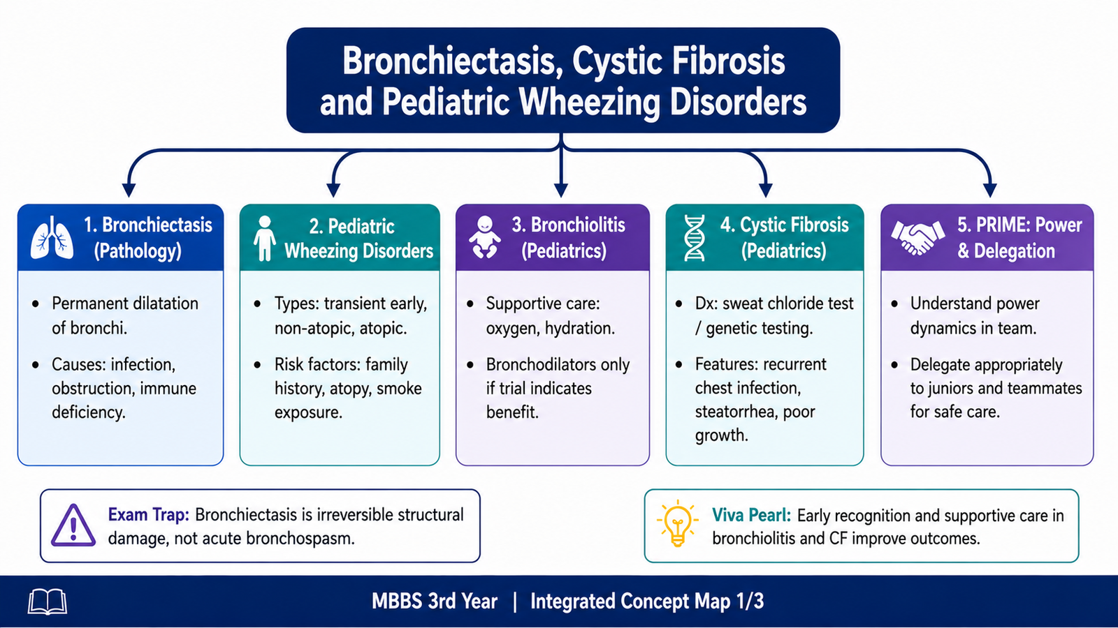
This topic connects three important respiratory problems encountered in childhood and general pathology. Bronchiectasis is permanent abnormal dilatation of bronchi caused by destruction of their walls, usually after repeated infection, obstruction or failure of mucus clearance. Cystic fibrosis is an inherited disorder of CFTR function that produces abnormally thick secretions and can lead to recurrent respiratory infection, bronchiectasis and gastrointestinal disease. Children may also present with wheezing, but not every wheezing child has asthma; the pattern and associated risk factors help predict persistence. Bronchiolitis is another important cause of wheezing in infants and is managed mainly with supportive care. The final section introduces safe power delegation within healthcare teams, as required by the PRIME learning outcome.
A. Bronchiectasis — Definition, Etiology and Risk Factors
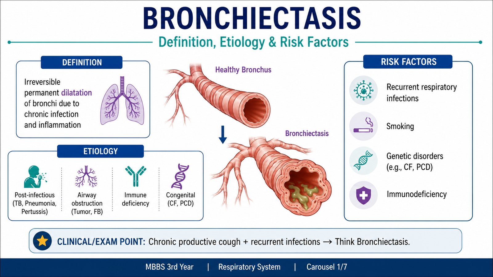
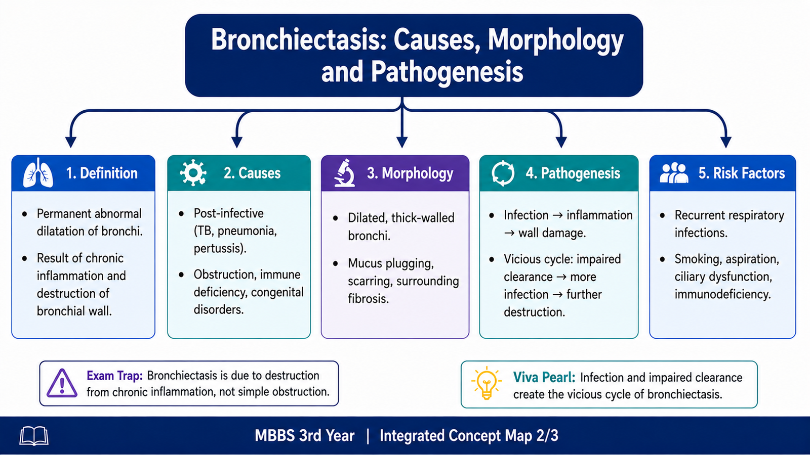
Bronchiectasis is permanent, abnormal dilatation of bronchi and bronchioles caused by destruction of the muscular and elastic components of their walls. The key word is permanent. Temporary widening of an airway during an acute illness is not bronchiectasis.
Bronchiectasis usually develops when normal clearance of bronchial secretions fails. Secretions remain within the airway, microorganisms multiply, inflammation develops and the bronchial wall is progressively injured. An underlying cause should therefore be sought, particularly in a child with recurrent respiratory infections.
Etiology
The major causes act through one of two closely related mechanisms: they either obstruct the airway or impair normal mucus clearance. Both promote retention of secretions and recurrent infection.
- Severe or recurrent respiratory infections: repeated inflammation may directly damage the bronchial wall.
- Bronchial obstruction: a foreign body, tumor or mucus plug prevents drainage distal to the obstruction.
- Cystic fibrosis: thick dehydrated mucus is difficult to clear and promotes recurrent infection.
- Primary ciliary dyskinesia: defective ciliary movement interferes with mucociliary clearance.
- Immunodeficiency: recurrent respiratory infections predispose to progressive airway injury.
- Congenital structural abnormalities: abnormal airway structure may impair drainage and increase susceptibility to infection.
Important Pediatric Risk Factors
In children, recurrent infection alone should not always be accepted as the final explanation. A disorder that repeatedly interferes with airway protection or clearance may be responsible.
- Recurrent or severe lower respiratory tract infections
- Cystic fibrosis
- Primary ciliary dyskinesia
- Immunodeficiency
- Airway obstruction, particularly an inhaled foreign body
- Repeated aspiration
B. Bronchiectasis — Pathogenesis and Morphology
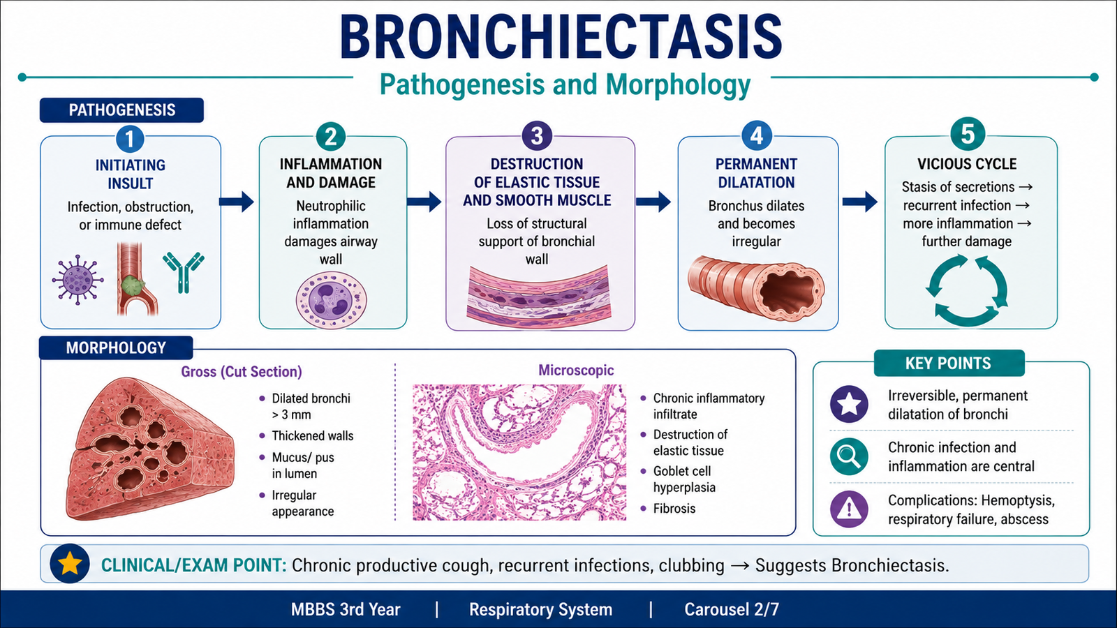
The pathogenesis of bronchiectasis is best understood as a self-perpetuating cycle. An initiating problem such as obstruction, abnormal mucus or defective ciliary clearance prevents effective removal of airway secretions. Retained secretions encourage bacterial growth and persistent inflammation.
Neutrophils and other inflammatory cells release enzymes and inflammatory mediators that damage the epithelium, smooth muscle and elastic tissue of the bronchial wall. Once these supporting tissues are destroyed, the bronchus becomes permanently widened. The widened airway then clears secretions even less effectively, so further infection and inflammation occur.
Gross Morphology
Grossly, the affected airways are markedly enlarged and may remain dilated almost to the pleural surface. The exact distribution depends on the underlying cause.
- Dilatation of bronchi and bronchioles
- Cylindrical, fusiform or saccular patterns of enlargement
- Thick mucopurulent secretions within affected airways
- Irregular thickening and fibrosis of chronically damaged bronchial walls
- Extension of dilated airways unusually close to the pleural surface
Microscopic Morphology
Microscopically, inflammation damages both the epithelial lining and the supporting structures of the bronchus. The appearance varies with disease activity and duration.
- Acute and chronic inflammatory cells within the bronchial wall
- Ulceration or loss of bronchial epithelium
- Destruction of smooth muscle and elastic tissue
- Fibrosis during chronic healing
- Purulent inflammatory material within the lumen
The destruction of muscle and elastic tissue explains why the airway cannot return to its original diameter. Fibrosis further distorts the airway and surrounding lung.
C. Pediatric Wheezing — Types of Wheezers and Risk of Persistent Asthm
Wheezing is a musical sound produced when air moves through narrowed airways. It is commonly heard during expiration because intrathoracic airways become relatively narrower during expiratory airflow. Wheezing is common in young children because their airways have a small diameter, so even modest mucosal swelling or mucus accumulation can substantially increase airway resistance.
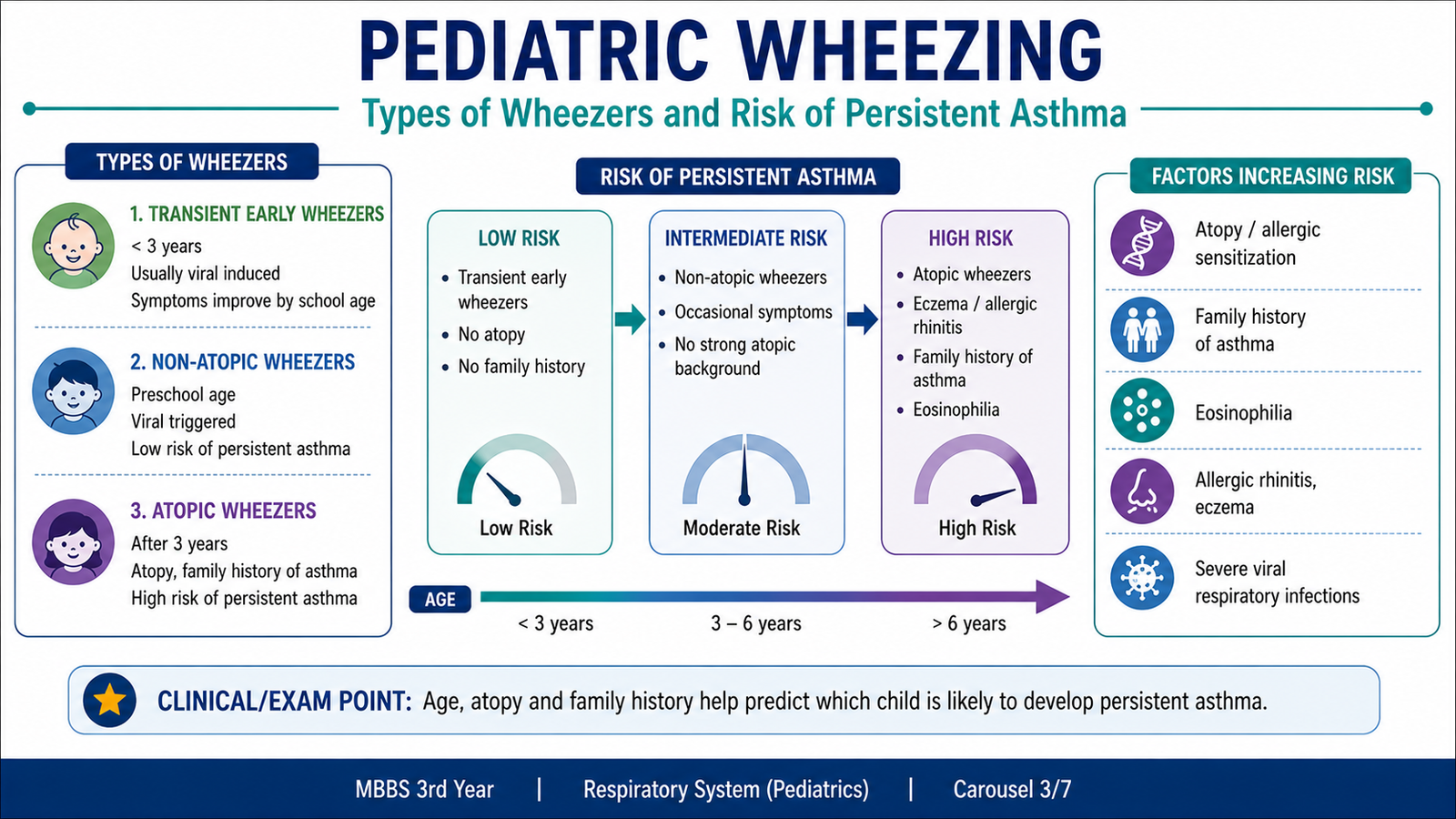
Not every child who wheezes has asthma. The pattern over time is important. Some children wheeze only during the first few years of life and later improve, whereas others continue to wheeze and have a greater likelihood of persistent asthma.
Types of Wheezing Patterns
| Pattern | Typical Course | Main Concept |
|---|---|---|
| Transient early wheeze | Begins in infancy or early childhood and later disappears. | Often relates to relatively small early-life airways and viral infections. |
| Persistent wheeze | Starts early and continues beyond early childhood. | More strongly associated with later asthma, especially when atopy is present. |
| Late-onset wheeze | Appears later rather than during infancy. | Atopic disease and asthma become more important associations. |
Risk Factors for Persistent Wheezing and Asthma
Persistence is more likely when wheezing is associated with an inherited or acquired tendency toward airway hyperresponsiveness and allergic inflammation rather than only temporary narrowing during viral infections.
- Parental asthma: indicates an inherited tendency toward asthma.
- Atopic dermatitis or eczema: suggests an atopic background.
- Allergic sensitization: increases the likelihood of persistent allergic airway disease.
- Wheezing apart from obvious respiratory infections: supports a tendency toward persistent airway hyperreactivity.
- Frequent or severe wheezing episodes: increase concern that symptoms may continue.
- Exposure to tobacco smoke: irritates the developing respiratory tract and contributes to recurrent respiratory symptoms.
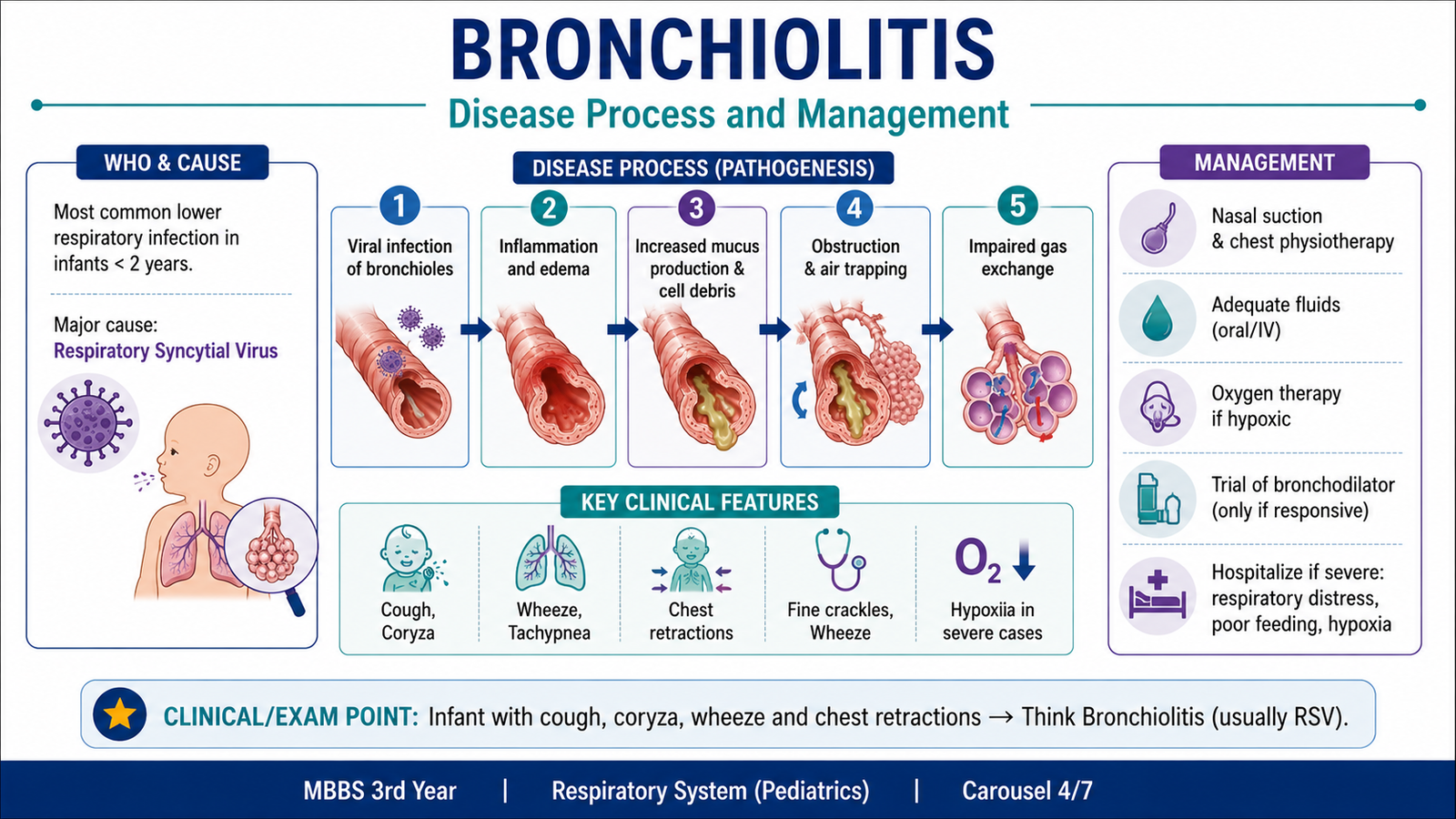
D. Bronchiolitis — Disease Process and Management
Bronchiolitis is an acute viral infection of the small airways that occurs mainly in infants and young children. Viral injury causes inflammation of the bronchiolar epithelium, edema of the airway wall, increased mucus production and shedding of epithelial cells into the lumen.
These changes markedly narrow small bronchioles. Because infant airways are already narrow, a relatively small amount of edema or mucus can produce a major increase in resistance to airflow. This explains the typical combination of cough, tachypnea, increased work of breathing, wheeze and sometimes crackles.
Management Principles
Management of typical bronchiolitis is mainly supportive. The important question is not simply whether the child is wheezing, but whether breathing, oxygenation and hydration are being maintained.
- Assess respiratory severity: look for tachypnea, chest recession, nasal flaring, grunting, apnea and exhaustion.
- Assess oxygenation: identify clinically important hypoxemia and provide supplemental oxygen when required.
- Maintain hydration: feeding may become difficult because infants must coordinate sucking, swallowing and breathing.
- Clear troublesome nasal secretions: excessive upper-airway secretions can interfere with feeding and breathing.
- Escalate respiratory support when necessary: worsening respiratory distress or respiratory failure requires higher-level supportive care.
Why Routine Asthma Medication Is Not the Main Treatment
In bronchiolitis, airway obstruction is produced mainly by edema, inflammatory debris and mucus in very small airways. This differs from classic asthma, where reversible bronchial smooth-muscle constriction is an important mechanism.
Therefore, bronchodilators and corticosteroids are not routinely the central therapy for uncomplicated bronchiolitis. Antibiotics do not treat the underlying viral infection and are reserved for situations in which a bacterial infection is also suspected or demonstrated.
E. Cystic Fibrosis — Core Defect and Diagnostic Criteria
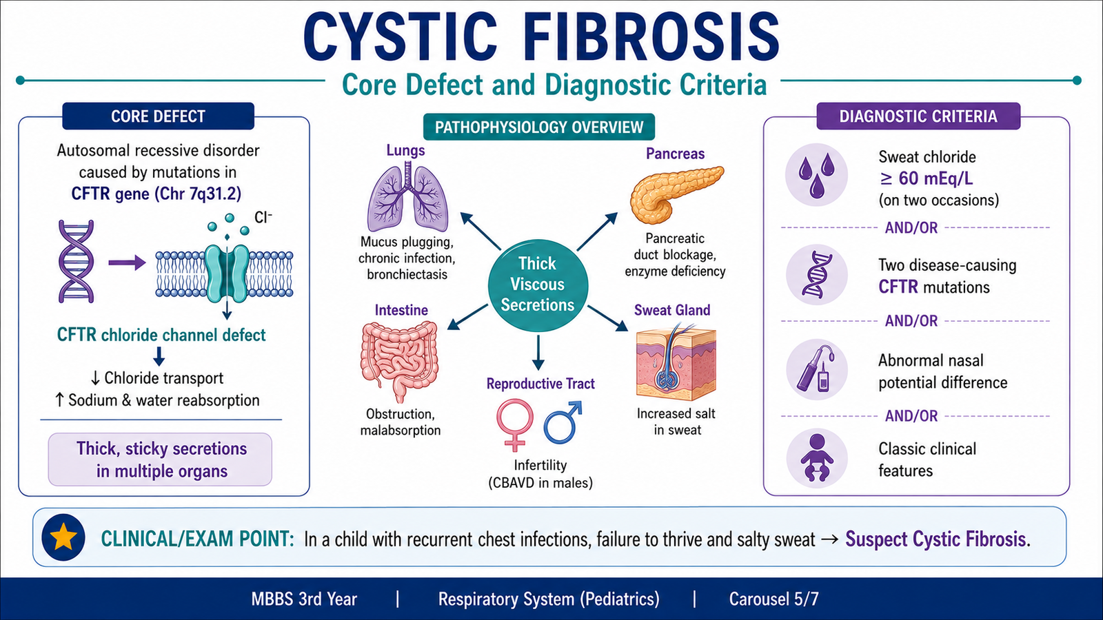
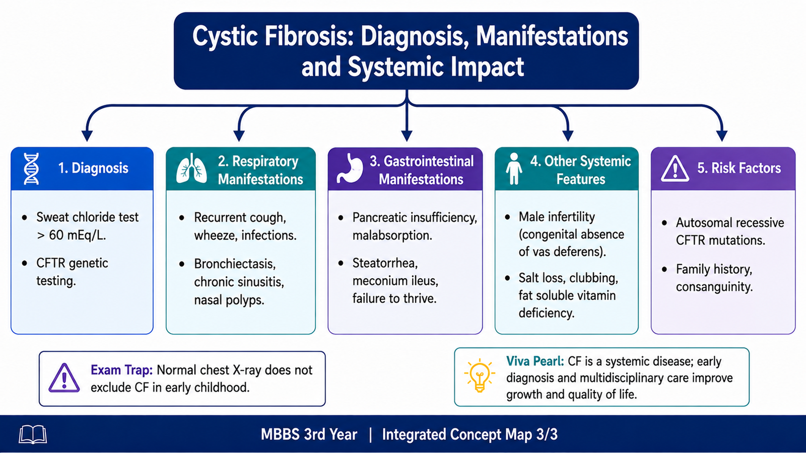
Cystic fibrosis (CF) is an inherited multisystem disorder caused by pathogenic abnormalities affecting the cystic fibrosis transmembrane conductance regulator (CFTR). CFTR is an epithelial ion channel that normally participates in chloride and bicarbonate transport and thereby influences movement of salt and water across epithelial surfaces.
When CFTR function is severely impaired, secretions at several epithelial surfaces become abnormally dehydrated and thick. The consequences differ between organs, but the central pattern is similar: thick secretions → obstruction → inflammation or tissue damage → organ dysfunction.
Diagnostic Approach
The diagnosis of cystic fibrosis depends on a compatible clinical setting or recognized risk together with objective evidence of abnormal CFTR function. A child may be investigated because of characteristic manifestations, a positive newborn screening result or a family history.
Evidence of CFTR Dysfunction
- Sweat chloride testing: increased sweat chloride provides important evidence of defective CFTR-mediated chloride handling.
- CFTR genetic testing: identification of disease-causing CFTR variants supports the diagnosis.
- Specialized CFTR functional testing: may be useful when clinical suspicion remains strong but routine tests are inconclusive.
Sweat testing is especially useful because sweat glands behave differently from airway glands. In sweat ducts, CFTR normally helps reabsorb chloride from sweat. When CFTR is defective, chloride and accompanying sodium remain in the sweat, producing the characteristic high salt content.
F. Cystic Fibrosis — Respiratory, Gastrointestinal and Other Systemic Manifestations
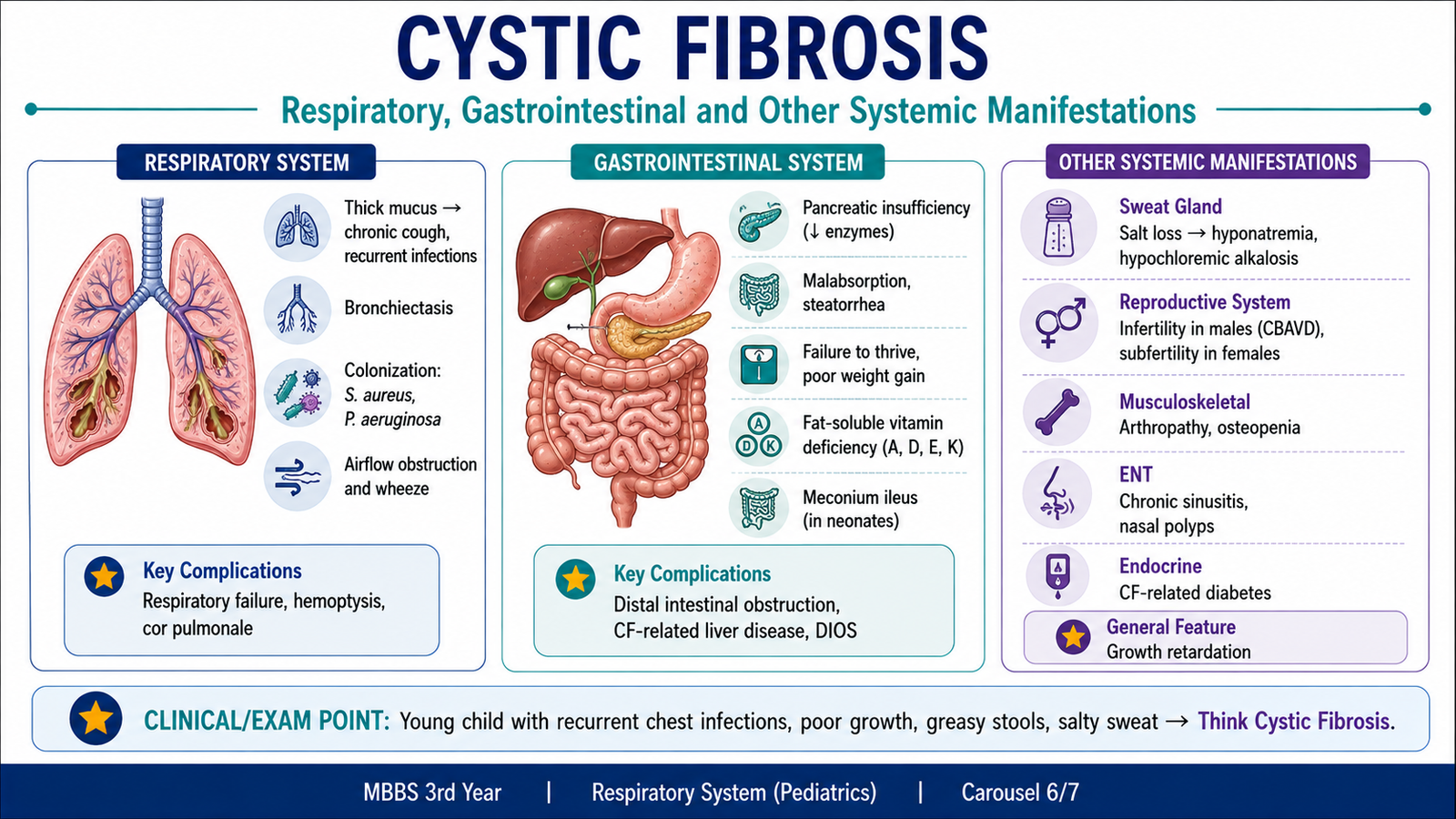
Cystic fibrosis affects several organs because CFTR is expressed in multiple epithelial tissues. The manifestations become easier to understand when each system is connected with the underlying abnormal secretion or ion transport.
Respiratory Manifestations
In the respiratory tract, dehydrated mucus becomes difficult for cilia to move. Mucus therefore remains within the airways and microorganisms are not cleared effectively. Recurrent infection produces persistent inflammation, and repeated inflammation progressively damages bronchial walls.
- Persistent or recurrent cough
- Thick respiratory secretions
- Recurrent lower respiratory tract infections
- Wheezing in some patients
- Progressive airway inflammation and damage
- Bronchiectasis as a major structural consequence of chronic airway disease
- Chronic sinus disease and nasal polyps may also occur.
The link between cystic fibrosis and bronchiectasis is direct: abnormal mucus clearance promotes infection; infection causes inflammation; inflammation destroys the supporting tissues of bronchi; and the bronchi become permanently dilated.
Gastrointestinal and Pancreatic Manifestations
In the pancreas, thick secretions obstruct pancreatic ducts. Digestive enzymes then fail to reach the intestinal lumen normally. This produces exocrine pancreatic insufficiency and impaired digestion, particularly of dietary fats.
- Exocrine pancreatic insufficiency
- Fat malabsorption
- Bulky, greasy stools
- Poor weight gain and growth failure
- Deficiency of fat-soluble vitamins when malabsorption is significant
- Meconium ileus may be an early manifestation in a newborn.
- Distal intestinal obstruction may occur later.
Hepatobiliary Manifestations
Thick secretions can obstruct small intrahepatic bile ducts. Persistent obstruction and inflammation may gradually produce fibrosis and chronic liver disease.
Sweat-Gland Manifestations
In sweat ducts, failure of normal chloride reabsorption leaves excessive chloride and sodium in sweat. This produces the well-known finding of salty sweat and explains why sweat chloride testing can demonstrate abnormal CFTR function.
Reproductive Manifestations
Male patients commonly have infertility because of abnormal development or absence of the vas deferens. This is another example of CF affecting epithelial and ductal structures outside the lungs.
| System | Main Abnormality | Important Manifestation |
|---|---|---|
| Respiratory | Thick poorly cleared mucus | Recurrent infection and bronchiectasis |
| Pancreas | Duct obstruction | Exocrine insufficiency and malabsorption |
| Intestine | Thick intestinal contents | Meconium ileus or later obstruction |
| Sweat glands | Reduced chloride reabsorption | Salty sweat |
| Hepatobiliary system | Small-duct obstruction | Fibrosis and chronic liver disease |
| Male reproductive tract | Abnormal vas deferens development | Infertility |
G. PRIME — Power Dynamics and Delegation in Healthcare Teams
Power dynamics describe the way authority, seniority, professional role, knowledge and influence affect interaction between members of a healthcare team. A clinical team naturally contains people with different responsibilities, but these differences should support organized patient care rather than prevent junior members from communicating important concerns.
A doctor should therefore use professional authority responsibly. Good leadership includes listening to team members, giving clear instructions, allowing appropriate questions and creating an environment in which patient-safety concerns can be raised.
Recognizing Power Dynamics
- Recognize differences in seniority, experience and professional authority.
- Communicate respectfully with both junior and senior team members.
- Encourage clarification when instructions are unclear.
- Allow appropriate concerns to be voiced, particularly when patient safety is involved.
- Avoid using hierarchy merely to silence questions or disagreement.
Delegating Powers and Tasks
Delegation means assigning an appropriate task and the authority required to perform it to another member of the team. Safe delegation requires the person delegating to consider both the nature of the task and the competence of the person receiving it.
- Select an appropriate task: the task should be suitable for delegation.
- Select an appropriate person: consider competence, training and experience.
- Give clear instructions: state what must be done and what result is expected.
- Define the limits of authority: clarify which decisions can be made independently and when senior advice is required.
- Provide appropriate supervision: more complex tasks or less experienced team members require closer oversight.
- Confirm completion and outcome: important tasks should be followed up rather than simply assumed to be complete.
 .
.Integrated Mechanism Flow
Bronchiectasis and cystic fibrosis are closely linked by failure of airway clearance. The following sequence connects the major respiratory concepts in this topic.
Important Comparison — Pediatric Wheezing Patterns
| Feature | Transient Early Wheeze | Persistent Wheeze | Late-Onset Wheeze |
|---|---|---|---|
| Onset | Infancy or early childhood | Early childhood | Later childhood |
| Course | Usually resolves as the child grows | Continues beyond early childhood | Develops after an initially symptom-free period |
| Atopy | Less prominent | Often important | Often important |
| Later asthma risk | Lower | Higher | Higher when associated with atopy |
⭐ AIM High-Yield Review
- Bronchiectasis is permanent abnormal dilatation of bronchi due to destruction of their muscular and elastic walls.
- The basic bronchiectasis cycle is impaired clearance → infection → inflammation → bronchial destruction → further impaired clearance.
- Important causes include recurrent infection, airway obstruction, cystic fibrosis, ciliary dysfunction and immunodeficiency.
- Gross bronchiectasis may be cylindrical, fusiform or saccular.
- Microscopy shows inflammation with destruction of bronchial epithelium, smooth muscle and elastic tissue, followed by fibrosis.
- ⭐ Wheezing in a child is a clinical sign and does not automatically mean asthma.
- Persistent wheezing is more strongly associated with atopy, parental asthma and wheezing outside viral infections.
- Bronchiolitis causes small-airway obstruction mainly through edema, mucus and epithelial debris.
- Management of uncomplicated bronchiolitis is mainly supportive, focusing on breathing, oxygenation and hydration.
- Cystic fibrosis results from abnormal CFTR function and produces abnormal epithelial salt and water transport.
- In CF lungs, dehydrated mucus causes impaired clearance, recurrent infection and eventually bronchiectasis.
- Pancreatic duct obstruction in CF causes exocrine insufficiency, malabsorption and poor growth.
- Meconium ileus is an important early gastrointestinal manifestation of CF.
- High salt content of sweat results from defective chloride reabsorption in sweat ducts and is useful diagnostically.
- Safe delegation requires the right task, the right person, clear authority, appropriate supervision and confirmation that the task was completed.
Bronchiectasis, Cystic Fibrosis and Pediatric Wheezing Disorders
Watch this pediatric respiratory teaching session to reinforce wheezing disorders, bronchiolitis and cystic fibrosis after completing the AIM Learning Material.