
Interstitial, Fibrosing and Occupational Lung Diseases
Topic Introduction
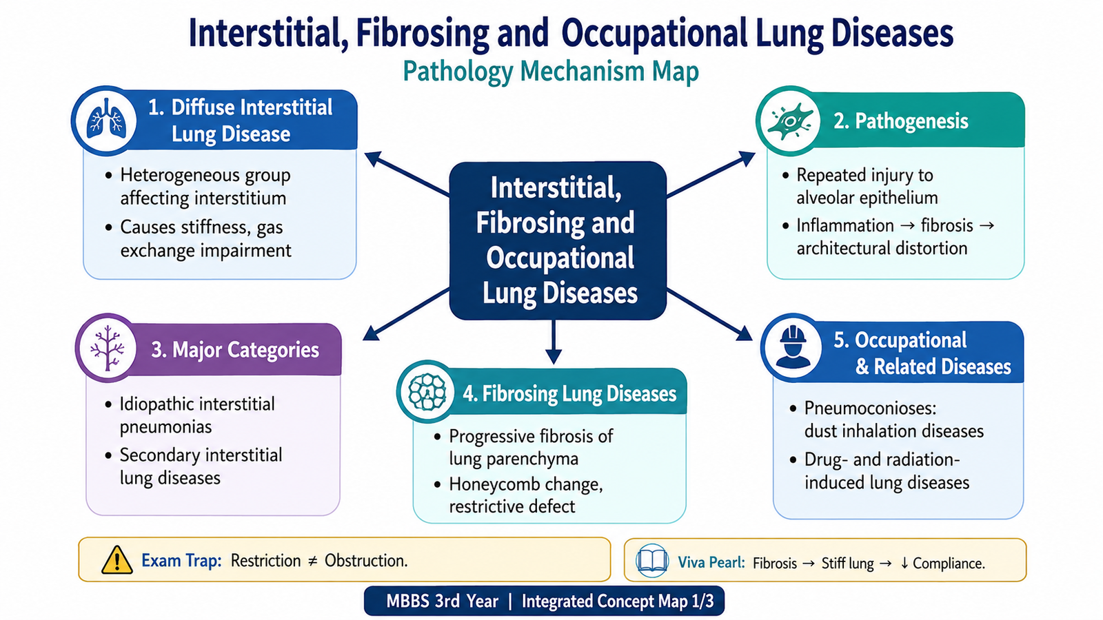
Interstitial lung diseases are a broad group of disorders in which the supporting tissue of the lung, particularly the alveolar walls and interstitium, becomes injured and often inflamed or fibrotic. Because the lung becomes stiff, these diseases mainly produce a restrictive pattern of functional impairment. Some arise without a known cause, whereas others result from inhaled occupational dusts, drugs or radiation. An important final pathway in many chronic cases is pulmonary fibrosis, in which normal thin alveolar walls are replaced by excessive collagen and distorted lung architecture. This chapter explains the basic concept and pathogenesis of diffuse interstitial lung disease, important fibrosing patterns, pneumoconiosis and its major types, their characteristic morphology, occupational control and prevention, and drug- and radiation-induced pulmonary disease.
A. Diffuse Interstitial Lung Disease — Definition and Major Categories
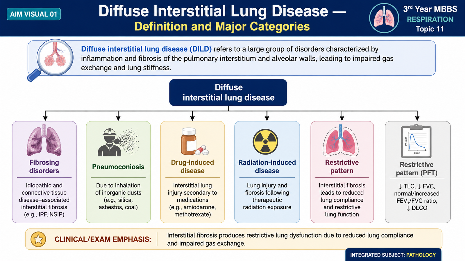
Diffuse interstitial lung disease (DILD), also called diffuse parenchymal lung disease, refers to a group of disorders characterized by widespread involvement of the pulmonary interstitium and, in many conditions, the alveolar walls and adjacent lung parenchyma. The interstitium normally provides a very thin supporting framework around the alveoli. When it becomes inflamed, thickened or fibrotic, the lungs lose compliance and oxygen must diffuse across a thicker barrier. This explains why these diseases are predominantly restrictive. A restrictive lung has difficulty expanding, so total lung capacity and other lung volumes tend to decrease. Thickening and destruction of the alveolar-capillary interface also impair gas transfer.
Major categories of chronic interstitial lung disease
Chronic interstitial lung diseases can be organized according to their dominant cause or pathological pattern. The classification helps students understand that several different initiating processes may ultimately produce similar fibrosis and respiratory impairment.
- Fibrosing disorders — particularly idiopathic pulmonary fibrosis and other fibrosing interstitial patterns.
- Occupational and environmental diseases — especially pneumoconioses caused by inhalation of mineral dusts.
- Drug-induced interstitial lung disease — produced by toxic or hypersensitivity effects of certain medicines.
- Radiation-induced lung disease — resulting from injury to lung tissue included in a radiation field.
- Other chronic interstitial disorders — diverse conditions in which inflammation or other tissue injury affects the pulmonary interstitium.
The exact initiating factor differs, but a common pathological theme is persistent injury of alveolar epithelial and interstitial structures followed by abnormal repair.
B. Pathogenesis of Diffuse Interstitial Lung Disease
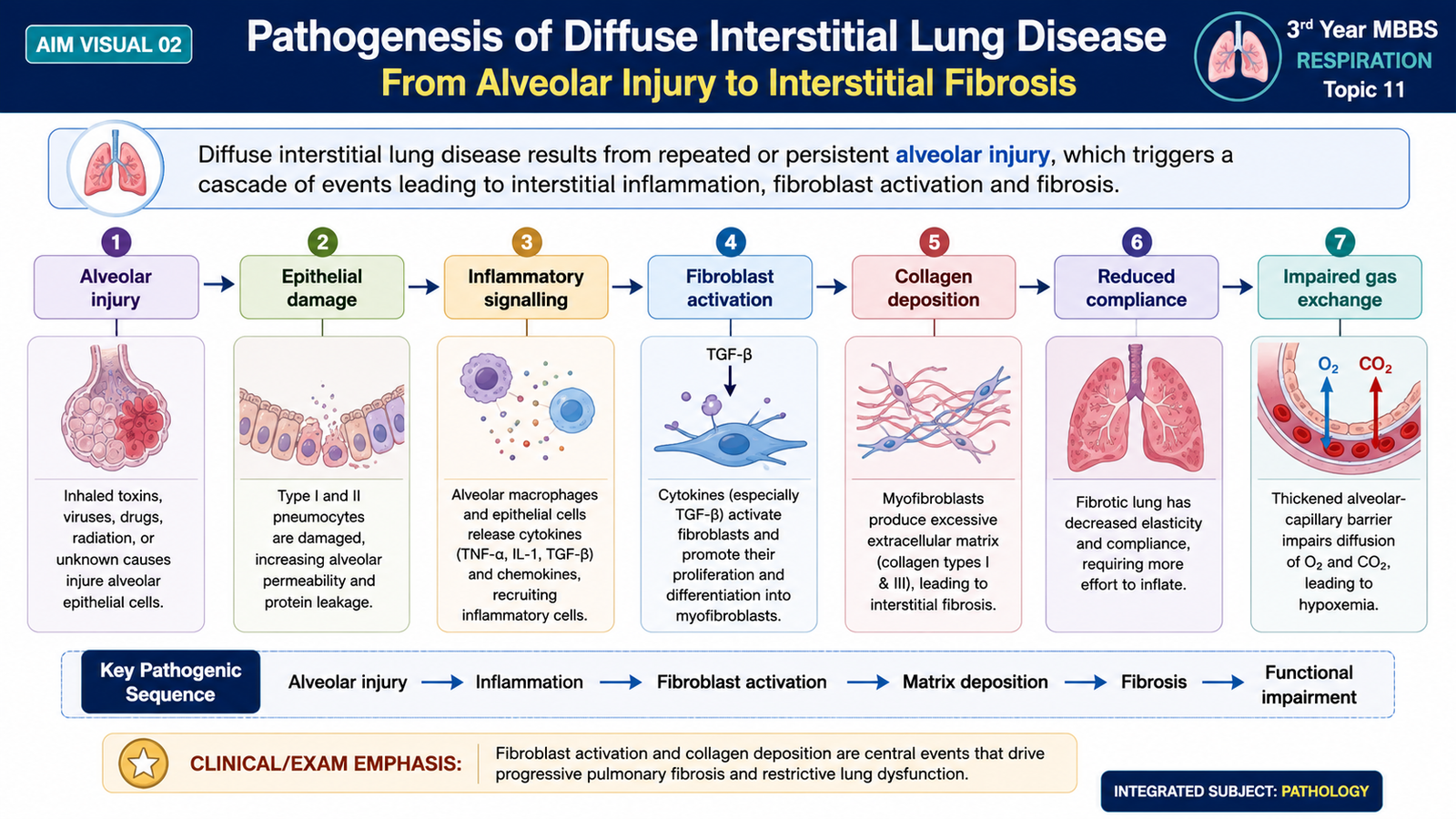
The central problem in many diffuse interstitial lung diseases is repeated or persistent injury to the alveolar epithelium and surrounding interstitium. The initiating injury may be caused by inhaled particles, toxic agents, radiation or an unknown stimulus. Damaged epithelial cells and activated inflammatory cells release mediators that stimulate fibroblasts. Fibroblasts then proliferate and produce extracellular matrix, especially collagen. In a normal response, repair stops after the damaged tissue has healed. In progressive interstitial fibrosis, however, repair becomes excessive or poorly regulated. Fibroblasts and myofibroblasts continue depositing collagen. The normally delicate alveolar walls become thick and stiff, and normal alveolar architecture is gradually distorted.
Two functional consequences are especially important. First, fibrosis reduces lung compliance, meaning that greater effort is required to expand the lungs. Second, thickening of the alveolar-capillary membrane increases the distance across which oxygen must diffuse. With progressive disease, destruction and remodelling of the pulmonary vascular bed may further increase the physiological burden.
General pathological progression
- Initial alveolar or interstitial injury.
- Inflammatory and reparative responses.
- Activation and proliferation of fibroblasts.
- Increasing extracellular matrix and collagen deposition.
- Distortion and loss of normal alveolar architecture.
- Advanced fibrosis with severe loss of functional gas-exchanging surface.
C. Fibrosing Lung Diseases
Fibrosing lung diseases are interstitial disorders in which excessive deposition of collagen progressively replaces or distorts normal lung tissue. The most important concept is that pulmonary fibrosis is not simply an accumulation of scar tissue. It represents abnormal repair after repeated microscopic injury, leading to loss of compliant alveolar tissue and progressive architectural remodelling.
Idiopathic Pulmonary Fibrosis and the Usual Interstitial Pneumonia Pattern
Idiopathic pulmonary fibrosis (IPF) is a chronic progressive fibrosing interstitial lung disease of unknown cause. Its characteristic pathological pattern is called usual interstitial pneumonia (UIP). The central mechanism is believed to involve repeated epithelial injury followed by abnormal fibroblastic repair rather than purely uncontrolled inflammation.
Pathogenesis
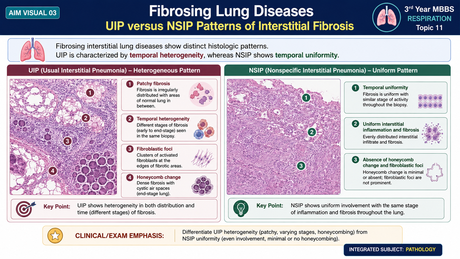
Repeated injury to alveolar epithelial cells promotes release of profibrotic mediators. These mediators stimulate fibroblasts and myofibroblasts, resulting in excessive extracellular matrix and collagen deposition. Different areas of lung are injured and repaired at different times. This produces one of the most characteristic features of UIP: temporal heterogeneity, meaning that early fibroblastic lesions and old dense scars can be present in the same specimen.
Gross morphology
- The lungs become firm because of extensive fibrosis.
- Fibrosis is often more marked in peripheral and subpleural areas.
- Advanced disease produces irregular cystic spaces separated by thick fibrous walls, creating a honeycomb appearance.
Microscopic morphology
- Patchy interstitial fibrosis rather than uniform involvement.
- Alternating areas of relatively preserved lung, active fibroblastic proliferation and dense old fibrosis.
- Fibroblastic foci, representing active sites of fibroblast and myofibroblast proliferation.
- Architectural distortion and cystically dilated residual airspaces in advanced disease.
The patchy distribution and coexistence of fibrosis of different ages distinguish UIP from more uniform fibrosing patterns.
Nonspecific Interstitial Pneumonia
Nonspecific interstitial pneumonia (NSIP) is another interstitial pattern characterized by more uniform involvement of the lung parenchyma. Unlike UIP, lesions are generally at a similar stage of evolution. Therefore, temporal uniformity is an important morphological clue. NSIP may show predominantly interstitial inflammation, predominantly fibrosis, or a mixture of the two. The key undergraduate distinction is that UIP is characteristically patchy and temporally heterogeneous, whereas NSIP tends to be more diffuse and temporally uniform.
| Feature | UIP / Idiopathic Pulmonary Fibrosis | NSIP |
|---|---|---|
| Distribution | Patchy | More uniform |
| Age of lesions | Different stages coexist | Similar stage |
| Fibroblastic foci | Characteristic | Less characteristic |
| Advanced change | Architectural distortion and honeycombing | Usually more uniform fibrosis |
D. Pneumoconiosis — Core Concept, Types and Pathogenesis
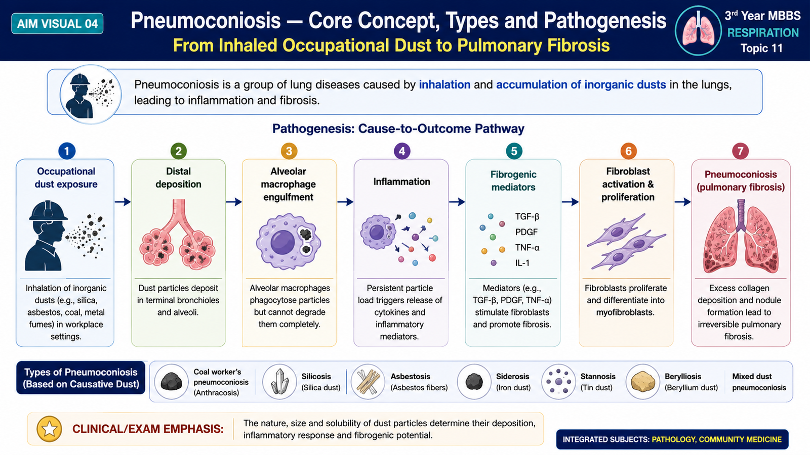
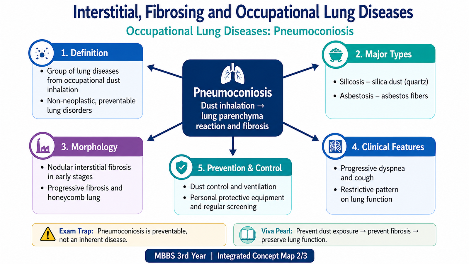
Pneumoconiosis refers to non-neoplastic lung disease caused by inhalation of mineral dusts, usually in an occupational setting. The severity of disease depends not simply on how much dust is inhaled but also on particle size, chemical properties, solubility, duration and intensity of exposure, and the effectiveness of pulmonary clearance mechanisms. Particles that are sufficiently small can reach the distal airspaces. Alveolar macrophages engulf these particles. Some dusts are relatively inert, whereas others directly injure macrophages or stimulate release of inflammatory and fibrogenic mediators. Persistent exposure and ineffective clearance may therefore lead to chronic inflammation, fibroblast activation and pulmonary fibrosis.
Major pneumoconioses
- Coal workers’ pneumoconiosis — associated with inhalation of coal dust.
- Silicosis — caused by inhalation of crystalline silica.
- Asbestosis — diffuse pulmonary interstitial fibrosis caused by inhalation of asbestos fibres.
Not all inhaled particles produce the same reaction. Silica is strongly fibrogenic because it damages macrophages and promotes repeated inflammatory activation. Asbestos fibres can persist in lung tissue and stimulate progressive fibrosis. Coal dust may accumulate within macrophages and produce a spectrum ranging from relatively minor pigment deposition to extensive fibrotic disease.
E. Morphology of the Major Pneumoconioses
The morphology of pneumoconiosis reflects the type of inhaled particle and the tissue reaction that it produces. Recognizing the characteristic distribution and microscopic appearance is important because coal dust, silica and asbestos do not produce identical lesions.
Coal Workers’ Pneumoconiosis
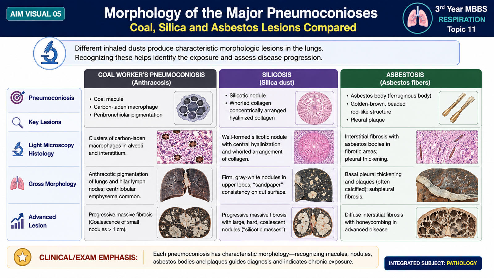
Coal dust particles are engulfed by alveolar macrophages. Dust-containing macrophages accumulate particularly around respiratory bronchioles. The disease forms a spectrum from simple lesions to extensive fibrosis.
Morphology
- Coal macules consist of carbon-laden macrophages around respiratory bronchioles.
- Small fibrotic nodules may develop with continued exposure.
- More advanced disease may produce large areas of dense, blackened fibrosis known as progressive massive fibrosis.
- Extensive fibrosis distorts lung architecture and markedly reduces functional lung tissue.
Silicosis
Silicosis develops after inhalation of crystalline silica particles. Macrophages ingest the particles, but silica can injure the macrophages and promote release of fibrogenic mediators. Repeated cell injury and activation drive the formation of characteristic fibrotic nodules.
Morphology
- Early lesions consist of small, pale-to-dark fibrotic nodules.
- Mature silicotic nodules contain concentrically arranged, whorled bundles of hyalinized collagen.
- Polarizable silica particles may be identified within lesions.
- Nodules commonly involve the upper portions of the lungs and may coalesce in advanced disease.
- Fibrosis may also involve hilar lymph nodes.
Asbestosis
Asbestosis is diffuse interstitial pulmonary fibrosis caused by inhalation of asbestos fibres. Fibres reaching the distal lung are taken up by macrophages but may persist because they cannot be effectively removed. Continued macrophage activation promotes fibrogenic signalling.
Morphology
- Diffuse interstitial fibrosis commonly begins in the lower lobes and subpleural regions.
- Fibrosis may progressively distort normal lung architecture.
- Asbestos bodies are the characteristic microscopic marker of exposure.
- These appear as golden-brown, beaded or fusiform structures with a translucent central core.
- Pleural fibrosis may occur, including discrete pleural plaques.
Asbestos bodies form when inhaled fibres become coated with iron-containing protein material. Their golden-brown appearance makes them an important pathological clue to asbestos exposure.
| Disease | Main exposure | Characteristic morphology | Important distribution/clue |
|---|---|---|---|
| Coal workers’ pneumoconiosis | Coal dust | Coal macules and carbon-laden macrophages | Progressive massive fibrosis in advanced disease |
| Silicosis | Crystalline silica | Whorled hyalinized collagen nodules | Predominantly upper lung involvement |
| Asbestosis | Asbestos fibres | Interstitial fibrosis and asbestos bodies | Lower/subpleural predominance; pleural plaques |
F. Control and Prevention of Pneumoconiosis
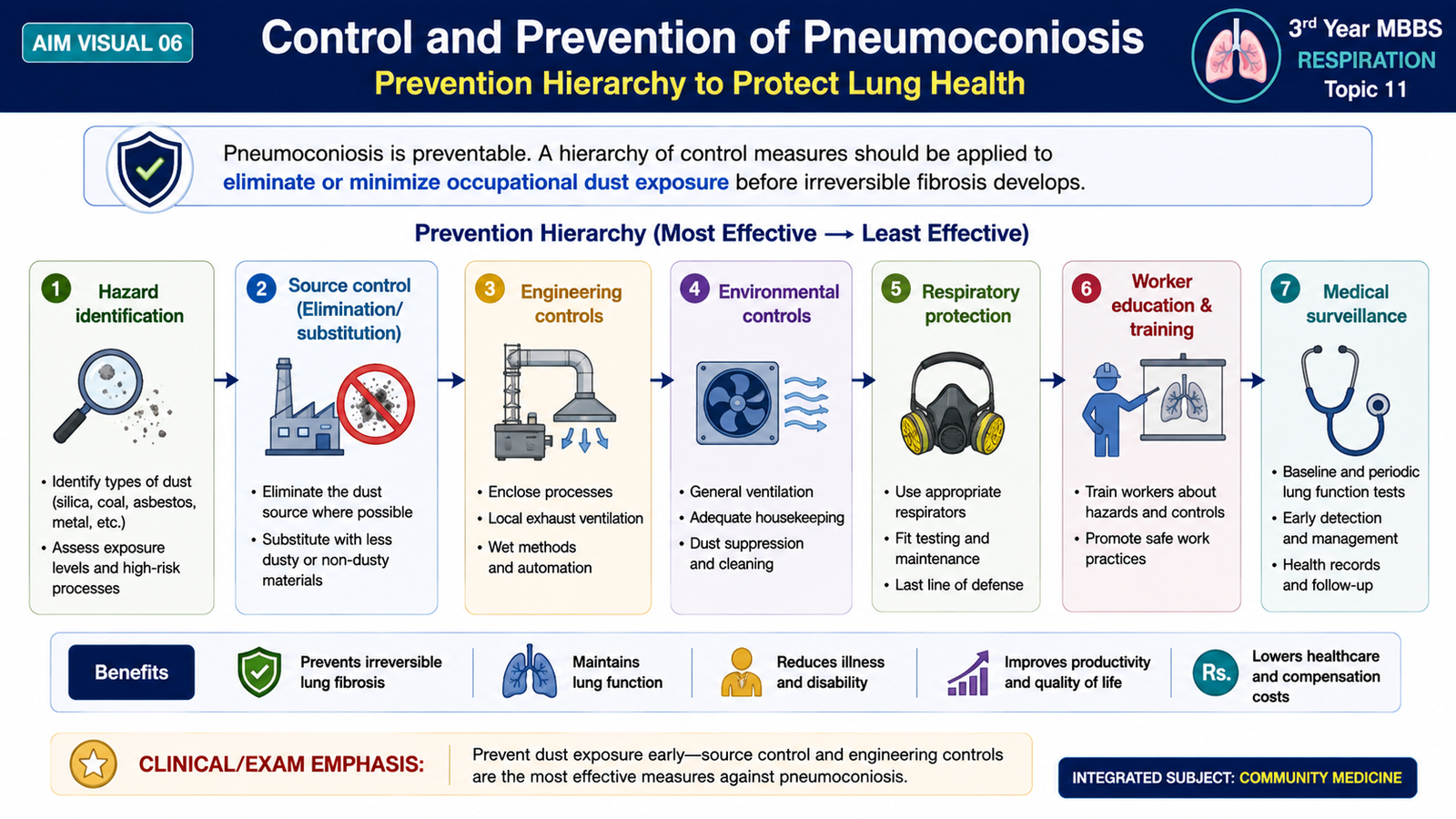
Pneumoconiosis is especially important in Community Medicine because many cases can be prevented by reducing occupational exposure before permanent lung fibrosis develops. Once marked fibrosis has formed, simply removing the worker from exposure cannot restore the lost normal lung architecture. Therefore, control at the workplace is more effective than relying only on treatment after disease becomes established.
Control of dust at the source
The first priority is to reduce the amount of hazardous dust generated or released into the working environment. Engineering controls act on the source or pathway of exposure and therefore protect groups of workers rather than depending only on individual behaviour.
- Substitute a less hazardous material or process where feasible.
- Enclose or isolate dust-producing machinery and processes.
- Use effective local exhaust ventilation.
- Use wet methods where suitable to reduce airborne dust formation.
- Maintain machinery and dust-control systems properly.
- Reduce unnecessary accumulation and re-suspension of workplace dust.
Worker-level protection
Personal protective measures are important when occupational exposure cannot be eliminated completely. They should support, not replace, effective control of the working environment.
- Use appropriate respiratory protective equipment when indicated.
- Provide occupational health education about dust hazards and safe work practices.
- Train workers in correct use and maintenance of protective equipment.
- Minimize unnecessary duration and intensity of hazardous exposure.
Medical surveillance
Occupational health surveillance aims to identify evidence of disease early and to assess whether workplace controls are adequate. It may include occupational history, appropriate clinical assessment and relevant investigations according to workplace risk and established occupational-health practice. When work-related lung disease is suspected, reducing further harmful exposure is important because continued inhalation may add to the accumulated lung burden and promote progression.
G. Drug- and Radiation-Induced Pulmonary Disease
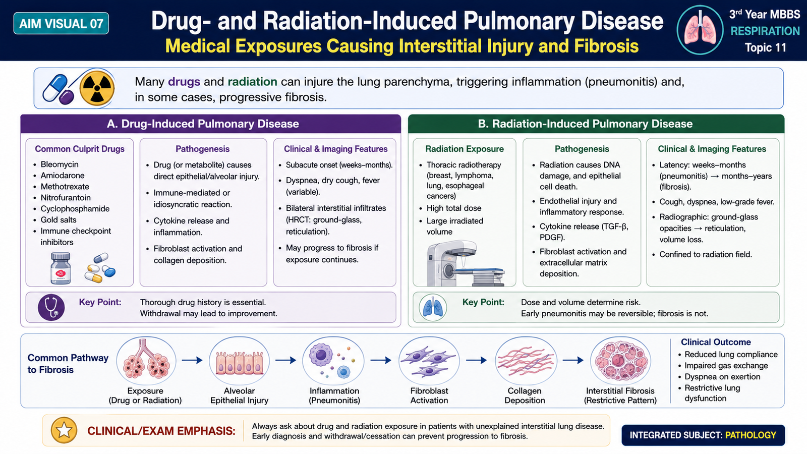
The lung can also be damaged by therapeutic agents and radiation. These forms of disease are important because the initiating exposure may be medically necessary, yet the resulting tissue injury can resemble other interstitial lung disorders. The essential concept is that toxic injury to alveolar epithelial or endothelial cells may trigger inflammation and, if sufficiently severe or persistent, lead to fibrosis.
Drug-induced pulmonary disease
Different drugs can damage the lungs through direct cellular toxicity, oxidative injury, hypersensitivity-type reactions or stimulation of inflammatory and fibrotic responses. The pathological pattern depends on the drug and the nature of the tissue reaction. Important examples associated with interstitial pulmonary injury include certain anticancer drugs, antiarrhythmic agents and other medicines capable of producing pneumonitis or fibrosis. At undergraduate level, the key principle is more important than memorizing a long list: a drug may injure alveolar structures, provoke inflammation and ultimately produce interstitial fibrosis.
Radiation-induced pulmonary disease
Radiation delivered to the thorax can injure alveolar epithelial cells, vascular endothelial cells and supporting interstitial tissue within the irradiated region. An earlier inflammatory phase is termed radiation pneumonitis. Persistent injury and abnormal repair may later produce radiation fibrosis. Fibrosis develops because tissue injury activates fibroblasts and promotes collagen deposition. The affected lung becomes thickened, stiff and less compliant. A useful diagnostic clue is that the anatomical distribution of injury generally corresponds to the lung tissue exposed to radiation.
Integrated Mechanism Flow
⭐ AIM High-Yield Review
- Diffuse interstitial lung disease primarily affects the interstitium, alveolar walls and adjacent lung parenchyma.
- Interstitial fibrosis produces a predominantly restrictive functional abnormality because the lungs become stiff and less compliant.
- Thickening of the alveolar-capillary interface contributes to impaired gas diffusion.
- A common fibrotic pathway is epithelial injury → fibroblast activation → excessive collagen deposition.
- Idiopathic pulmonary fibrosis characteristically shows the usual interstitial pneumonia pattern.
- ⭐ UIP shows patchy fibrosis, temporal heterogeneity and fibroblastic foci.
- Advanced UIP may produce honeycomb change.
- NSIP is generally more temporally uniform than UIP.
- Pneumoconiosis results from inhalation of mineral dusts, commonly through occupational exposure.
- Coal workers’ pneumoconiosis is characterized by carbon-laden macrophages and coal macules; severe disease may progress to massive fibrosis.
- ⭐ Silicosis classically produces nodules containing concentric whorls of hyalinized collagen.
- ⭐ Asbestosis shows interstitial fibrosis and characteristic asbestos bodies; lower and subpleural lung involvement is typical.
- Control of pneumoconiosis should focus first on reducing hazardous dust exposure at its source and through engineering measures.
- Drug-induced pulmonary injury can progress from cellular injury or inflammation to interstitial fibrosis.
- Radiation injury may produce an inflammatory pneumonitis followed by fibrosis through abnormal reparative collagen deposition.