
This chapter follows the supplied KMU learning outcomes and builds tuberculosis from the organism and disease mechanism to diagnosis, treatment and prevention. First understand the disease process and drug logic; then use the AIM High-Yield Review for rapid revision.
Pulmonary Tuberculosis and Granulomatous Lung Diseases
An integrated approach to pulmonary tuberculosis, Mycobacterium tuberculosis, antituberculous pharmacotherapy, drug resistance, sarcoidosis, hypersensitivity pneumonitis, pulmonary eosinophilia and tuberculosis prevention and control.
Topic Introduction
Pulmonary tuberculosis (TB) is a chronic infectious disease caused mainly by Mycobacterium tuberculosis. It is especially important because the organism can survive inside macrophages, provoke a granulomatous immune response, remain latent for years and later reactivate. Understanding TB therefore requires linking microbiology with immune-mediated tissue injury, characteristic pathological lesions, clinical and radiological findings, laboratory diagnosis and multidrug treatment.
This topic also includes important non-tuberculous disorders that may produce granulomatous or inflammatory lung disease, particularly sarcoidosis, hypersensitivity pneumonitis and pulmonary eosinophilia. Finally, because TB spreads between people and treatment failure can create drug resistance, prevention, contact tracing, treatment adherence and public-health control form an essential part of the topic. The supplied curriculum specifically integrates Pathology, Pharmacology, Microbiology, Community Medicine, Medicine and Family Medicine.
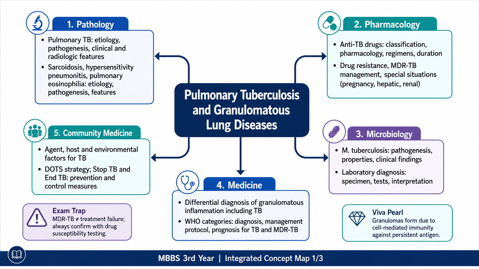
A. Mycobacterium tuberculosis — Properties, Transmission and Pathogenesis
Mycobacterium tuberculosis is a slender, aerobic, non-spore-forming bacillus with a lipid-rich cell wall containing large amounts of mycolic acids. This unusual wall makes the organism relatively resistant to ordinary staining and allows it to retain certain dyes even after treatment with acid-alcohol; therefore, it is described as an acid-fast bacillus. The lipid-rich wall also contributes to slow growth and helps the organism resist destruction within host phagocytic cells.
Important microbiological properties
- Acid fast: demonstrated by acid-fast staining techniques.
- Obligate aerobe: this helps explain the preference of reactivation TB for well-oxygenated regions such as the lung apices.
- Slow growing: conventional culture requires substantially more time than culture of ordinary bacteria.
- Intracellular survival: bacilli can persist within macrophages.
- Cell-mediated immunity is central: both protection and much of the tissue injury in TB arise from the host T-cell response.
Transmission and entry
Pulmonary TB is transmitted mainly through airborne droplet nuclei generated by a person with infectious pulmonary or laryngeal disease. Very small particles can remain suspended in air and reach the terminal airways and alveoli when inhaled. Transmission is favored by prolonged close contact, poor ventilation and a high bacillary burden in the source patient.
Pathogenesis
After inhalation, bacilli reach alveoli and are phagocytosed by alveolar macrophages. Instead of being immediately destroyed, some organisms survive and multiply intracellularly. Infected macrophages may carry organisms through lymphatics to regional lymph nodes. During the first few weeks, the host develops a T-cell-mediated immune response against mycobacterial antigens.
Activated CD4+ T cells produce cytokines, particularly interferon-gamma, that increase the ability of macrophages to kill intracellular organisms. Macrophages transform into epithelioid cells and may fuse to form multinucleated giant cells. Aggregates of these cells surrounded by lymphocytes form a granuloma. In TB, the centre commonly undergoes caseous necrosis.
This immune response is beneficial because it limits spread, but it is also responsible for tissue destruction. Thus, the classic tuberculous lesion is produced not simply by direct bacterial damage but by the interaction between persistent bacilli and the host cell-mediated immune response.
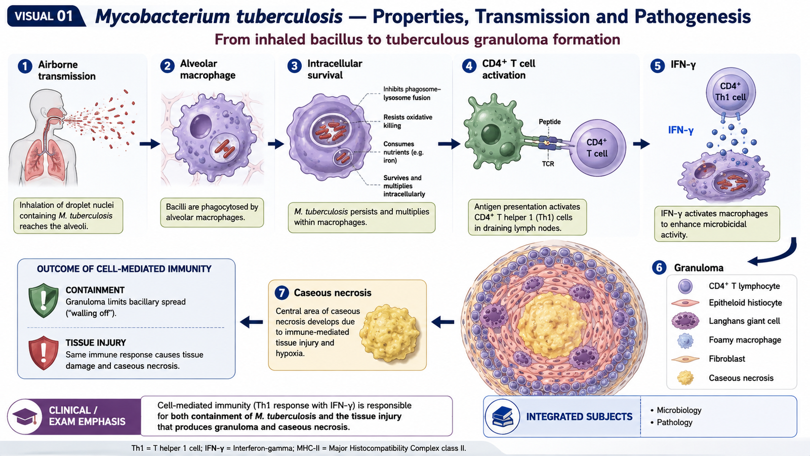
B. Pulmonary Tuberculosis — Types, Pathology and Morphology
The pathological appearance of pulmonary TB depends on whether infection is occurring for the first time, whether previously contained infection has reactivated, and how effective the host immune response is. The two classic patterns are primary tuberculosis and secondary or reactivation tuberculosis.
Primary pulmonary tuberculosis
Primary TB develops in a person who has not previously mounted an effective immune response to M. tuberculosis. The initial pulmonary lesion is usually a small area of gray-white inflammatory consolidation called a Ghon focus, classically located in the lower part of the upper lobe or upper part of the lower lobe near the pleura.
Bacilli drain through lymphatics to hilar lymph nodes. The combination of a Ghon focus and involved draining hilar lymph nodes is called the Ghon complex. Healing may occur by fibrosis and calcification. A healed and calcified primary complex is sometimes termed a Ranke complex.
Secondary or reactivation pulmonary tuberculosis
Secondary TB usually develops in a previously sensitized person, most commonly because dormant bacilli reactivate when immune control weakens, although reinfection can also occur. Because the host is already sensitized, the inflammatory and tissue-destructive response is generally more marked.
The classic lesion is located near the apex of one or both upper lobes. The high oxygen tension of the apical lung favors growth of this aerobic organism. Caseation and tissue destruction may progress to form an irregular pulmonary cavity.
Gross morphology
- Areas of gray-white or yellow-white consolidation.
- Central soft, cheesy caseous necrosis.
- Fibrosis in healing lesions.
- Cavitation in progressive secondary pulmonary TB.
- Pleural involvement may occur when lesions extend toward the lung surface.
Microscopic morphology
The characteristic lesion is the caseating granuloma. It consists of central amorphous granular necrotic material surrounded by epithelioid macrophages, multinucleated giant cells and lymphocytes. Langhans-type giant cells may show nuclei arranged toward the peripheral part of the cell, although this appearance is not specific for TB.
Progressive and disseminated disease
If immune containment fails, bacilli may spread through airways, lymphatics or blood. Bronchogenic spread produces additional pulmonary lesions. Hematogenous dissemination can produce numerous tiny lesions in multiple organs, creating miliary tuberculosis. Progressive pulmonary destruction may also cause fibrosis, cavitation, hemoptysis and impairment of respiratory function.
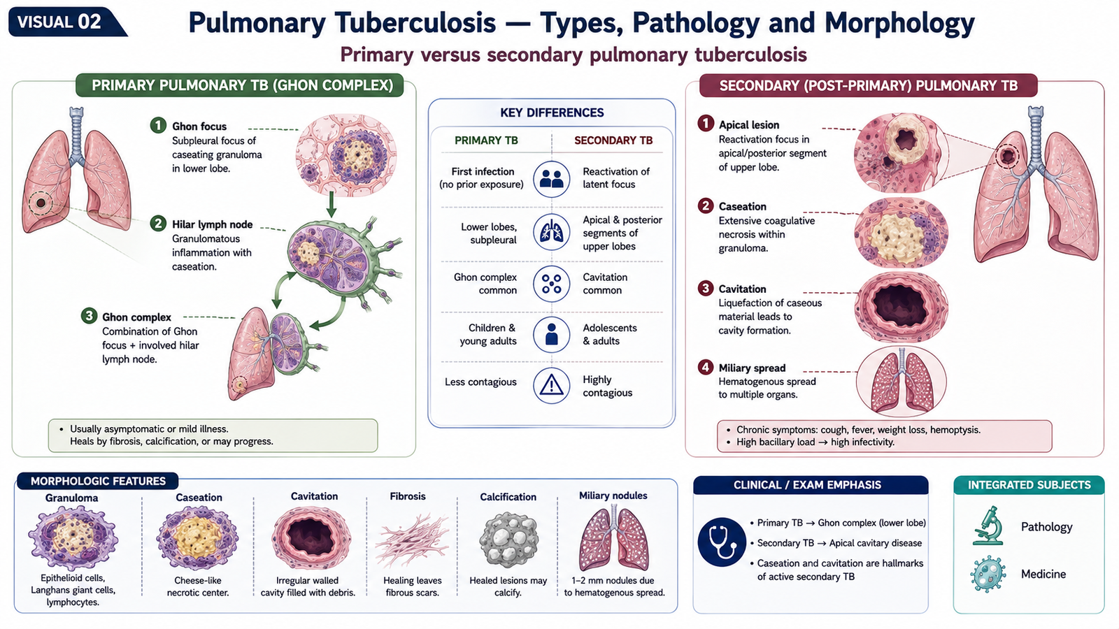
C. Pulmonary Tuberculosis — Clinical Features, Radiology and Complications
Clinical presentation depends on the extent and activity of disease. Primary infection may be asymptomatic or produce mild illness, whereas active secondary pulmonary TB more often produces constitutional symptoms together with respiratory complaints.
Clinical features
- Persistent cough, with or without sputum production.
- Fever, often low-grade and prolonged.
- Night sweats.
- Loss of appetite and weight loss.
- Fatigue and general weakness.
- Chest discomfort or pleuritic pain when pleura is involved.
- Hemoptysis, particularly when cavitary disease damages pulmonary vessels.
The constitutional features arise largely from sustained inflammation and cytokine activity. Hemoptysis occurs when inflammatory destruction, cavitation or erosion of vessels allows blood to enter the airways.
Radiological features
Imaging findings should always be interpreted with the clinical and microbiological picture because no single radiological pattern proves TB.
- Primary disease: parenchymal focus with hilar or mediastinal lymph-node enlargement may be seen.
- Secondary disease: upper-lobe or apical infiltrates are characteristic.
- Cavitation: suggests destructive active disease and often corresponds to a high bacillary burden.
- Miliary pattern: numerous tiny nodules distributed through both lungs indicate hematogenous dissemination.
- Healing: fibrosis and calcification may remain as residual evidence of previous infection.
Important complications
- Massive or recurrent hemoptysis.
- Pleural effusion or pleural disease.
- Extensive fibrosis and permanent lung damage.
- Bronchiectatic change in damaged lung.
- Disseminated or miliary TB.
- Spread to meninges, bones, kidneys and other organs.
- Secondary colonization of a residual cavity by fungi such as Aspergillus.
 .
.D. Laboratory Diagnosis, Investigations and Differential Diagnosis
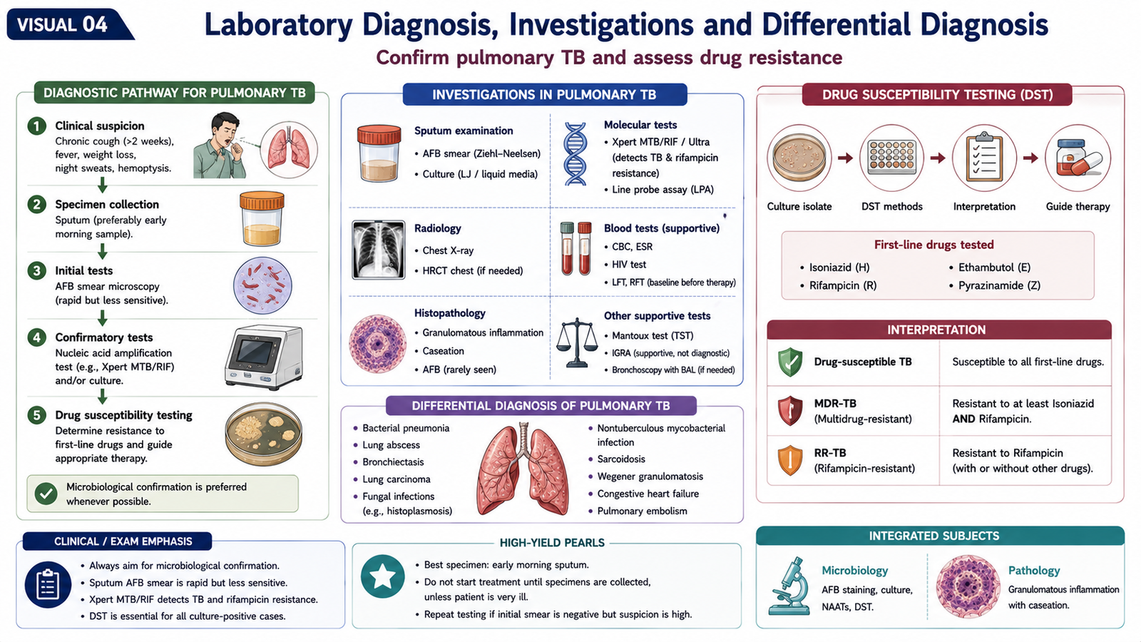
Diagnosis of active pulmonary TB requires integration of symptoms, imaging and direct evidence of the organism whenever possible. A patient may have a strongly suggestive clinical picture, but microbiological testing is particularly important because it confirms disease and helps detect drug resistance.
Specimen selection
For pulmonary disease, sputum is the preferred specimen when the patient can produce it. Other respiratory samples may be required when sputum cannot be obtained or when results remain inconclusive despite strong clinical suspicion.
Major laboratory approaches
- Acid-fast microscopy: demonstrates acid-fast bacilli and can rapidly support a diagnosis, but it cannot reliably distinguish M. tuberculosis from all other mycobacteria.
- Rapid molecular testing: detects mycobacterial genetic material and can identify important resistance markers, especially rifampicin resistance, much faster than conventional culture.
- Culture: provides definitive microbiological isolation and enables more complete drug-susceptibility testing, although growth is slow.
- Drug-susceptibility testing: determines whether an isolate is susceptible or resistant to important antituberculous drugs and is essential when resistant TB is suspected.
Immunological tests
Tuberculin skin testing and interferon-gamma release assays indicate immune sensitization to mycobacterial antigens. They may support identification of TB infection, but they do not by themselves prove active pulmonary disease. Active TB must be evaluated with clinical, radiological and microbiological evidence.
Histopathology
Tissue biopsy may show caseating granulomatous inflammation. However, granulomas are not unique to TB, so morphology must be correlated with microbiological evidence and the clinical setting.
Differential diagnosis of granulomatous inflammation
Granulomatous inflammation in the lung has both infectious and non-infectious causes. Important considerations include:
- Tuberculosis and other mycobacterial infections.
- Deep fungal infections.
- Sarcoidosis.
- Hypersensitivity pneumonitis.
- Exposure-related granulomatous disorders such as chronic beryllium disease.
- Certain granulomatous vasculitic disorders.
The presence of necrosis increases suspicion for infection, particularly TB, but is not absolutely specific. Conversely, non-necrotizing granulomas suggest sarcoidosis in the correct clinical setting, but infection still needs to be excluded. Modern diagnostic approaches to sarcoidosis therefore require a compatible presentation, granulomatous pathology when tissue confirmation is needed, and exclusion of alternative causes. :contentReference[oaicite:1]{index=1}
E. First-Line Antituberculous Drugs
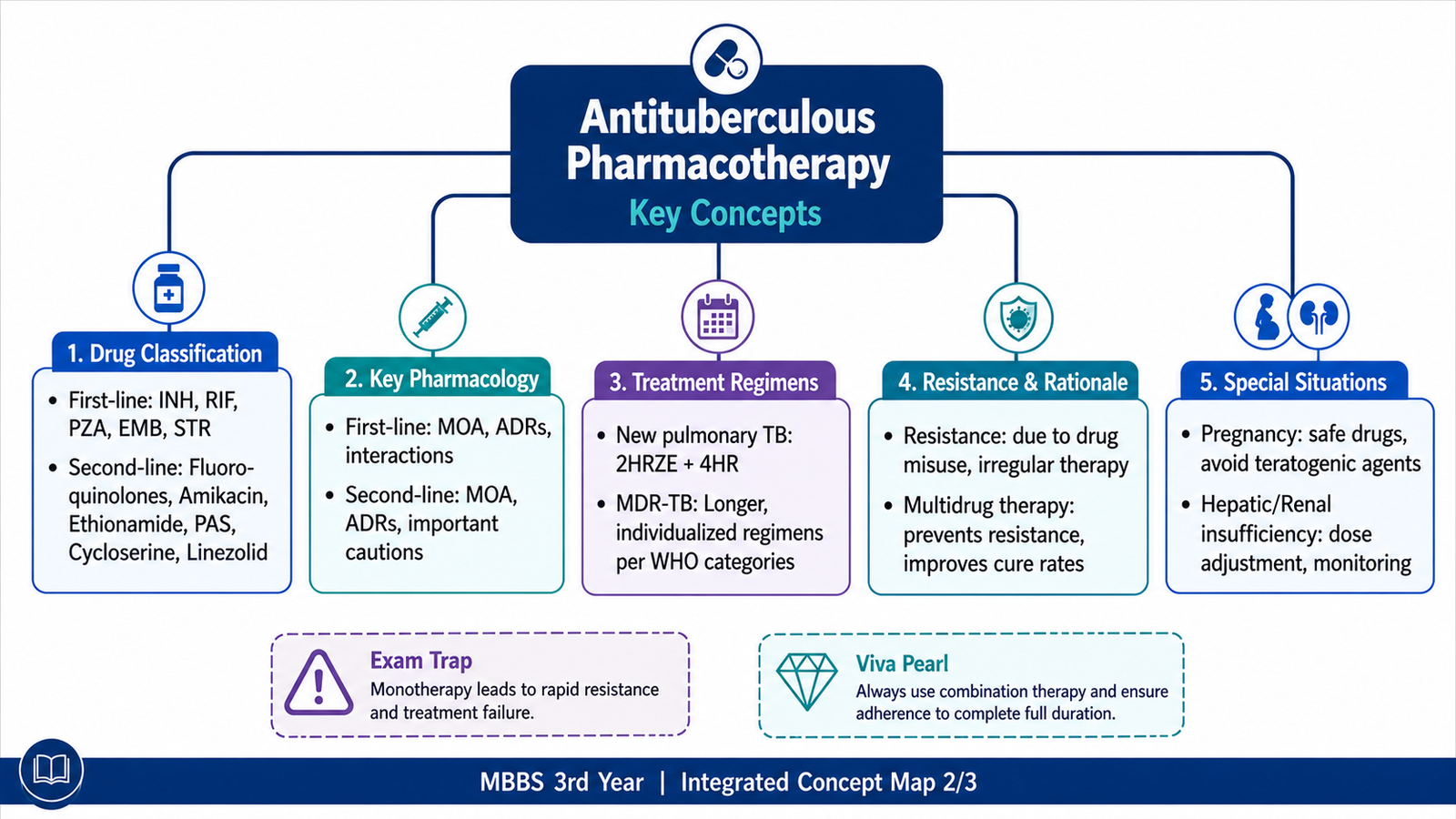
The major first-line drugs used for drug-susceptible TB are isoniazid, rifampicin, pyrazinamide and ethambutol. Their mechanisms differ, which is precisely why combining them is effective: different drugs attack different bacterial targets and reduce the probability that naturally resistant bacilli will survive treatment.
| Drug | Main mechanism | Important role | Characteristic adverse effects |
|---|---|---|---|
| Isoniazid (H) | Prodrug activated within the bacillus; inhibits synthesis of mycolic acids required for the mycobacterial cell wall. | Strong bactericidal activity against actively multiplying bacilli. | Hepatotoxicity; peripheral neuropathy from functional pyridoxine deficiency. |
| Rifampicin (R) | Inhibits bacterial DNA-dependent RNA polymerase and therefore RNA synthesis. | Powerful bactericidal and sterilizing activity. | Hepatotoxicity, orange-red discoloration of body fluids and major drug interactions from hepatic enzyme induction. |
| Pyrazinamide (Z) | Prodrug converted to pyrazinoic acid; particularly active against bacilli in acidic intracellular or inflammatory environments. | Important sterilizing action during the initial phase of therapy. | Hepatotoxicity and hyperuricemia; arthralgia may occur. |
| Ethambutol (E) | Inhibits arabinosyl transferases involved in synthesis of the mycobacterial cell wall. | Helps protect the regimen against emergence of resistance while susceptibility is being established. | Optic neuritis with reduced visual acuity and impaired red-green colour discrimination. |
Isoniazid: important pharmacological points
Isoniazid enters mycobacteria and is activated by a bacterial catalase-peroxidase enzyme. Activated metabolites interfere with enzymes involved in mycolic-acid synthesis. Because mycolic acids are essential components of the mycobacterial cell wall, inhibition rapidly affects actively dividing bacilli.
Isoniazid may interfere with pyridoxine metabolism, producing peripheral neuropathy, particularly in susceptible patients. Pyridoxine supplementation is therefore important in groups at increased risk, including pregnancy and certain patients with nutritional, renal or neurological vulnerability.
Rifampicin: important pharmacological points
Rifampicin blocks transcription by binding bacterial DNA-dependent RNA polymerase. It is particularly important because of its bactericidal and sterilizing activity. A major pharmacological issue is enzyme induction, which can reduce concentrations of many co-administered drugs. This makes interaction checking essential.
Pyrazinamide and ethambutol
Pyrazinamide contributes strongly during the early phase of therapy by acting in environments where some persistent bacilli remain metabolically active. Ethambutol has a different cell-wall target and is especially useful in preventing selection of resistant organisms before full susceptibility information is available.

F. Drug Resistance, Second-Line Drugs and MDR/RR-TB
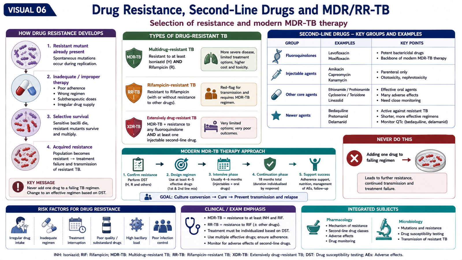
M. tuberculosis develops clinically important resistance mainly through spontaneous chromosomal mutations followed by selection of resistant organisms. Resistance is not produced because the bacillus deliberately adapts to a drug. Instead, resistant mutants may already exist within a large bacterial population. If treatment is inadequate, interrupted or effectively reduced to a single active drug, susceptible organisms are killed while resistant organisms survive and multiply.
Important resistance terminology
- RR-TB: tuberculosis resistant to rifampicin.
- MDR-TB: resistance to at least both rifampicin and isoniazid.
- Pre-XDR-TB: rifampicin-resistant/MDR disease with additional resistance to a fluoroquinolone.
- XDR-TB: more extensive resistance involving rifampicin, a fluoroquinolone and additional key drugs used for resistant disease.
Current WHO terminology and treatment selection are based on the actual drug-resistance pattern rather than the old practice of assigning all patients to fixed numerical retreatment categories. WHO currently defines MDR-TB as resistance to both rifampicin and isoniazid and prioritizes modern all-oral regimens for eligible patients with MDR/RR-TB. :contentReference[oaicite:2]{index=2}
Important drugs used in resistant TB
Fluoroquinolones — levofloxacin and moxifloxacin: inhibit bacterial DNA gyrase and interfere with DNA replication. They are highly important components of many resistant-TB regimens.
Bedaquiline: inhibits mycobacterial ATP synthase, reducing production of cellular energy. It is an important modern drug for MDR/RR-TB. QT-interval prolongation is an important safety concern.
Linezolid: binds the 50S ribosomal subunit and inhibits bacterial protein synthesis. Important toxicities include bone-marrow suppression, peripheral neuropathy and optic neuropathy, particularly with prolonged exposure.
Pretomanid: is a nitroimidazole-class antituberculous drug used as part of specific multidrug regimens for resistant TB rather than as monotherapy.
Clofazimine: has antimycobacterial activity and is incorporated into several resistant-TB regimens. Skin discoloration and gastrointestinal effects are notable adverse effects.
Cycloserine/terizidone: interferes with peptidoglycan synthesis. Neuropsychiatric toxicity and seizures are clinically important cautions.
Delamanid: is another newer agent used in selected drug-resistant TB regimens; QT prolongation is an important concern.
Other agents: ethionamide/prothionamide, para-aminosalicylic acid and, in selected circumstances, amikacin may be considered when designing regimens according to susceptibility, previous exposure, toxicity and available effective oral drugs.
WHO’s 2025 treatment framework prioritizes all-oral regimens for drug-resistant TB. The main duration categories are approximately 6-month regimens, 9-month regimens in appropriate patients, and longer individualized regimens when shorter options cannot be used. :contentReference[oaicite:3]{index=3}
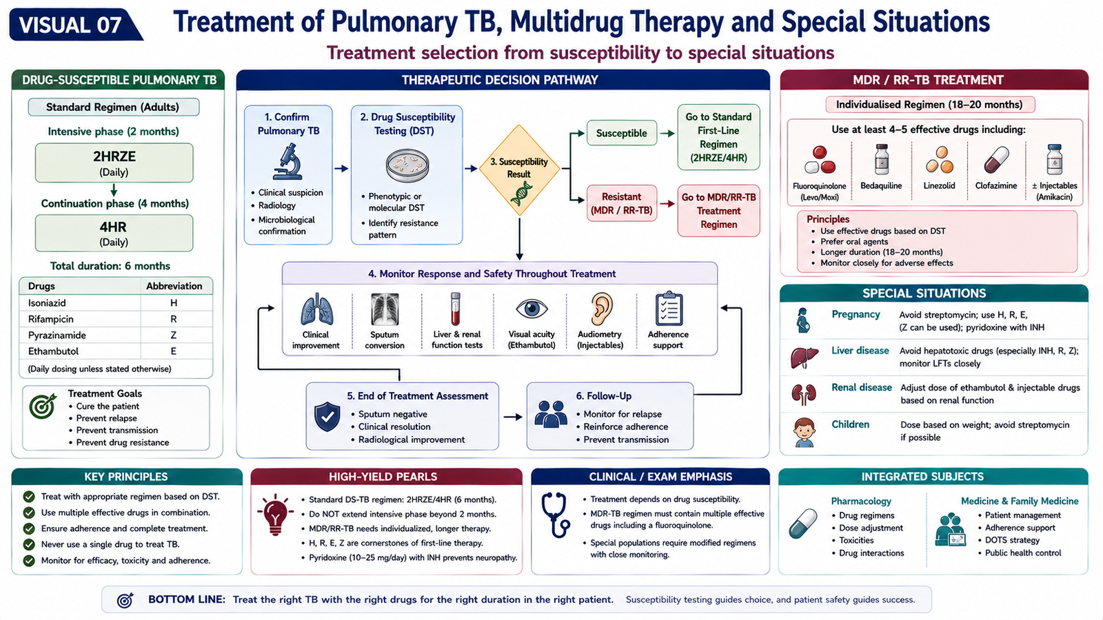
G. Treatment of Pulmonary TB, Multidrug Therapy and Special Situations
The aim of TB treatment is not only to improve symptoms. Effective therapy must rapidly reduce the number of viable organisms, eliminate persistent bacilli that could cause relapse, prevent emergence of drug resistance and interrupt transmission. For these reasons, active TB is treated with a combination of effective drugs for an adequate duration.
Why multidrug therapy is essential
A pulmonary cavity can contain a very large bacterial population. Within this population, a small number of bacilli may possess spontaneous resistance to one drug. Monotherapy would remove susceptible organisms and allow the resistant subpopulation to dominate. The chance that one organism is naturally resistant to several unrelated drugs simultaneously is far lower. Combining drugs therefore makes resistance much less likely while also allowing drugs with different activity against rapidly multiplying and more persistent bacilli to work together.
Newly diagnosed drug-susceptible pulmonary TB
A well-established WHO regimen for drug-susceptible TB is 6 months: an initial 2-month phase with isoniazid, rifampicin, pyrazinamide and ethambutol followed by a 4-month continuation phase with isoniazid and rifampicin, written as 2HRZE/4HR. Daily administration is used. :contentReference[oaicite:4]{index=4}
WHO also recognizes a shorter 4-month rifapentine–moxifloxacin-containing regimen for eligible patients, but the traditional 2HRZE/4HR regimen remains a core undergraduate regimen and is widely used. :contentReference[oaicite:5]{index=5}
Management principles in MDR/RR-TB
MDR/RR-TB should be managed according to rapid resistance testing, prior drug exposure and susceptibility to key agents. It should never be managed simply by adding one new drug to a failing regimen. Current WHO guidance prioritizes eligible patients for short all-oral regimens. A major 6-month option is BPaLM—bedaquiline, pretomanid, linezolid and moxifloxacin. Other 6-month all-oral options are also available, while approximately 9-month regimens or longer individualized regimens are used when appropriate. :contentReference[oaicite:6]{index=6}
Pregnancy
Active TB during pregnancy requires treatment because untreated maternal disease presents substantial risk to both mother and fetus. WHO states that the standard 6-month drug-susceptible TB regimen can be used during pregnancy. Pyridoxine supplementation is recommended when isoniazid is used. Drug-resistant TB in pregnancy needs specialist selection of drugs because the safety profile of individual agents must be considered carefully. :contentReference[oaicite:7]{index=7}
Hepatic insufficiency
Isoniazid, rifampicin and especially pyrazinamide can cause hepatotoxicity. Pre-existing liver disease therefore requires careful assessment of baseline hepatic function, the severity of liver disease and the number of hepatotoxic drugs that can safely be used. Significant symptoms or biochemical evidence of drug-induced liver injury during treatment requires prompt clinical review rather than routine continuation without assessment.
Renal insufficiency
Isoniazid and rifampicin are mainly handled hepatically and generally present fewer dosing problems in renal impairment than drugs that depend substantially on renal elimination. Ethambutol and pyrazinamide require particular attention in significant renal impairment, and the dosing interval or regimen may need adjustment according to renal function and specialist guidance. Pyridoxine is also important in patients receiving isoniazid when renal failure increases the risk of neuropathy.
Monitoring response
Clinical improvement, microbiological response, adherence and adverse effects should all be monitored. Failure to improve should trigger assessment for poor adherence, incorrect diagnosis, malabsorption, major drug interaction or drug resistance rather than automatic extension or random alteration of treatment. WHO emphasizes rapid testing for resistance when treatment failure is suspected. :contentReference[oaicite:8]{index=8}
H. Sarcoidosis
Sarcoidosis is a multisystem inflammatory disorder characterized pathologically by non-necrotizing granulomas. The exact initiating cause is not established. The disease appears to result from an exaggerated cell-mediated immune response to one or more unidentified antigens in genetically susceptible individuals.
Pathogenesis
Antigen-presenting cells activate predominantly CD4+ T lymphocytes. Cytokines recruit and activate macrophages, which differentiate into epithelioid histiocytes and form granulomas. Persistent inflammation may eventually stimulate fibrosis in some patients.
Morphology
The characteristic microscopic lesion is a compact, well-formed, non-necrotizing granuloma composed mainly of epithelioid macrophages and multinucleated giant cells. Small amounts of central fibrinoid necrosis may occasionally occur, so the word “non-necrotizing” is more accurate than assuming that necrosis can never be present.
Within giant cells, inclusions such as asteroid bodies or laminated calcium-containing Schaumann bodies may be seen, but neither is diagnostic by itself. In pulmonary sarcoidosis, granulomas characteristically follow lymphatic routes, including bronchovascular bundles, interlobular septa and subpleural regions. :contentReference[oaicite:9]{index=9}
Clinical features
- Dry cough and progressive breathlessness.
- Chest discomfort.
- Constitutional symptoms such as fatigue, fever or weight loss in some patients.
- Bilateral hilar lymph-node enlargement.
- Extrapulmonary involvement may affect skin, eyes, lymph nodes and other organs.
Radiological features
Bilateral hilar lymphadenopathy is a classic radiological clue. Pulmonary parenchymal disease may show a reticulonodular or nodular pattern, often with a perilymphatic distribution. Advanced disease may produce fibrosis and architectural distortion.
Clinical course
The course is variable. Some patients have spontaneous resolution, whereas others develop persistent disease and progressive pulmonary fibrosis. Diagnosis requires correlation of a compatible clinical-radiological presentation with granulomatous pathology when necessary and exclusion of alternative granulomatous diseases, especially infection such as tuberculosis. :contentReference[oaicite:10]{index=10}

I. Hypersensitivity Pneumonitis and Pulmonary Eosinophilia
Hypersensitivity pneumonitis
Hypersensitivity pneumonitis (HP) is an immune-mediated inflammatory disease of the lung caused by repeated inhalation of an antigen to which the person has become sensitized. The antigens are commonly encountered in occupational, environmental or domestic settings. The important point is that this is not a simple immediate allergic reaction: repeated antigen exposure produces inflammation centred on the small airways and pulmonary interstitium.
Pathogenesis
Inhaled antigen reaches distal airways and alveoli. In a sensitized host, both immune-complex and T-cell-mediated mechanisms contribute to inflammation. Repeated exposure produces lymphocytic interstitial inflammation and poorly formed non-necrotizing granulomas, usually with a bronchiolocentric distribution. Persistent inflammation may eventually cause fibrosis.
Clinical and radiological features
- Cough and breathlessness related to relevant environmental or occupational exposure.
- Acute or subacute episodes may follow significant antigen exposure.
- Chronic disease may present with progressive exertional dyspnea.
- HRCT may show ground-glass change, centrilobular nodules, mosaic attenuation and air trapping.
- Fibrotic disease may show architectural distortion and other features of chronic interstitial lung injury.
Exposure history is therefore a major diagnostic clue. A pattern of ground-glass centrilobular nodules and air trapping is particularly compatible with HP in the appropriate setting. :contentReference[oaicite:11]{index=11}
Pulmonary eosinophilia
Pulmonary eosinophilia refers to a group of disorders in which eosinophils accumulate within the lungs, often together with increased eosinophils in peripheral blood. It is a pattern rather than a single disease. Causes include allergic disorders, parasitic infection, drug reactions and primary eosinophilic pneumonias.
Pathogenesis
Th2-type immune responses promote production of cytokines such as interleukin-5, which supports eosinophil production, recruitment and survival. Activated eosinophils release inflammatory mediators and granule proteins that damage airway and alveolar tissues.
Clinical and radiological features
- Cough, dyspnea and fever may occur.
- Wheeze is common when eosinophilic disease is associated with asthma or allergic airway disease.
- Peripheral-blood eosinophilia may be present, depending on the specific disorder.
- Pulmonary infiltrates may be transient, migratory or persistent according to the underlying cause.
- Bronchoalveolar lavage demonstrating marked eosinophilia can strongly support an eosinophilic lung disorder in the proper clinical context. :contentReference[oaicite:12]{index=12}
The diagnostic approach therefore asks two questions: Is there genuine pulmonary eosinophilic inflammation? and what is causing it? Drug history, travel or parasitic exposure, asthma/allergic history and systemic features help identify the underlying disorder.

J. Tuberculosis Epidemiology, Prevention, DOTS, Contact Tracing and Continuing Care
TB control requires more than prescribing effective drugs to an individual patient. Because pulmonary TB is transmitted through the air and incomplete treatment promotes ongoing transmission and drug resistance, the public-health response must identify infectious patients early, ensure completion of effective treatment and evaluate people who have been significantly exposed.
Agent, host and environmental factors
Agent factors include the infectivity of the source case, bacillary burden, drug susceptibility and ability of M. tuberculosis to persist within the host.
Host factors influencing progression from infection to active disease include impaired cell-mediated immunity, HIV infection, undernutrition, diabetes, immunosuppressive therapy, extremes of age and other conditions that weaken immune control.
Environmental factors include prolonged close exposure, overcrowding, poor ventilation, poverty and barriers to timely diagnosis and treatment. TB is transmitted through the air from people with pulmonary disease, making ventilation and rapid treatment of infectious cases particularly important. :contentReference[oaicite:13]{index=13}
DOTS strategy
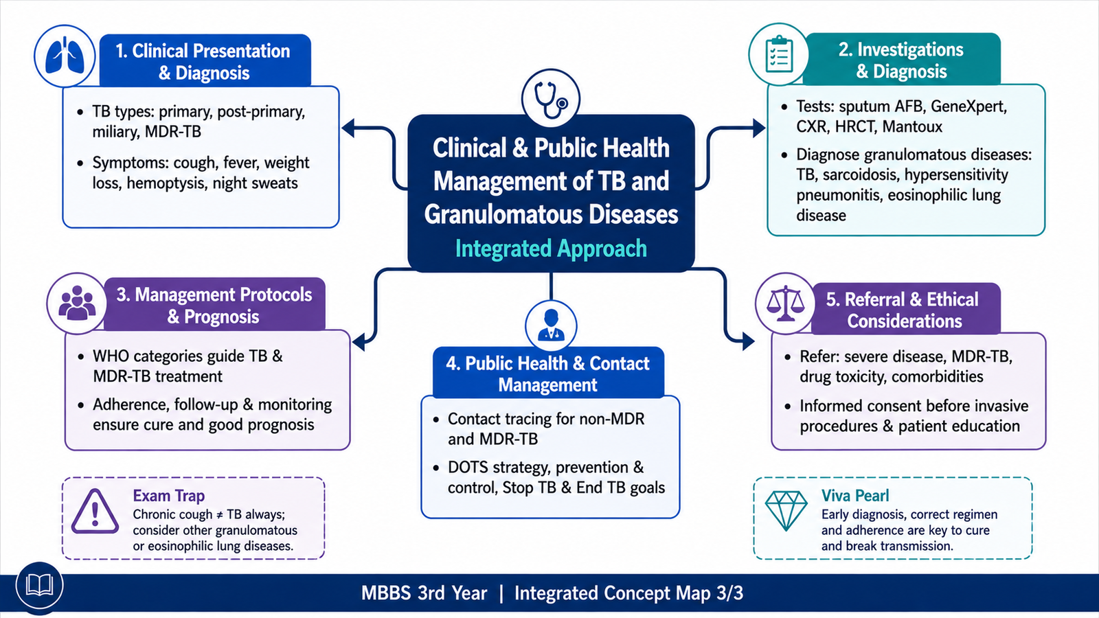
DOTS means Directly Observed Treatment, Short-course. It was developed as a comprehensive TB-control strategy rather than merely a person watching a patient swallow tablets. Its major principles include political and health-system commitment, reliable diagnosis, standardized effective treatment with patient support, an uninterrupted drug supply and systematic recording and evaluation of outcomes.
The modern concept of TB care remains strongly patient-centred. Treatment support should help patients complete therapy rather than functioning as punishment or surveillance. WHO emphasizes supervision and support because completion of the full multidrug course is essential for cure and prevention of acquired resistance. :contentReference[oaicite:14]{index=14}
Prevention and control
- Early recognition and diagnosis of active pulmonary TB.
- Prompt initiation of an effective multidrug regimen.
- Appropriate respiratory infection-control measures.
- Good ventilation in healthcare and high-risk environments.
- BCG vaccination according to national immunization policy.
- Detection and management of TB infection in eligible high-risk contacts.
- Screening of populations at particularly high risk according to public-health programmes.
- Detection of drug resistance so ineffective regimens are not continued.
- Continuous treatment support to reduce interruption and loss to follow-up.
Stop TB and End TB
Global TB-control strategies progressively expanded from directly observed treatment toward broader goals that include prevention, universal access to diagnosis and effective treatment, management of drug-resistant TB, patient-centred care, stronger health systems, research and reduction of the social conditions that sustain transmission. WHO’s current global approach is organized around the End TB Strategy, with the ultimate goal of markedly reducing TB illness, deaths and transmission. TB remains preventable and curable, but control depends on detecting and treating people who would otherwise continue the chain of infection. :contentReference[oaicite:15]{index=15}
Contact tracing
Contact tracing begins by identifying people who have had significant exposure to a patient with infectious pulmonary TB. Household and other close contacts are prioritized because cumulative exposure is greatest. Contacts are assessed for symptoms and risk factors and then investigated according to age, immune status and local TB policy.
The purpose is to identify two groups: contacts who already have active TB disease, who require full diagnostic assessment and treatment, and contacts who have TB infection without active disease, some of whom may benefit from preventive treatment after active TB has been excluded.
When the index patient has MDR/RR-TB, contact investigation is especially important. Any active TB detected in a close contact must be assessed rapidly for drug resistance because the contact may have acquired the same resistant strain. Preventive management of contacts of drug-resistant cases depends on the resistance profile and current specialist/public-health guidance.
Primary and continuing care
At primary-care level, the clinician should recognize a possible TB presentation, arrange appropriate sputum and molecular testing, assess relevant contacts and facilitate treatment adherence. Continuing care includes monitoring clinical response, adverse drug effects, treatment completion and barriers that could lead to interruption.
When specialist referral is important
- Suspected or confirmed MDR/RR-TB.
- Failure to improve or persistent microbiological positivity suggesting treatment failure or resistance.
- Major drug toxicity or inability to construct an effective standard regimen.
- Significant liver or renal disease complicating drug selection.
- Complex pregnancy-related treatment decisions, especially resistant TB.
- Severe complications such as major hemoptysis or extensive pulmonary destruction.
- Diagnostic uncertainty, particularly when TB must be differentiated from another granulomatous lung disease.
Integrated Mechanism Flow — Pulmonary Tuberculosis
Important Comparison — Major Granulomatous and Related Lung Diseases
| Feature | Tuberculosis | Sarcoidosis | Hypersensitivity Pneumonitis | Pulmonary Eosinophilia |
|---|---|---|---|---|
| Primary driver | M. tuberculosis | Unknown antigen/immune dysregulation | Repeated inhaled antigen | Eosinophilic inflammation from several possible causes |
| Inflammatory pattern | Caseating granulomas | Well-formed non-necrotizing granulomas | Bronchiolocentric lymphocytic inflammation with poorly formed granulomas | Eosinophil-rich inflammation |
| Major clue | Microbiological evidence of TB | Bilateral hilar lymphadenopathy | Relevant exposure history | Blood/BAL eosinophilia |
| Typical imaging tendency | Upper-zone disease/cavitation in secondary TB | Hilar adenopathy, perilymphatic nodules | Ground glass, centrilobular nodules, mosaic attenuation/air trapping | Variable pulmonary infiltrates |
| Key diagnostic principle | Demonstrate organism and resistance where possible | Exclude infectious and other granulomatous causes | Link exposure with compatible lung findings | Identify both eosinophilia and its underlying cause |
⭐ AIM High-Yield Review
- M. tuberculosis is an acid-fast aerobic bacillus whose lipid-rich cell wall contains mycolic acids.
- TB pathology results largely from cell-mediated immunity against organisms surviving inside macrophages.
- ⭐ The hallmark lesion is a caseating granuloma composed of epithelioid macrophages, giant cells and lymphocytes surrounding central caseous necrosis.
- Ghon focus + involved hilar lymph nodes = Ghon complex in primary TB.
- ⭐ Secondary TB characteristically favors the lung apices and commonly causes caseation and cavitation.
- A sputum molecular test can rapidly detect TB and important drug-resistance markers; culture remains important for confirmation and susceptibility assessment.
- ⭐ First-line drug hallmark toxicities: isoniazid—neuropathy/hepatotoxicity; rifampicin—enzyme induction and orange body fluids; pyrazinamide—hyperuricemia/hepatotoxicity; ethambutol—optic neuritis.
- Standard drug-susceptible pulmonary TB can be treated with 2HRZE/4HR, giving a total duration of 6 months. :contentReference[oaicite:16]{index=16}
- Multidrug therapy prevents selection of naturally resistant bacterial mutants and attacks different bacillary populations.
- MDR-TB means resistance to at least isoniazid and rifampicin; current management is based on resistance testing and appropriate multidrug regimens rather than adding one drug to failing therapy. :contentReference[oaicite:17]{index=17}
- ⭐ Sarcoidosis classically produces non-necrotizing granulomas and bilateral hilar lymphadenopathy; infection must be excluded.
- Hypersensitivity pneumonitis strongly depends on a relevant inhalational exposure history and may show centrilobular ground-glass nodules and air trapping.
- Pulmonary eosinophilia is a pattern of eosinophil-rich lung inflammation, not one single disease.
- Contact tracing aims to identify both undiagnosed active TB and contacts with infection who may benefit from preventive intervention.
- ⭐ Successful TB control depends on early diagnosis + effective multidrug treatment + adherence support + contact investigation + prevention of transmission.
🎥 Video Learning — Pulmonary Tuberculosis
Watch this video to reinforce the pathogenesis, pathology, clinical features, diagnosis and treatment principles of pulmonary tuberculosis.