
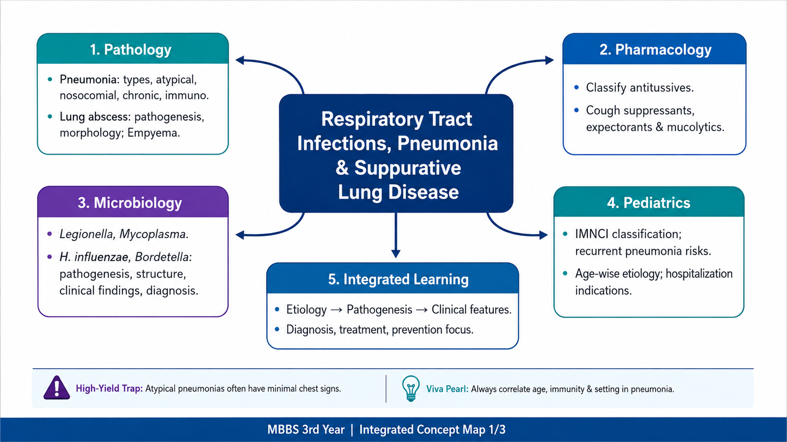
This chapter follows the supplied KMU learning outcomes and builds respiratory infection concepts in a logical sequence. First understand how different infections produce different patterns of lung injury, then revise the organisms, cough pharmacology and childhood pneumonia points highlighted at the end.
Topic 2 — Respiratory Tract Infections, Pneumonia and Suppurative Lung Disease
Understand the major patterns of pneumonia, important respiratory pathogens, lung abscess and empyema, drugs used to control cough, and the recognition of pneumonia in children.
Topic Introduction
Respiratory infections range from relatively localized airway infections to pneumonia involving the gas-exchanging parts of the lung. Pneumonia develops when microorganisms reach the lower respiratory tract, overcome normal pulmonary defenses and produce inflammation within the lung. The pattern of disease depends on the organism, the route of infection and the condition of the host. Some infections produce acute consolidation, whereas others produce atypical interstitial inflammation, chronic destructive disease or suppuration such as lung abscess and empyema. In this chapter, these pathological patterns are connected with four important respiratory pathogens, the pharmacology of cough-control medicines and the essential pediatric approach to childhood pneumonia.
A. Pneumonia: Core Concept, Etiology and Major Patterns
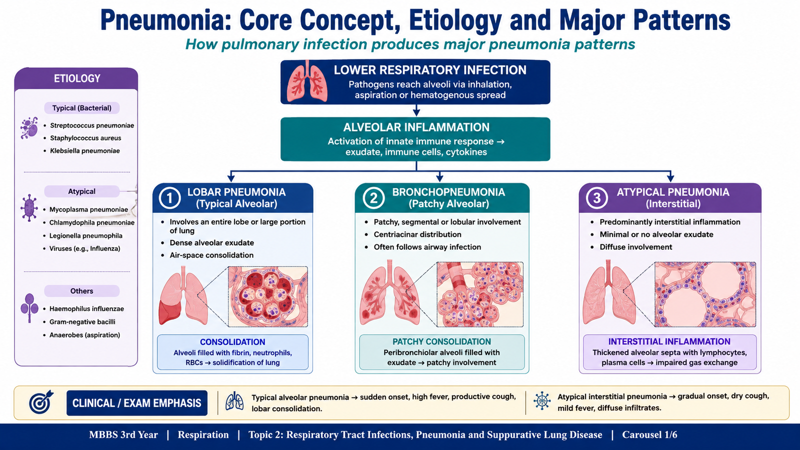
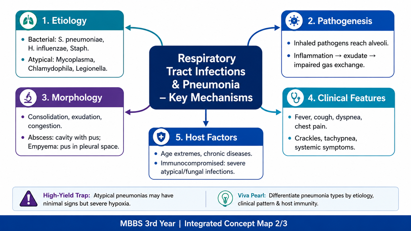
Pneumonia is an infection of the lung parenchyma in which microorganisms produce inflammation involving the alveoli, surrounding interstitium, or both. Normally, the lower respiratory tract is protected by the cough reflex, mucociliary clearance, alveolar macrophages and immune defenses. Pneumonia develops when these defenses are overwhelmed or impaired.
How pneumonia develops
Organisms usually reach the lungs by inhalation of infectious droplets, microaspiration of organisms colonizing the upper airway, or less commonly through the bloodstream. Once organisms reach the distal airways and alveoli, they multiply and trigger an inflammatory response. Increased vascular permeability allows protein-rich fluid and inflammatory cells to enter the alveoli. When alveolar spaces become filled with inflammatory exudate instead of air, the affected lung becomes consolidated. This interferes with normal gas exchange and contributes to breathlessness and hypoxemia.
Microorganism reaches lower respiratory tract → multiplication → inflammatory response → alveolar/interstitial injury → inflammatory exudate → impaired gas exchange and clinical pneumonia
Community-acquired pneumonia
Community-acquired pneumonia (CAP) is pneumonia acquired outside a hospital or healthcare setting. It can be understood broadly as typical bacterial pneumonia and atypical pneumonia. The distinction reflects differences in organisms, pathological pattern and clinical presentation rather than simply disease severity.
Typical bacterial pneumonia
Typical bacterial infection commonly produces an acute inflammatory exudate within alveoli. Neutrophils, fibrin and edema fluid fill the air spaces, producing consolidation. Important bacterial causes include Streptococcus pneumoniae and Haemophilus influenzae, among others.
Two classical morphological patterns are important:
- Lobar pneumonia: consolidation involves a large portion of a lobe or an entire lobe in a relatively uniform pattern.
- Bronchopneumonia: infection produces patchy areas of suppurative consolidation centered around bronchi and bronchioles, often involving more than one lobe.
Lobar pneumonia: morphological evolution
The classical stages describe how an untreated lobar bacterial pneumonia may evolve. They are useful for understanding the relationship between inflammation and gross appearance.
- Congestion: the lung is heavy and red because of vascular engorgement and intra-alveolar fluid. Few neutrophils and many organisms may be present.
- Red hepatization: alveoli become packed with neutrophils, fibrin and red blood cells. The lung becomes red, firm and liver-like.
- Gray hepatization: red cells break down while fibrin and inflammatory cells remain. The affected lung becomes gray-brown and firm.
- Resolution: the intra-alveolar exudate is enzymatically digested and removed by macrophages, expectoration or lymphatic drainage, allowing normal architecture to be restored when healing is uncomplicated.
Clinical features
Acute bacterial pneumonia commonly causes fever, cough, sputum production, malaise and breathlessness. Pleuritic chest pain may occur when inflammation reaches the pleural surface. Consolidated alveoli transmit sound differently from normal air-filled lung and may therefore produce characteristic examination findings.
Community-acquired atypical pneumonia
Atypical pneumonia is characterized mainly by inflammation of the alveolar walls and pulmonary interstitium rather than dense neutrophilic filling of alveolar spaces. Important causes include Mycoplasma pneumoniae and Legionella pneumophila, as well as respiratory viruses.
The onset may be less abrupt than typical bacterial pneumonia. Fever, headache, malaise and a relatively dry cough are common. Because the inflammatory process is predominantly interstitial, extensive dense lobar consolidation is less characteristic.
| Feature | Typical bacterial pneumonia | Atypical pneumonia |
|---|---|---|
| Main site of inflammation | Alveolar spaces | Alveolar septa/interstitium |
| Exudate | Prominent neutrophilic exudate | Less prominent alveolar exudate |
| Cough | Often productive | Often dry |
| Examples | S. pneumoniae, H. influenzae | Mycoplasma, Legionella, viruses |
B. Nosocomial, Chronic and Immunocompromised-Host Pneumonia
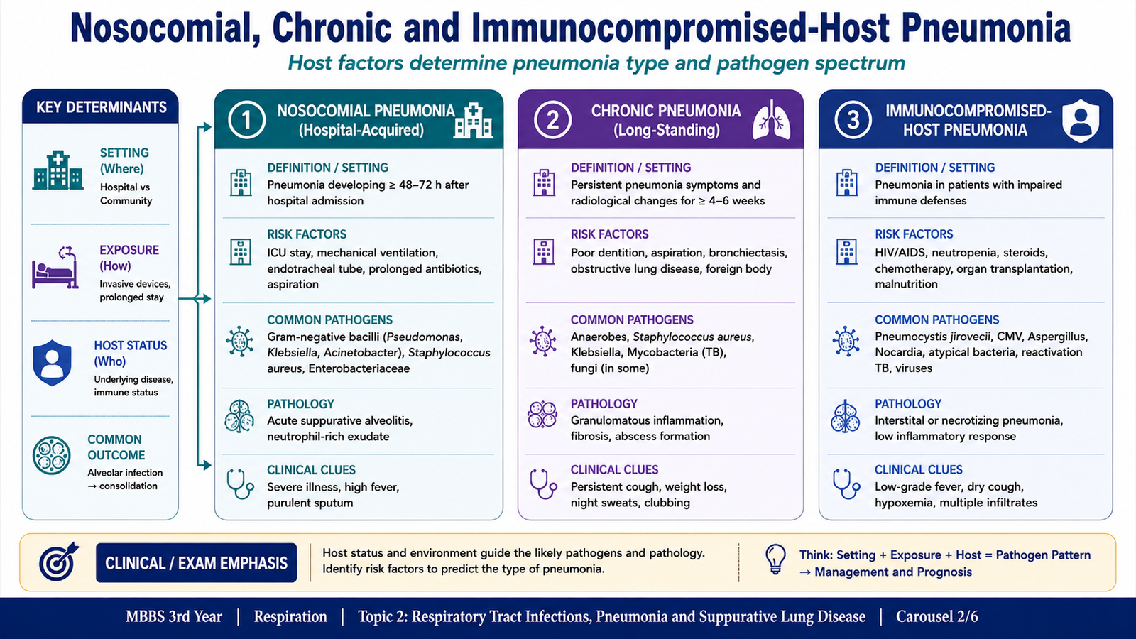
Not all pneumonias arise in otherwise healthy people in the community. The environment in which infection is acquired and the patient’s immune status strongly influence both the organisms involved and the pattern of disease. Hospitalized, chronically ill and immunocompromised patients are particularly vulnerable because normal respiratory defenses may be impaired.
Nosocomial pneumonia
Nosocomial pneumonia, or hospital-acquired pneumonia, develops in a patient during hospitalization and is not already incubating at admission. Hospitalized patients may acquire colonization of the oropharynx by potentially pathogenic organisms. Aspiration of these organisms into the lower airways can then initiate infection.
Important predisposing factors include severe underlying illness, impaired consciousness, prolonged hospitalization and mechanical ventilation. Endotracheal tubes are particularly important because they bypass normal upper-airway defenses, interfere with mucociliary clearance and provide a surface on which microorganisms can persist.
Hospital-acquired infections are often caused by gram-negative bacilli such as Klebsiella, Enterobacter, Escherichia coli and Pseudomonas, while Staphylococcus aureus is another important pathogen.
Clinical features include new or worsening fever, cough, purulent respiratory secretions, breathlessness and evidence of a new pulmonary infiltrate in the appropriate clinical setting.
Chronic pneumonia
Chronic pneumonia refers to persistent pulmonary infection in which the inflammatory response and tissue injury continue over a prolonged period. It is classically associated with organisms that are difficult for the host to eradicate, particularly mycobacteria and certain fungi.
Instead of the predominantly acute neutrophilic reaction seen in many bacterial pneumonias, chronic infections may produce granulomatous inflammation. Activated macrophages accumulate and may transform into epithelioid cells, often accompanied by lymphocytes and sometimes giant cells. Persistent inflammation can cause tissue destruction, necrosis and fibrosis.
Clinical features tend to develop more gradually and may include prolonged cough, fever, malaise and weight loss, depending on the causative infection and extent of lung involvement.
Pneumonia in the immunocompromised host
Patients with impaired immunity are susceptible not only to common respiratory pathogens but also to opportunistic organisms that rarely cause serious disease in immunocompetent people. The type of infection depends on which component of immunity is defective.
Important causes can include bacteria, fungi, viruses and opportunistic organisms such as Pneumocystis jirovecii. Because the normal inflammatory response may be weak, clinical and radiological findings may sometimes be less typical despite significant infection.
C. Lung Abscess and Empyema
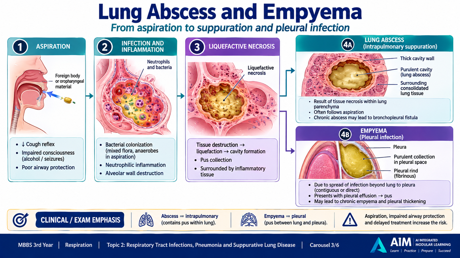
Pulmonary infection can occasionally progress beyond ordinary pneumonia and produce localized suppuration. Two important complications are lung abscess, in which pus and necrotic tissue form a cavity within the lung, and empyema, in which pus accumulates within the pleural space.
Lung abscess
A lung abscess is a localized area of suppurative destruction of lung tissue resulting in formation of a cavity containing necrotic material and pus. The central event is tissue necrosis caused by severe infection.
Pathogenesis
Aspiration of infected material is an important mechanism. Material containing bacteria enters the lower respiratory tract, especially when normal protective reflexes are impaired. Infection then produces intense neutrophilic inflammation. Enzymes released during suppuration destroy the lung tissue, causing liquefactive necrosis and cavity formation.
Other pathways include severe necrotizing pneumonia, bronchial obstruction followed by secondary infection, septic emboli reaching the lungs through the bloodstream, and direct spread from an adjacent infection.
Morphology
Grossly, an abscess appears as a localized cavity containing purulent, necrotic material. It may be single or multiple depending on the mechanism of infection. Aspiration-related abscesses tend to occur in lung regions determined by gravity and body position during aspiration.
Microscopically, the center contains necrotic debris and numerous neutrophils. The surrounding lung shows acute inflammation. With persistence, granulation tissue and later fibrosis may develop around the cavity.
Clinical course
Patients commonly develop fever, cough and production of abundant purulent sputum. Foul-smelling sputum can occur particularly when anaerobic organisms are involved. A cavity may become visible radiologically, and communication with a bronchus may produce an air-fluid level.
Important complications include extension into the pleural cavity, hemorrhage from damaged vessels, dissemination of infection and healing with fibrosis.
Empyema
Empyema is the accumulation of pus within the pleural cavity. It commonly develops when infection from an adjacent pneumonia or lung abscess extends through the visceral pleura into the pleural space.
The pleural inflammatory response produces a protein-rich exudate containing many neutrophils. Fibrin deposition may create adhesions and loculated collections. If the process persists, organization and fibrosis can produce pleural thickening and may restrict lung expansion.
D. Important Respiratory Pathogens
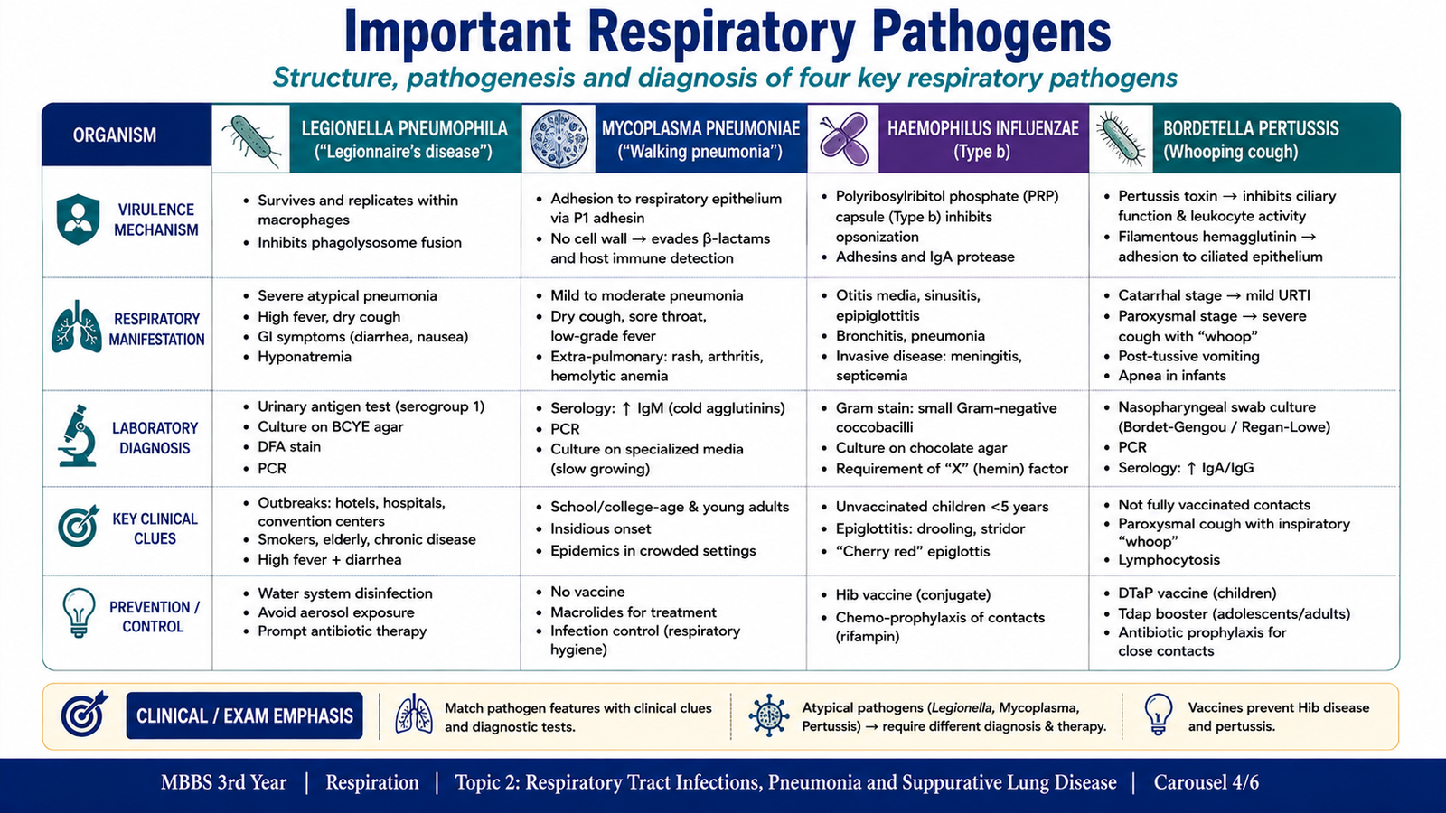
Several organisms produce characteristic respiratory infections because of their structure, mode of transmission and interaction with respiratory epithelium. For this topic, four organisms are especially important: Legionella pneumophila, Mycoplasma pneumoniae, Haemophilus influenzae and Bordetella pertussis. Understanding their biological characteristics helps explain both their clinical presentation and their laboratory diagnosis.
1. Legionella pneumophila
Legionella pneumophila is a gram-negative bacillus associated with aquatic environments and contaminated water systems. Infection is acquired mainly by inhalation of contaminated aerosols rather than ordinary person-to-person respiratory spread.
After reaching the lower respiratory tract, the organism is taken up by alveolar macrophages. Instead of being efficiently destroyed, it can survive and multiply intracellularly by interfering with normal intracellular killing. This produces inflammatory lung injury and pneumonia.
Clinical disease may present as pneumonia with fever, cough and systemic illness. Extrapulmonary features such as gastrointestinal symptoms may accompany the respiratory infection.
Laboratory diagnosis may involve respiratory specimens for culture on specialized media, antigen detection, nucleic-acid-based testing, or other appropriate microbiological methods. The organism does not grow well on ordinary routine bacteriological media.
2. Mycoplasma pneumoniae
Mycoplasma pneumoniae is a very small bacterium that characteristically lacks a cell wall. Because it has no peptidoglycan cell wall, it does not stain reliably with the Gram stain and drugs whose action depends on inhibiting cell-wall synthesis are ineffective against it.
The organism attaches closely to respiratory epithelial cells and interferes with normal ciliary function. Local epithelial injury and the host immune response contribute to inflammation, producing an atypical pattern of pneumonia.
Patients commonly have fever, malaise and persistent cough. The disease may develop gradually and is often associated with community spread among people in close contact.
Laboratory diagnosis commonly relies on nucleic-acid detection or serological evidence rather than routine Gram stain and conventional bacterial culture.
3. Haemophilus influenzae
Haemophilus influenzae is a small, pleomorphic gram-negative coccobacillus. Some strains possess a polysaccharide capsule, whereas others are non-encapsulated. The organism colonizes the human upper respiratory tract and spreads through respiratory secretions.
Colonization is followed by local multiplication when respiratory defenses are impaired. Non-encapsulated strains are particularly important in mucosal respiratory infections. In susceptible patients, infection may extend into the lower respiratory tract and cause pneumonia.
Clinical manifestations depend on the site and strain involved and can include respiratory tract infection and pneumonia.
For laboratory diagnosis, appropriate respiratory or other clinically relevant specimens are examined. The organism is fastidious and requires enriched culture conditions containing essential growth factors. Identification may also use modern antigen or molecular methods where appropriate.
4. Bordetella pertussis
Bordetella pertussis is a small gram-negative coccobacillus that causes pertussis, or whooping cough. It spreads through respiratory droplets and attaches to ciliated epithelial cells of the respiratory tract.
After attachment, bacterial virulence factors interfere with normal respiratory epithelial function. Pertussis toxin alters cellular signaling through increased cyclic AMP activity, while other bacterial factors contribute to epithelial injury and impaired mucociliary clearance. Accumulated secretions and airway irritation help produce the characteristic severe coughing episodes.
The illness classically progresses from an early catarrhal phase with nonspecific upper-respiratory symptoms to a paroxysmal phase characterized by repeated bouts of cough. A characteristic inspiratory “whoop” may occur, particularly in children.
Nasopharyngeal material is the appropriate type of specimen because the organism colonizes the respiratory epithelium. Laboratory diagnosis may use nucleic-acid testing or culture on specialized media.
| Organism | Key structural/biological clue | Major pathogenic clue | Diagnostic clue |
|---|---|---|---|
| Legionella | Gram-negative bacillus | Intracellular multiplication in macrophages | Special culture requirements; antigen/molecular detection |
| Mycoplasma | No cell wall | Attachment to respiratory epithelium and ciliary injury | Molecular testing or serology |
| H. influenzae | Pleomorphic gram-negative coccobacillus | Respiratory colonization followed by local infection | Fastidious growth on enriched media |
| B. pertussis | Small gram-negative coccobacillus | Ciliary attachment and toxin-mediated dysfunction | Nasopharyngeal specimen; molecular testing/culture |
E. Antitussives, Expectorants and Mucolytic Agents
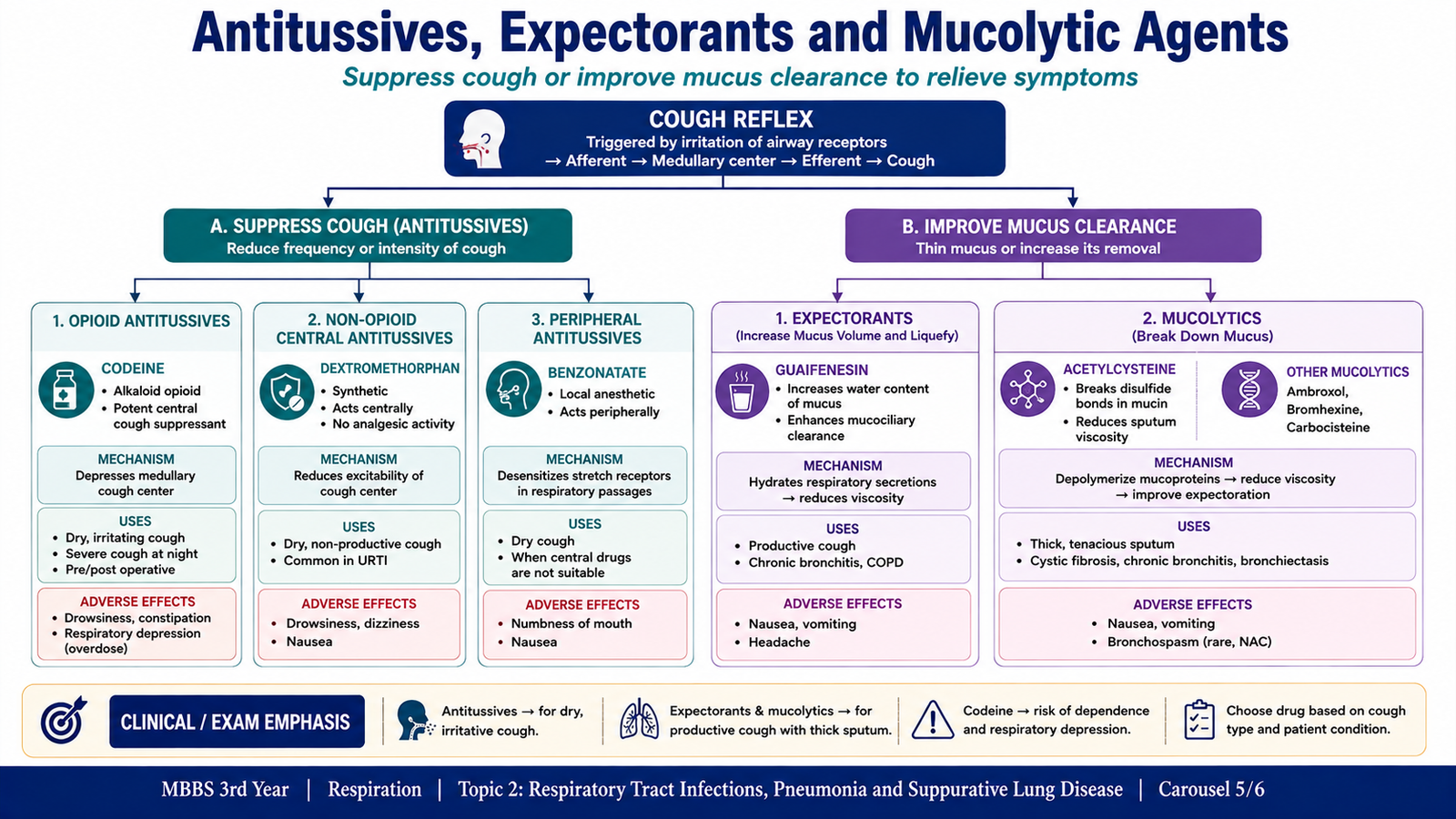
Cough is an important protective reflex that helps clear secretions and foreign material from the respiratory tract. Drug therapy should therefore be understood according to its purpose. Antitussives suppress the cough reflex, whereas expectorants and mucolytics aim to make respiratory secretions easier to remove.
Classification of antitussives
Antitussive drugs can be classified according to whether they mainly act centrally on the cough center or act through peripheral mechanisms.
- Centrally acting opioid antitussives: codeine.
- Centrally acting non-opioid antitussives: dextromethorphan.
- Peripherally acting cough suppressants: agents that reduce stimulation of sensory pathways involved in cough, such as benzonatate.
Codeine
Codeine is an opioid that suppresses cough by acting centrally on neural pathways controlling the cough reflex. The result is a reduction in cough frequency.
Its opioid activity also explains important adverse effects, including sedation, constipation and respiratory depression, particularly when excessive opioid effects occur. Dependence is another disadvantage associated with opioid use.
Codeine → central opioid action → suppression of cough center activity → reduced cough
Dextromethorphan
Dextromethorphan is a centrally acting non-opioid cough suppressant. It reduces the cough reflex without producing the same typical analgesic and dependence profile as therapeutic opioid antitussives. It is therefore commonly used when suppression of troublesome nonproductive cough is desired.
Adverse effects may include dizziness, drowsiness and gastrointestinal upset. Excessive exposure can produce important central nervous system effects. Interaction with drugs that increase serotonergic activity can also be clinically important.
Expectorants
Expectorants are intended to facilitate removal of respiratory secretions. Guaifenesin is a commonly recognized example. By promoting easier clearance of mucus, expectorants are used when the aim is to assist productive coughing rather than completely suppress it.
Mucolytic agents
Mucolytics reduce the viscosity of thick mucus so that secretions can be cleared more easily. Acetylcysteine breaks disulfide bonds within mucus glycoproteins. This disrupts the structure responsible for mucus viscosity and makes the secretion less thick.
Other mucolytic agents, such as bromhexine or its metabolite ambroxol, can also facilitate clearance of respiratory secretions.
Choosing between suppression and clearance
The pharmacological goal depends on the nature of the cough. A troublesome dry, nonproductive cough may benefit from suppression. In contrast, when substantial secretions need to be removed, excessive suppression of the cough reflex can interfere with airway clearance. In such situations, facilitating mucus clearance is more logical.
F. Childhood Pneumonia: IMNCI Classification, Etiology and Hospitalization
Pneumonia is particularly important in children because clinical deterioration can occur rapidly, especially in young infants or children with underlying risk factors. The pediatric approach therefore emphasizes simple clinical recognition, assessment of severity and identification of children who need referral or hospitalization.
IMNCI classification
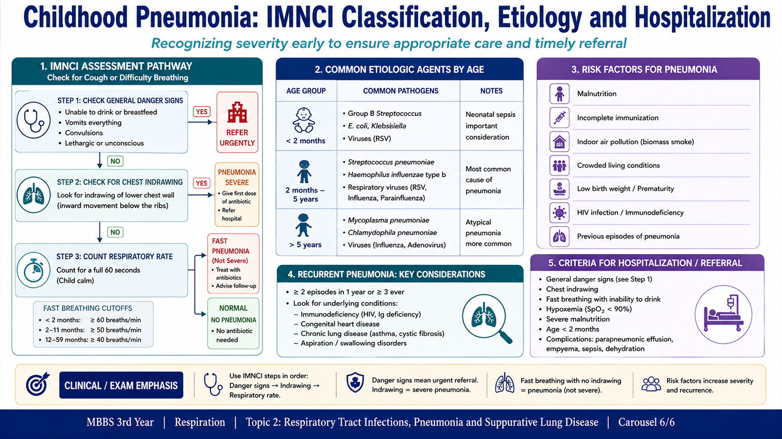
The Integrated Management of Neonatal and Childhood Illnesses (IMNCI) approach classifies a child with cough or difficult breathing according to easily recognizable clinical signs. The purpose is to identify severe illness quickly without depending on complex investigations.
In the commonly taught IMNCI framework, important findings include fast breathing, chest indrawing and general danger signs. These findings help distinguish children who can be managed at a lower level of care from those requiring urgent referral or hospital assessment.
| Clinical pattern | Interpretation | Main implication |
|---|---|---|
| Cough/difficult breathing with fast breathing | Pneumonia | Requires appropriate pneumonia management and follow-up |
| Chest indrawing and/or severe clinical features | More severe disease | Needs careful assessment and referral/hospital care when indicated |
| General danger signs | Severe illness | Urgent referral or hospital assessment |
General danger signs indicate that the child may not be able to maintain normal feeding, ventilation or neurological function. Examples include inability to drink or breastfeed, persistent vomiting, convulsions, lethargy or unconsciousness, and severe respiratory compromise.
Etiological agents according to age
The likely cause of pneumonia changes with age because exposure patterns and immune defenses change as the child grows.
- Neonates: organisms acquired around birth are important, including group B streptococci and gram-negative enteric organisms.
- Infants and young children: respiratory viruses are common, while bacterial pathogens such as Streptococcus pneumoniae and Haemophilus influenzae are important causes.
- School-age children and adolescents: typical bacterial pathogens remain important, while Mycoplasma pneumoniae becomes a particularly relevant cause of community-acquired atypical pneumonia.
Risk factors for recurrent pneumonia
Repeated episodes of pneumonia should raise the possibility that normal airway protection, lung structure or host immunity is abnormal. Identifying the underlying factor is therefore more important than simply labeling each episode as an isolated infection.
Important predisposing factors include:
- aspiration due to swallowing dysfunction or gastroesophageal reflux;
- congenital abnormalities affecting the respiratory tract;
- airway obstruction or an inhaled foreign body;
- impaired mucociliary clearance;
- immune deficiency;
- chronic pulmonary disease;
- neuromuscular disorders that impair effective coughing and secretion clearance;
- poor nutritional status and other conditions that reduce host defense.
When hospitalization should be considered
Hospitalization is required when pneumonia is severe enough that safe treatment and observation cannot be provided at home. The decision is based on respiratory status, general condition, ability to maintain hydration and feeding, age, complications and underlying disease.
Important indications include:
- severe respiratory distress;
- hypoxemia or cyanosis;
- general danger signs or altered consciousness;
- inability to drink or feed adequately;
- significant dehydration;
- suspected complications such as pleural infection;
- serious underlying disease or immunocompromise;
- very young age with significant illness;
- failure to improve or clinical deterioration with appropriate initial management.
Integrated Mechanism Flow
⭐ AIM High-Yield Review
- Pneumonia is infection of lung parenchyma producing alveolar, interstitial or mixed inflammation.
- Typical bacterial pneumonia mainly produces intra-alveolar neutrophilic exudate and consolidation.
- Lobar pneumonia classically progresses through congestion, red hepatization, gray hepatization and resolution.
- Atypical pneumonia predominantly involves the alveolar septa and interstitium; Mycoplasma and Legionella are important causes.
- Hospital-acquired pneumonia is favored by severe illness, aspiration, impaired defenses and airway instrumentation.
- Chronic pneumonia may produce persistent inflammation, tissue destruction and granulomatous inflammation.
- Immunocompromised patients are susceptible to both ordinary and opportunistic pulmonary pathogens.
- ⭐ A lung abscess is a suppurative necrotic cavity within lung parenchyma; empyema is pus within the pleural space.
- Mycoplasma pneumoniae has no cell wall, explaining its poor Gram staining and resistance to drugs directed at bacterial cell-wall synthesis.
- Legionella can multiply within macrophages and requires specialized laboratory approaches.
- Bordetella pertussis attaches to ciliated respiratory epithelium and produces toxin-mediated dysfunction leading to paroxysmal cough.
- Codeine and dextromethorphan suppress cough centrally, whereas mucolytics and expectorants facilitate secretion clearance.
- ⭐ Acetylcysteine reduces mucus viscosity by disrupting disulfide bonds in mucus glycoproteins.
- In childhood pneumonia, fast breathing, chest indrawing and general danger signs help determine disease severity in the IMNCI approach.
- Severe respiratory distress, hypoxemia, danger signs, inability to feed, complications or serious underlying disease are important reasons for hospital assessment.