
Topic 8 — Bronchial Asthma and Its Pharmacotherapy
Bronchial asthma is a chronic inflammatory airway disorder characterized by variable narrowing of the bronchi, bronchial hyperresponsiveness and episodes of respiratory symptoms. Understanding asthma requires connecting airway pathology with clinical presentation, spirometry, pharmacological control, acute treatment and prevention.
1. Topic Introduction
Bronchial asthma is a chronic inflammatory disease of the airways in which susceptible bronchi become unusually responsive to different stimuli. Exposure to a trigger may therefore produce bronchial smooth-muscle contraction, mucosal edema and increased mucus secretion. Together, these changes narrow the airways and produce wheeze, breathlessness, chest tightness and cough. Unlike fixed airflow obstruction, asthma typically shows variability over time and may improve spontaneously or after treatment. In this chapter, you will learn the types and pathogenesis of asthma, airway morphology, clinical recognition and spirometry, the major anti-asthma drug classes, treatment of acute and chronic disease, status asthmaticus, epidemiological and occupational factors, prevention and indications for specialist referral.
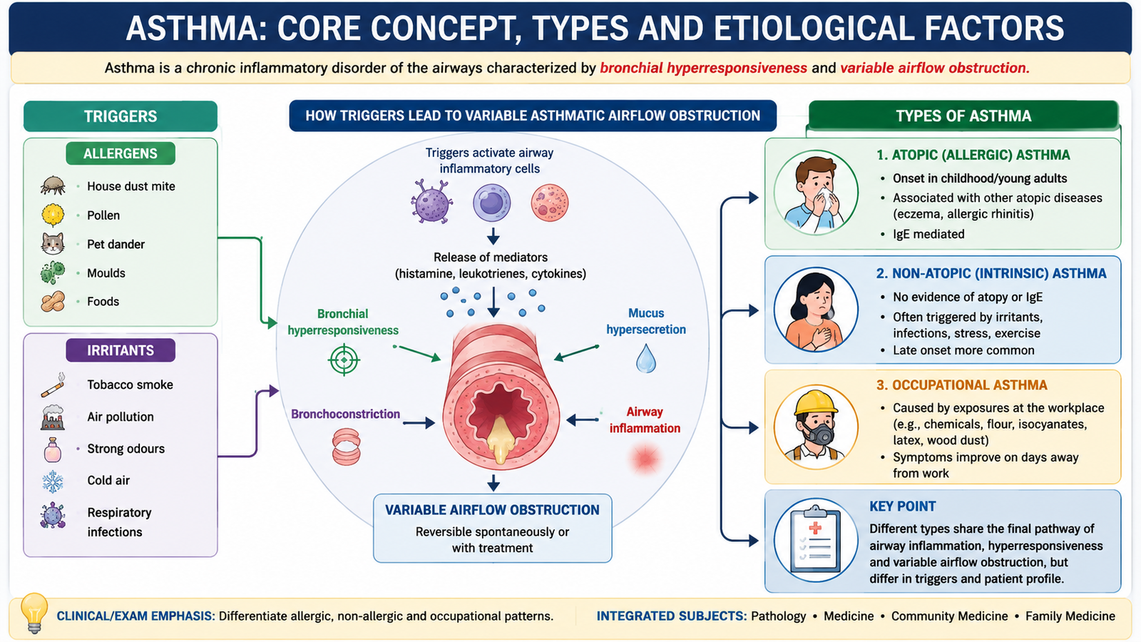
A. Asthma: Core Concept, Types and Etiological Factors
Asthma is best understood as a disorder in which chronic airway inflammation creates bronchial hyperresponsiveness. The airways then narrow excessively when exposed to stimuli that may cause little or no response in a healthy person. This narrowing is usually variable and at least partly reversible. The disease therefore reflects an interaction between a susceptible host and environmental or occupational triggers.
Definition
Bronchial asthma is a chronic inflammatory disorder of the airways associated with bronchial hyperresponsiveness and variable airflow obstruction. Clinically, it causes recurrent episodes of wheezing, breathlessness, chest tightness and cough.
Major Types of Asthma
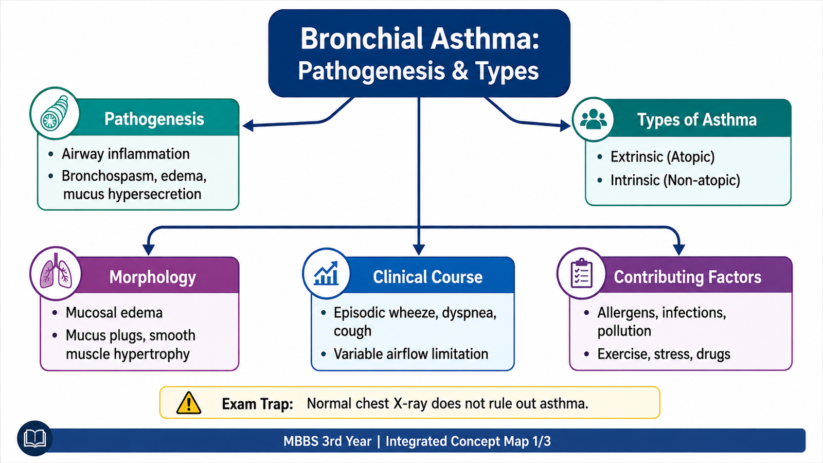
Asthma can arise through different initiating pathways. The older distinction between atopic and non-atopic asthma remains useful for understanding pathogenesis, although individual patients may show overlapping features.
| Feature | Atopic Asthma | Non-Atopic Asthma |
|---|---|---|
| Typical association | Allergic sensitization | No obvious allergen sensitization |
| IgE relationship | Often IgE-mediated | IgE mechanism is less prominent |
| Common triggers | Environmental allergens | Respiratory infections, pollutants, exercise, cold air or irritants |
| Underlying principle | Allergic immune response produces airway inflammation | Airway hyperresponsiveness occurs without a dominant allergic mechanism |
Other Important Clinical Forms
- Drug-associated asthma: certain susceptible patients develop bronchospasm after particular medicines, especially drugs that alter arachidonic-acid mediator balance or promote bronchoconstriction.
- Occupational asthma: repeated exposure to sensitizing substances or irritants at work produces or aggravates asthma.
- Exercise-related bronchoconstriction: physical exertion may provoke temporary airway narrowing in susceptible individuals.
Risk and Contributing Factors
The development and expression of asthma depend on both host susceptibility and environmental exposure. Important contributing factors include a personal or family tendency toward allergic disease, exposure to allergens, tobacco smoke and other irritants, occupational sensitizers, respiratory infections and environmental pollution. The importance of individual factors varies from patient to patient.
B. Pathogenesis and Morphology of Bronchial Asthma
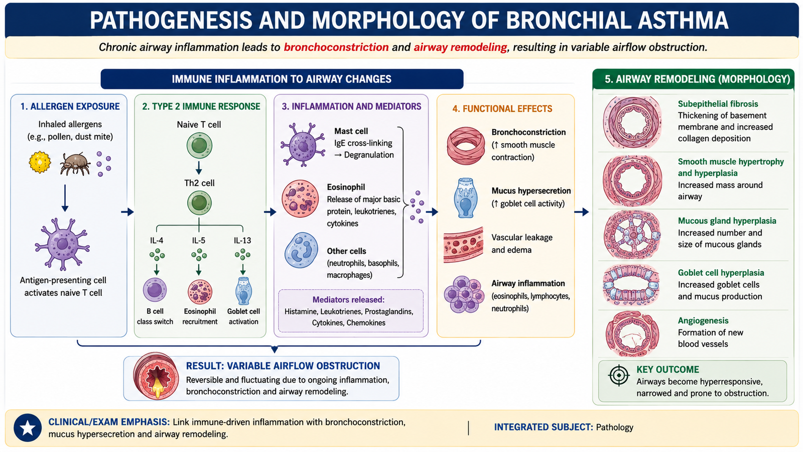
The central pathological process in asthma is persistent airway inflammation associated with exaggerated bronchial responsiveness. During an attack, narrowing is produced by smooth-muscle contraction, mucosal edema and mucus. Repeated inflammation may eventually produce structural alteration of the airway wall, often termed airway remodeling.
Atopic Asthma: Immune Mechanism
Atopic asthma commonly begins when inhaled allergens are presented to helper T lymphocytes. A type 2 helper T-cell response develops. Cytokines produced during this response promote IgE formation, eosinophilic inflammation and other changes that favor airway hyperresponsiveness.
Allergen exposure → type 2 helper T-cell response → IgE production and eosinophil recruitment → mast-cell sensitization and airway inflammation → bronchial hyperresponsiveness → bronchoconstriction, edema and mucus secretion.
IgE binds to mast cells. On subsequent exposure, the allergen interacts with sensitized mast cells and causes release of mediators. These mediators produce rapid bronchoconstriction and increase vascular permeability. Inflammatory cells, especially eosinophils, contribute to a later phase of inflammation and airway injury.
Major Cellular Participants
- Mast cells release mediators that produce bronchoconstriction and promote inflammation.
- Eosinophils release inflammatory proteins and mediators that can damage airway epithelium and maintain hyperresponsiveness.
- T lymphocytes coordinate allergic inflammation through cytokine production.
- Airway epithelial cells participate in inflammatory signaling and may become injured during persistent disease.
- Bronchial smooth muscle contracts excessively in response to stimuli and may become hypertrophied in long-standing disease.
Why the Airway Narrows
Three major processes act together. First, contraction of bronchial smooth muscle rapidly decreases airway diameter. Second, inflammation increases vascular permeability and produces mucosal edema, further reducing the lumen. Third, increased mucus secretion can partially obstruct already narrowed bronchi. Because airway resistance rises markedly as the airway becomes narrower, even a relatively small reduction in airway diameter may significantly impair expiratory airflow.
Airway Remodeling
Recurrent or persistent inflammation can lead to structural changes in the bronchial wall. These changes contribute to more persistent airflow limitation in some patients and help explain why longstanding poorly controlled asthma may become less completely reversible.
- Thickening beneath the bronchial epithelium due to increased collagen deposition.
- Hypertrophy and hyperplasia of bronchial smooth muscle.
- Enlargement of mucus-producing glands and increased goblet cells.
- Persistent inflammatory-cell infiltration.
Morphology
Grossly, severe asthma may show overexpanded lungs together with plugging of bronchi and bronchioles by thick mucus. Microscopically, the characteristic pattern reflects chronic inflammation and remodeling of the airway wall.
- Eosinophil-rich inflammatory infiltrate.
- Thickened subepithelial basement-membrane region due to fibrosis.
- Hypertrophy and hyperplasia of bronchial smooth muscle.
- Goblet-cell hyperplasia and increased mucus production.
- Increased submucosal mucus glands.
- Mucus plugs containing shed epithelial cells and inflammatory material may be present.
Asthma is not simply episodic bronchial smooth-muscle spasm. Chronic airway inflammation and structural remodeling are fundamental components of the disease.
C. Clinical Presentation, Clinical Course and Severity
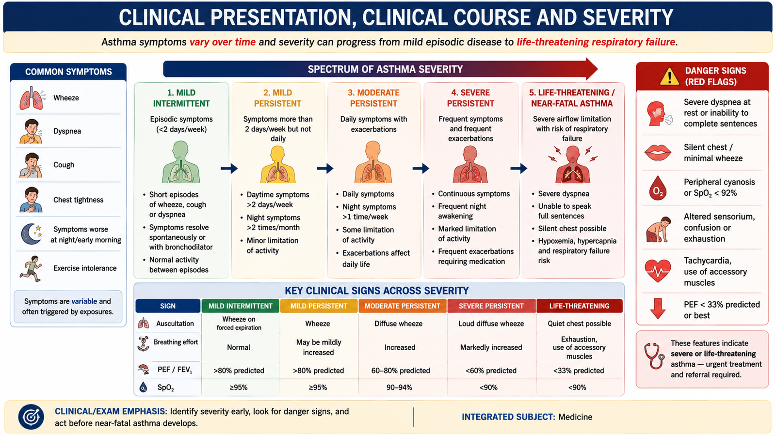
Asthma commonly presents as recurrent episodes rather than constant symptoms. The characteristic clinical pattern is variation: symptoms may worsen after exposure to a trigger and improve spontaneously or after bronchodilator treatment. Between episodes, some patients may have few or no symptoms, whereas others with more persistent disease remain symptomatic.
Typical Clinical Features
- Wheezing: produced by airflow through narrowed intrathoracic airways.
- Breathlessness: occurs because airway resistance increases and expiration becomes difficult.
- Chest tightness: often accompanies bronchoconstriction.
- Cough: may occur with or without obvious wheeze and can be prominent at night or after exposure to a trigger.
- Prolonged expiration: reflects difficulty emptying the lungs through narrowed airways.
Symptoms may be triggered or worsened by allergens, respiratory infections, smoke, environmental irritants, cold air, exercise, certain medicines or occupational exposure. A pattern of episodic symptoms related to identifiable triggers is therefore an important diagnostic clue.
During a More Severe Attack
As bronchial obstruction becomes more marked, the patient must generate greater respiratory effort to move air through narrowed airways. Tachypnea, difficulty speaking normally, use of accessory respiratory muscles and increasing respiratory distress may develop.
Clinical Severity Categories
Acute asthma can be clinically described as mild, moderate, life-threatening or near-fatal according to the overall degree of respiratory compromise. These categories are based on the patient’s clinical state and objective evidence of airflow or gas-exchange impairment rather than on wheeze alone.
| Category | General Clinical Pattern |
|---|---|
| Mild | Symptoms are present but respiratory distress is limited and the patient remains relatively comfortable. |
| Moderate | More obvious airflow obstruction and increased work of breathing are present. |
| Life-threatening | Severe obstruction produces marked respiratory compromise, exhaustion, altered gas exchange or other danger signs. |
| Near-fatal | Respiratory failure develops or ventilatory support becomes necessary. |
Status Asthmaticus
Status asthmaticus refers to a severe and persistent asthma exacerbation that does not respond adequately to usual initial bronchodilator treatment. Persistent airflow obstruction may produce worsening ventilation, fatigue and eventually respiratory failure. It is therefore a medical emergency.
A patient with increasing exhaustion, altered consciousness, markedly reduced air entry, cyanosis or evidence of respiratory failure requires urgent escalation of care. A quiet chest in a severely breathless patient may represent extremely poor airflow rather than improvement.
D. Diagnosis, Spirometry and Risk Assessment
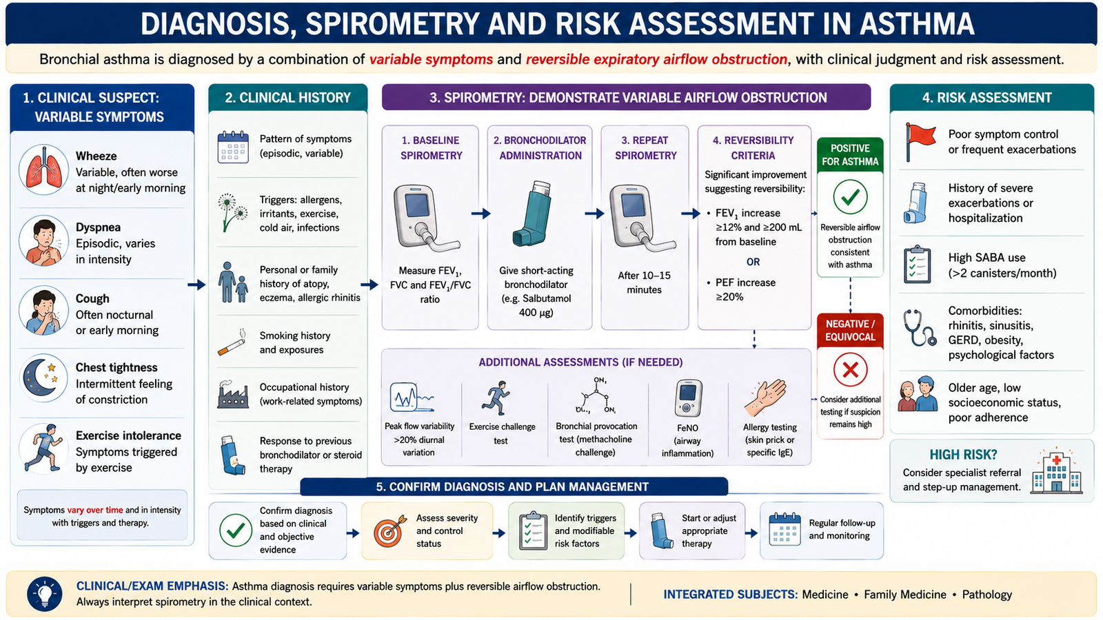
The diagnosis of asthma combines a compatible clinical history with evidence that airflow obstruction varies over time. No single symptom is diagnostic. The strongest clinical pattern is recurrent respiratory symptoms that change in intensity, occur after typical triggers and are accompanied by demonstrable variable expiratory airflow limitation.
History
Ask about the pattern of wheeze, cough, breathlessness and chest tightness. Variation over time is important. Symptoms may be worse during the night, after exercise, during respiratory infections, after allergen exposure or in particular environments such as the workplace.
Relevant history also includes previous severe attacks, emergency treatment, hospital admission, adherence to therapy, inhaler technique, smoking and environmental exposure, occupational exposure and associated allergic conditions.
Examination
Examination may be normal between attacks. During symptomatic periods, expiratory wheeze and prolonged expiration may be present. In acute severe disease, assess respiratory effort, ability to speak, air entry, mental state and signs suggesting impaired oxygenation or respiratory failure.
Spirometry
Spirometry measures how much air a patient can exhale and how rapidly it can be expelled. In an obstructive airway disorder, the amount of air expelled during the early part of forced expiration falls disproportionately compared with the total forced vital capacity.
In asthma, the important concept is variability or reversibility of airflow obstruction. When expiratory airflow improves after administration of a bronchodilator, the finding supports asthma in the appropriate clinical setting. Normal spirometry at one visit does not automatically exclude asthma because the disease is variable and the patient may be tested between symptomatic episodes.
Compatible symptoms → identify obstructive expiratory pattern → assess variability or bronchodilator reversibility → relate the result to the clinical history → support or reconsider the diagnosis.
Additional Investigations
Additional testing is selected according to the clinical situation. Assessment may include measures of expiratory airflow variability, evidence of allergic sensitization and investigations aimed at excluding another explanation when the presentation is atypical. The purpose is to confirm variable airway obstruction and identify clinically relevant contributing factors rather than order extensive investigations routinely.
Risk Assessment
Risk assessment is not limited to the patient’s symptoms today. A patient may have relatively few daily symptoms but still be at risk of a serious future exacerbation. Important concerns include previous severe attacks, poor adherence, incorrect inhaler technique, ongoing exposure to tobacco smoke or occupational triggers, and failure to obtain adequate control despite appropriate treatment.
When Specialist Referral Is Needed
Specialist review should be considered when the diagnosis remains uncertain, symptoms remain poorly controlled despite appropriate therapy, severe or recurrent exacerbations occur, an occupational cause is suspected, treatment-related problems are difficult to manage or features suggest another significant respiratory disorder.
E. Pharmacological Treatment: Major Drug Classes
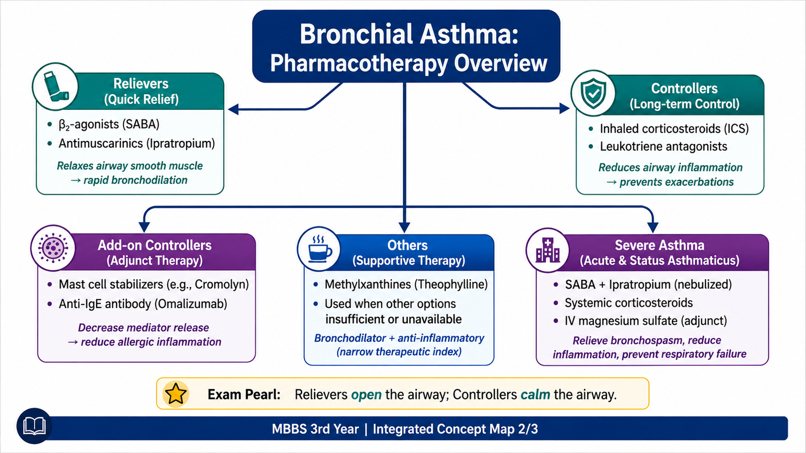
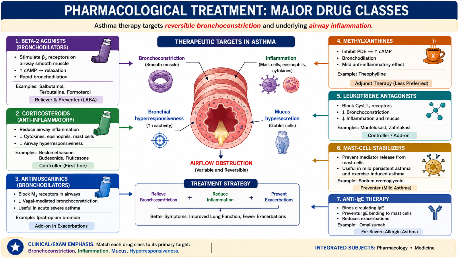
Asthma pharmacotherapy targets two major problems. Bronchodilators reverse or reduce airway smooth-muscle contraction, while anti-inflammatory drugs suppress the underlying airway inflammation and reduce future symptoms and exacerbations. Some additional drugs target specific inflammatory mediators or pathways in selected patients.
1. Beta-2 Adrenergic Agonists
Beta-2 agonists stimulate beta-2 adrenergic receptors on bronchial smooth muscle. Receptor activation increases intracellular cyclic AMP, which promotes smooth-muscle relaxation and therefore bronchodilation.
Short-acting agents such as salbutamol produce relatively rapid bronchodilation and are useful when quick reversal of bronchoconstriction is required. Longer-acting agents provide more prolonged bronchodilation and are used within long-term treatment strategies rather than as substitutes for control of the underlying inflammation.
Adverse effects arise from adrenergic stimulation and may include tremor, palpitations and tachycardia. Metabolic effects such as reduction in serum potassium may occur, particularly with greater systemic exposure.
2. Corticosteroids
Corticosteroids are the most important anti-inflammatory drugs in asthma. They act through intracellular glucocorticoid receptors and alter gene transcription, reducing the production of inflammatory cytokines and suppressing inflammatory-cell activity. They also reduce airway edema, mucus production and bronchial hyperresponsiveness.
Inhaled corticosteroids deliver anti-inflammatory treatment directly to the airways and are central to long-term control. Local adverse effects may include oral candidiasis and dysphonia. Systemic corticosteroids are used when a stronger systemic anti-inflammatory effect is required, particularly in significant acute exacerbations, but prolonged systemic exposure produces substantially more adverse effects.
3. Antimuscarinic Drugs
Parasympathetic stimulation promotes bronchial smooth-muscle contraction through muscarinic receptors. Antimuscarinic drugs block this cholinergic pathway and therefore reduce bronchoconstriction. Ipratropium is a commonly recognized short-acting example, while longer-acting agents provide more prolonged muscarinic blockade.
Because inhaled antimuscarinic agents have limited systemic absorption, their adverse effects are generally less prominent than those of systemic anticholinergic drugs. Dry mouth may occur.
4. Methylxanthines
Theophylline is the classic methylxanthine used in obstructive airway disease. Its pharmacological actions include phosphodiesterase inhibition and antagonism of adenosine-mediated effects. These mechanisms contribute to bronchodilation.
Theophylline has a narrow therapeutic index, so excessive concentrations may produce important toxicity. Gastrointestinal symptoms, tremor, insomnia and tachycardia may occur, while severe toxicity can cause dangerous cardiac arrhythmias and seizures. Its metabolism is also affected by important drug interactions, which contributes to variability in blood concentrations.
5. Leukotriene Pathway Inhibitors
Leukotrienes contribute to bronchoconstriction, mucus production, vascular permeability and airway inflammation. Drugs such as montelukast block leukotriene receptors and thereby reduce leukotriene-mediated airway effects. They are administered systemically rather than by inhalation.
A related approach is inhibition of leukotriene synthesis. These medicines are not direct rapid bronchodilators and are mainly used as controller therapy in selected patients.
6. Mast-Cell Stabilizers
Mast-cell stabilizing drugs such as cromolyn reduce mediator release from mast cells and can help prevent stimulus-induced bronchoconstriction. They are preventive rather than rescue drugs because they do not rapidly reverse established bronchospasm.
Their systemic absorption is limited when administered to the respiratory tract. They therefore have relatively few systemic adverse effects, although local irritation may occur.
7. Anti-IgE Therapy
Omalizumab is a monoclonal antibody directed against IgE. By binding circulating IgE, it reduces the amount of IgE available to interact with high-affinity IgE receptors on mast cells and other inflammatory cells. This decreases IgE-dependent allergic activation.
Anti-IgE therapy is intended for selected patients with significant allergic asthma rather than routine use in every patient. Because it is a biologic treatment, hypersensitivity reactions are an important safety consideration.
| Class | Main Target | Main Benefit | Important Point |
|---|---|---|---|
| Beta-2 agonists | Beta-2 receptor | Bronchodilation | Rapid symptom relief with short-acting agents |
| Corticosteroids | Inflammatory gene expression | Suppress airway inflammation | Core controller therapy |
| Antimuscarinics | Muscarinic receptors | Reduce cholinergic bronchoconstriction | Useful bronchodilator class |
| Methylxanthines | Phosphodiesterase/adenosine pathways | Bronchodilation | Narrow therapeutic index |
| Leukotriene antagonists | Leukotriene pathway | Reduce mediator effects | Controller, not rapid rescue therapy |
| Mast-cell stabilizers | Mast-cell mediator release | Prevent trigger-related response | Preventive rather than rescue drugs |
| Anti-IgE antibody | IgE | Reduce allergic activation | For selected allergic asthma |
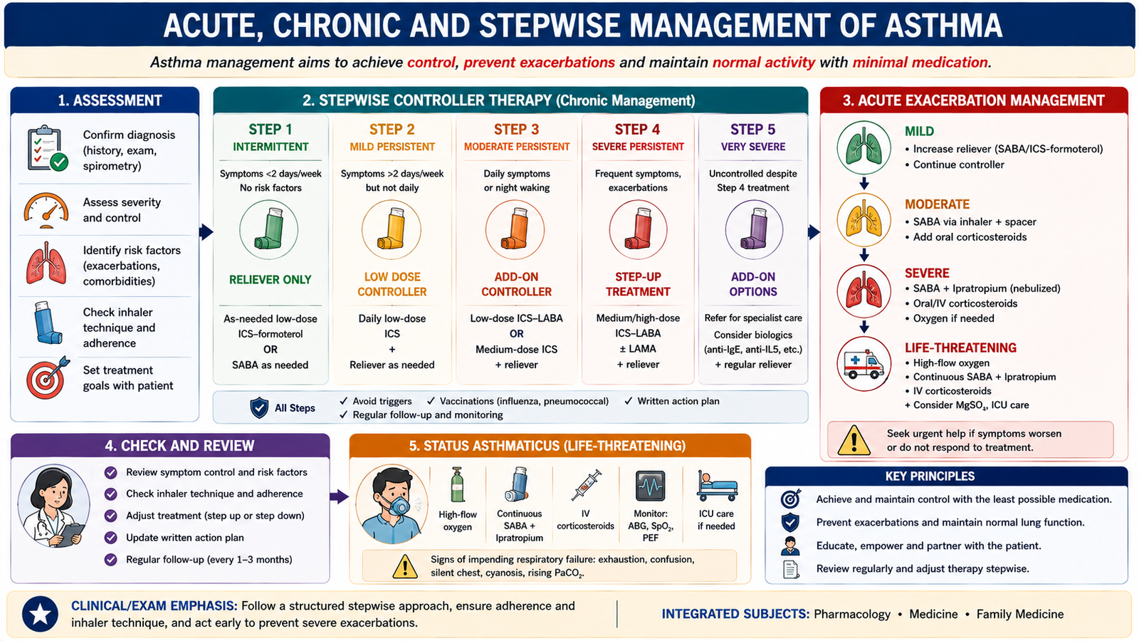
F. Acute, Chronic and Stepwise Management of Asthma
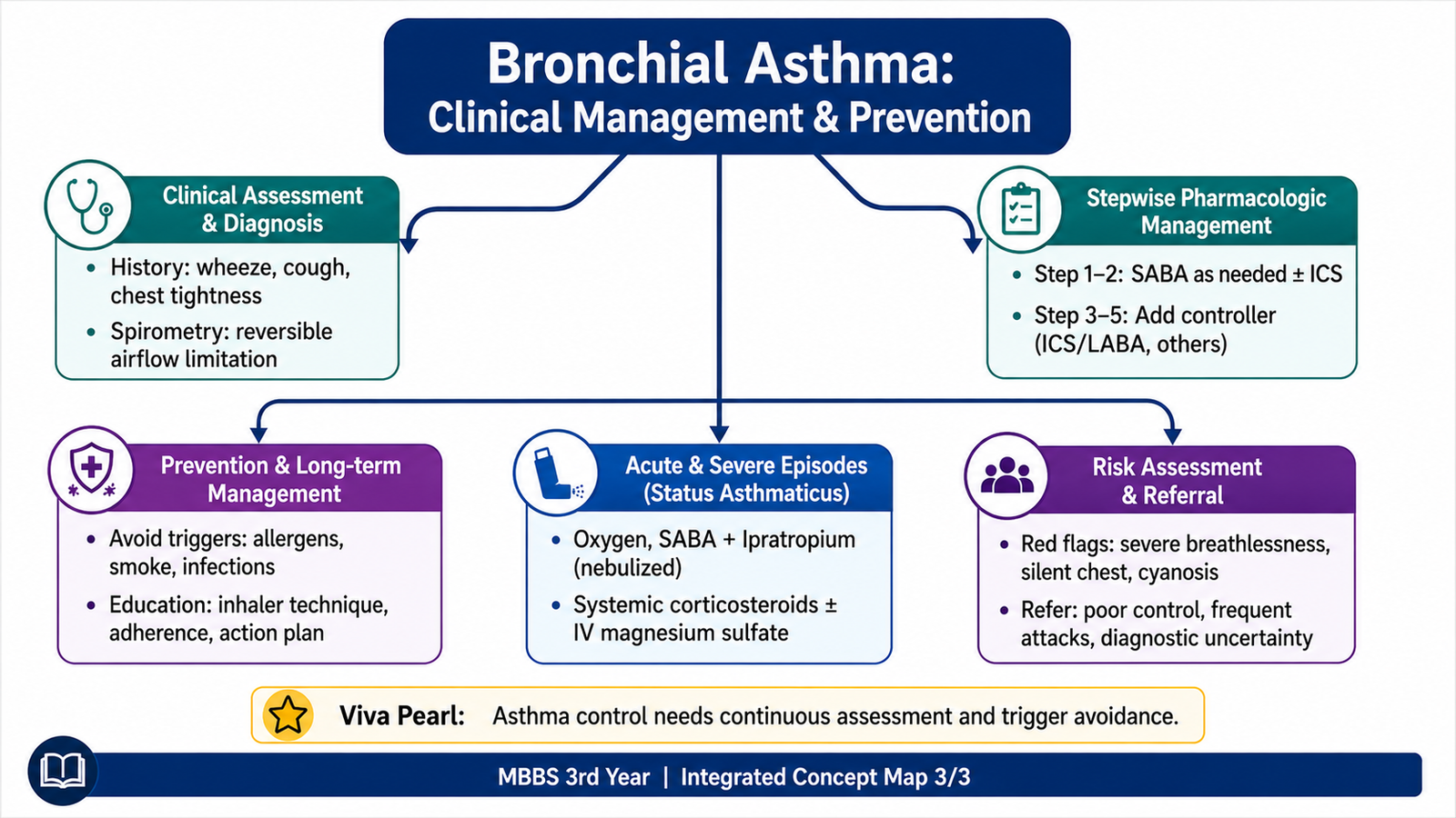
Asthma treatment must address both immediate bronchoconstriction and the chronic inflammatory process responsible for future attacks. Management therefore combines appropriate medication with assessment of control, correct inhaler technique, adherence, avoidance of relevant triggers and patient education.
Management of an Acute Exacerbation
The first priority during an acute attack is to assess severity while promptly treating bronchial obstruction and impaired oxygenation. Rapid-acting inhaled bronchodilator therapy is used to reverse bronchoconstriction. More severe attacks require systemic anti-inflammatory treatment because airway inflammation cannot be adequately controlled by bronchodilation alone.
An inhaled antimuscarinic bronchodilator may provide additional bronchodilation in a more significant acute attack. Failure to improve, progressive exhaustion or evidence of respiratory failure requires urgent specialist and hospital-level management.
Status Asthmaticus
Status asthmaticus is managed as a severe persistent exacerbation. Repeated or continuous bronchodilator treatment, systemic corticosteroid therapy, oxygenation support and close reassessment are required. The clinician must look actively for deterioration because a patient who becomes fatigued may lose the ability to maintain adequate ventilation.
Falling respiratory effort in a severely obstructed patient may indicate exhaustion rather than recovery. Altered consciousness, worsening gas exchange or respiratory failure requires immediate escalation and ventilatory support when necessary.
Long-Term Control
Long-term treatment aims to minimize symptoms, maintain normal activity, reduce exacerbations and prevent progressive airway consequences while using an appropriate amount of treatment. Because chronic airway inflammation is central to asthma, anti-inflammatory controller treatment is fundamental when persistent disease is present.
Stepwise Pharmacological Principle
The stepwise approach means that treatment intensity is adjusted according to the patient’s level of control, current symptoms and future risk. Therapy is increased when adequate control is not achieved after checking correct diagnosis, adherence, inhaler technique and ongoing trigger exposure. When control has remained satisfactory, unnecessary treatment intensity should be avoided.
As asthma becomes more difficult to control, treatment generally moves from a simpler inhaled anti-inflammatory strategy toward combinations of controller medicines, commonly involving inhaled corticosteroid therapy with additional bronchodilator or other controller treatment. Selected patients with persistent severe allergic disease may require targeted biologic treatment.
Before Increasing Pharmacotherapy
- Confirm that the symptoms are compatible with asthma.
- Check whether prescribed medicines are being taken consistently.
- Observe inhaler technique and correct errors.
- Identify smoking, allergens, irritants or occupational exposures.
- Assess whether another condition is contributing to poor control.
Long-Term Patient Education
Education is part of treatment rather than an optional extra. Patients should understand the difference between medicines used mainly for rapid relief and those used to control airway inflammation. They should also know how to use their inhaler correctly, recognize worsening symptoms, avoid known triggers where practical and seek medical care promptly when danger signs develop.
Physical activity should generally be encouraged when asthma is appropriately controlled. If exercise repeatedly provokes symptoms, asthma control and preventive therapy should be reassessed rather than advising unnecessary long-term physical inactivity.
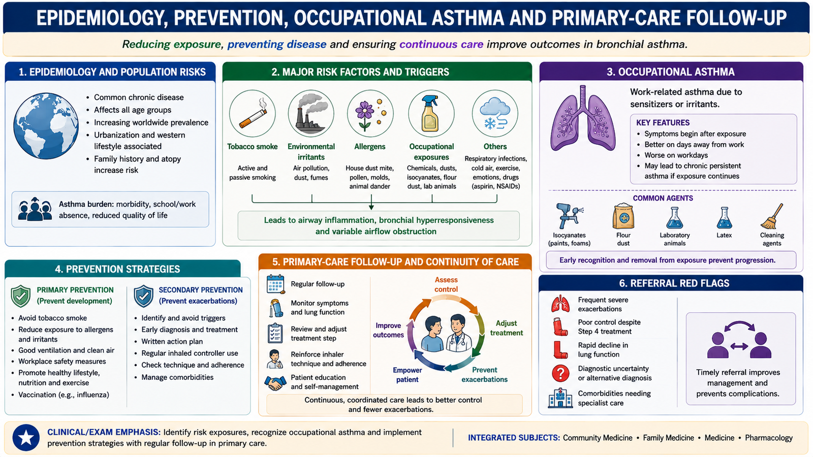
G. Epidemiology, Prevention, Occupational Asthma and Primary-Care Follow-Up
Asthma is an important chronic respiratory disorder affecting both children and adults. Its occurrence and severity are influenced by the interaction between host susceptibility and environmental exposures. From a community and family-medicine perspective, the major goals are to identify modifiable risk factors, reduce harmful exposure, recognize occupational disease, maintain long-term control and identify patients who need specialist care.
Epidemiological and Population Risk Factors
Asthma occurs in populations with different patterns of allergen exposure, air quality, smoking exposure, occupational hazards and socioeconomic conditions. Important individual determinants include genetic susceptibility and atopic tendency, while environmental determinants influence whether disease develops or becomes clinically active.
- Family or personal tendency toward allergic disease.
- Exposure to indoor or outdoor allergens in susceptible individuals.
- Tobacco smoke and other respiratory irritants.
- Air pollution.
- Occupational sensitizers and irritants.
- Respiratory infections as triggers of exacerbation.
Primary Prevention
Primary prevention aims to reduce factors that contribute to development of disease before asthma becomes established. In practice, this involves reducing avoidable exposure to tobacco smoke, harmful occupational agents and unnecessary respiratory irritants while promoting healthier air environments.
Secondary Prevention and Control
Once asthma is present, prevention focuses on reducing symptoms, exacerbations and complications. This requires early recognition, appropriate controller therapy, adherence, correct inhaler technique, trigger reduction and regular assessment of control.
Occupational Asthma
Occupational asthma is asthma caused or aggravated by exposure encountered in the workplace. The responsible exposure may act as a sensitizer or an irritant. Suspicion should increase when symptoms begin after a new occupational exposure, worsen during working periods and improve away from work.
Prevention requires identification of the responsible workplace agent and reduction or elimination of exposure when possible. Appropriate workplace controls, protective measures and occupational-health assessment are important because continued exposure may worsen disease.
Symptoms that repeatedly worsen at work and improve during time away from the workplace should prompt careful assessment of occupational exposure.
Primary-Care Follow-Up
General practice plays a major role in continuing asthma care. Follow-up should assess symptom control, recent exacerbations, inhaler technique, treatment adherence, trigger exposure and risk of future severe attacks. The aim is to detect deteriorating control before an emergency occurs.
Guideline-Based Care
Asthma should be managed according to recognized evidence-based asthma guidelines using a structured approach to diagnosis, assessment of control, inhaled therapy, stepwise adjustment, patient education and referral. The exact treatment plan should be individualized rather than based on symptoms alone.
Referral Red Flags
- Uncertain diagnosis or atypical clinical features.
- Repeated severe exacerbations or hospital presentations.
- Poor control despite appropriate treatment and good technique.
- Suspected occupational asthma requiring further assessment.
- Features suggesting life-threatening or near-fatal asthma.
- Requirement for advanced or targeted therapy beyond routine primary-care management.
3. Integrated Mechanism Flow
Mast-cell, T-cell and eosinophilic inflammatory response
Bronchial hyperresponsiveness
Smooth-muscle contraction + mucosal edema + mucus secretion
Variable expiratory airflow obstruction
Wheeze, breathlessness, chest tightness and cough
Repeated inflammation may produce airway remodeling, while appropriate anti-inflammatory treatment reduces disease activity and future exacerbation risk.
4. Important Comparison — Reliever Effect vs Controller Effect
| Feature | Predominantly Reliever/Bronchodilator Effect | Controller/Anti-inflammatory Effect |
|---|---|---|
| Main problem targeted | Acute airway smooth-muscle narrowing | Chronic airway inflammation |
| Main immediate effect | Bronchodilation | Reduction of inflammatory activity |
| Clinical purpose | Improve bronchoconstriction and symptoms | Improve long-term control and reduce exacerbation risk |
| Typical example | Beta-2 agonist bronchodilator | Inhaled corticosteroid |
| Exam distinction | Opening the airway does not remove the underlying inflammation | Controlling inflammation addresses a central disease mechanism |
5. AIM High-Yield Review
- ⭐ Asthma is characterized by chronic airway inflammation, bronchial hyperresponsiveness and variable airflow obstruction.
- Atopic asthma commonly involves a type 2 helper T-cell response, IgE, mast cells and eosinophils.
- Bronchial narrowing results from smooth-muscle contraction + mucosal edema + increased mucus.
- Airway remodeling includes subepithelial fibrosis, smooth-muscle enlargement and increased mucus-producing structures.
- Typical symptoms are episodic wheeze, breathlessness, chest tightness and cough.
- ⭐ Variable or reversible expiratory airflow limitation on spirometry strongly supports asthma in the appropriate clinical setting.
- A quiet chest in a severely breathless patient can indicate dangerously poor airflow.
- Beta-2 agonists relax bronchial smooth muscle mainly through increased cyclic AMP.
- ⭐ Inhaled corticosteroids target the underlying airway inflammation and are central controller drugs.
- Antimuscarinic agents reduce cholinergic bronchoconstriction.
- Theophylline has a narrow therapeutic index; severe toxicity may produce arrhythmias and seizures.
- Leukotriene antagonists reduce leukotriene-mediated bronchoconstriction and inflammation but are not rapid rescue bronchodilators.
- Omalizumab targets IgE and is used in selected allergic asthma.
- Before stepping up long-term treatment, check diagnosis, adherence, inhaler technique and ongoing trigger exposure.
- ⭐ Occupational asthma should be suspected when symptoms repeatedly worsen during workplace exposure and improve away from work.
Bronchial Asthma — Pathophysiology, Diagnosis and Treatment
Watch this video after completing the learning material to reinforce airway inflammation, bronchoconstriction, clinical features, diagnosis and treatment of asthma.