
Topic 7 — Chronic Obstructive Pulmonary Disease and Emphysema
Topic Introduction
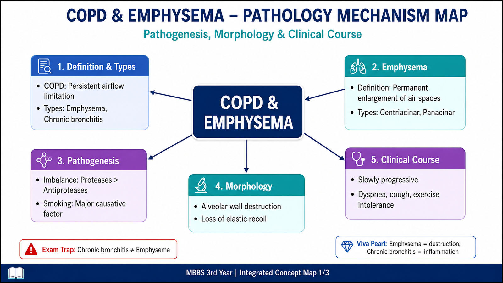
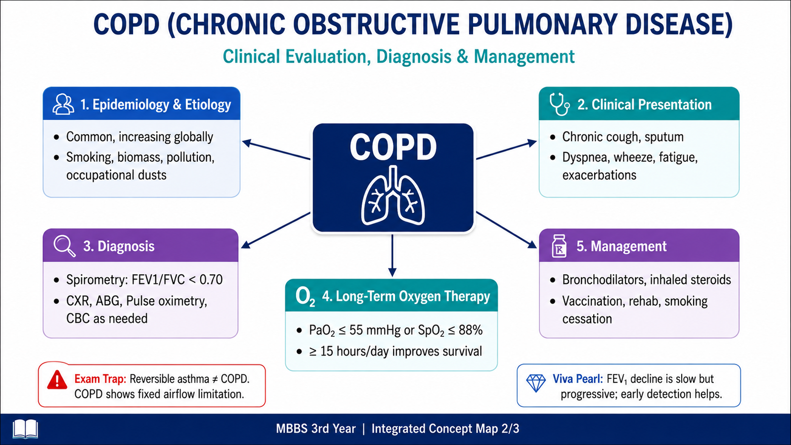
Chronic obstructive pulmonary disease, or COPD, is a chronic respiratory disorder in which airflow through the lungs becomes persistently limited. The obstruction develops mainly because of disease in the small airways, destruction of alveolar walls, or a combination of both. Two important pathological patterns associated with COPD are chronic bronchitis and emphysema. Smoking is the major preventable cause, although other inhaled pollutants and some inherited factors can contribute. In this chapter, you will learn the definitions and pathological changes of obstructive lung disease, the mechanisms and morphology of emphysema and chronic bronchitis, the clinical presentation and investigation of COPD, and the principles of treatment, long-term oxygen therapy, home care and referral.
A. Obstructive Pulmonary Disease and COPD: Core Concepts, Epidemiology and Etiology
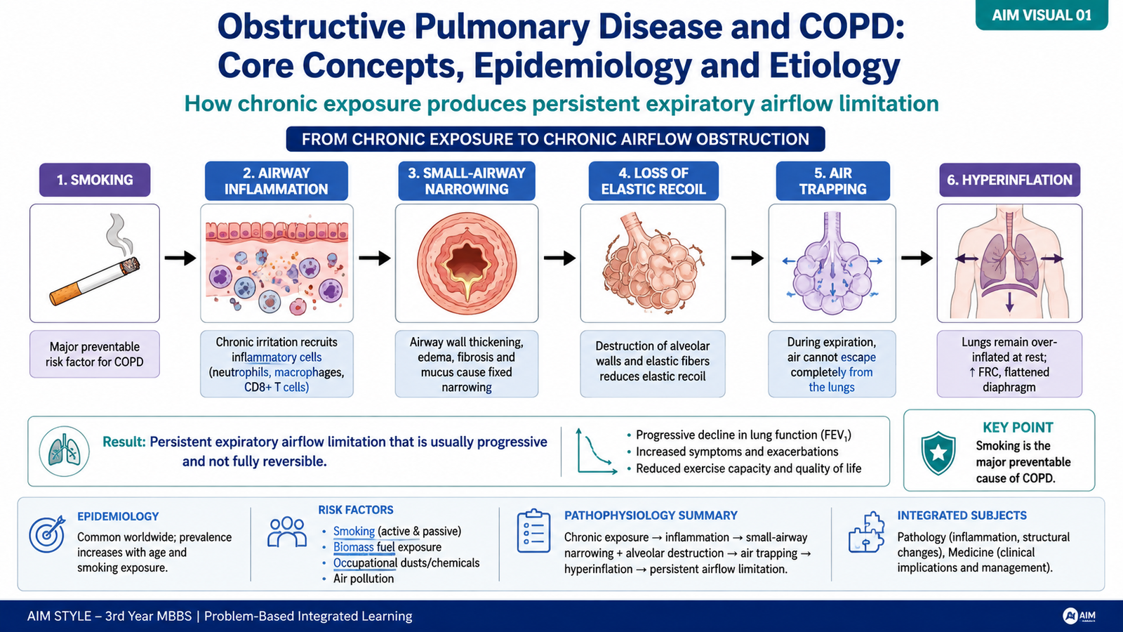
Obstructive pulmonary diseases are disorders in which airflow is limited because resistance to movement of air through the respiratory tract is increased. The patient therefore has particular difficulty moving air out of the lungs during expiration. Important obstructive diseases include chronic bronchitis, emphysema, asthma and bronchiectasis. COPD refers mainly to chronic, persistent airflow limitation produced by abnormalities of the airways and/or alveoli, most commonly resulting from long-term exposure to harmful inhaled particles and gases.
What makes COPD obstructive?
Normal expiration depends on open airways and the elastic recoil of lung tissue. In COPD, narrowing of small airways increases resistance, while emphysematous destruction reduces the elastic force that normally helps keep small airways open during expiration. Air therefore leaves the lungs more slowly and may become trapped.
Epidemiology and major causes
COPD is a common chronic respiratory disease, particularly among adults with prolonged exposure to tobacco smoke or other respiratory irritants. The likelihood of developing clinically important disease increases with the intensity and duration of exposure, but susceptibility differs between individuals.
- Cigarette smoking: the most important preventable risk factor.
- Biomass-fuel smoke and indoor pollution: repeated exposure can cause chronic airway injury.
- Occupational dusts, fumes and chemicals: prolonged exposure may contribute to airflow limitation.
- Air pollution: may aggravate respiratory disease and contributes to cumulative inhalational exposure.
- Genetic susceptibility: severe alpha-1 antitrypsin deficiency is an important inherited cause of emphysema.
Smoking does not produce disease merely by filling the lungs with smoke. It causes persistent inflammation, increases oxidative stress, disturbs protective antiprotease mechanisms and promotes structural remodeling of small airways. These changes explain why airflow obstruction can persist even after an acute irritant exposure has ended.
B. Emphysema: Types, Pathogenesis, Morphology and Clinical Course
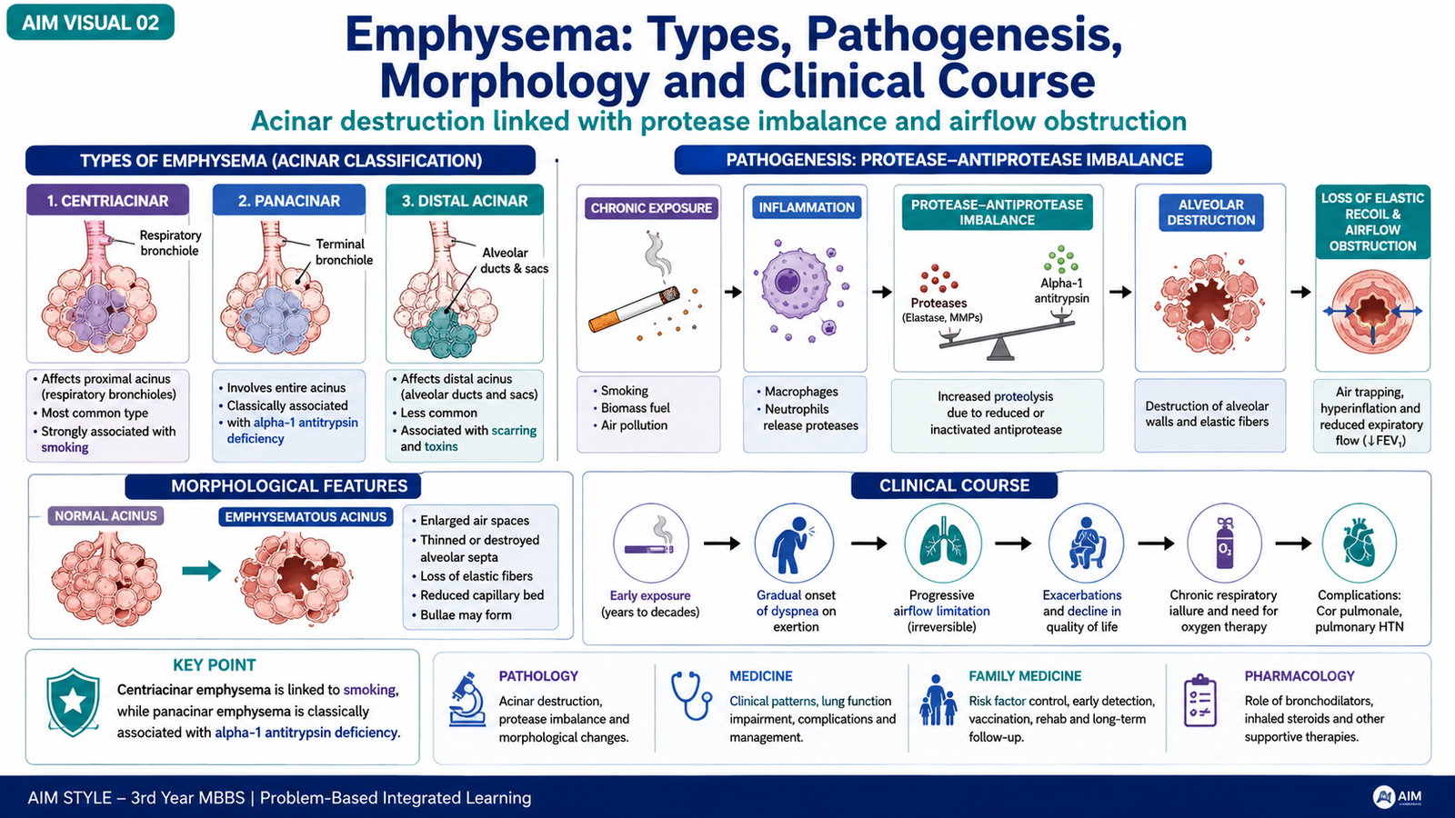
Emphysema is a pathological condition characterized by permanent enlargement of airspaces distal to the terminal bronchioles, accompanied by destruction of their walls without prominent fibrosis. The central problem is therefore loss of alveolar tissue rather than simply excess air inside otherwise normal lungs. Destruction of alveolar septa reduces the surface area available for gas exchange and also removes elastic support from small airways.
Types of emphysema
The types are classified according to which part of the pulmonary acinus is predominantly affected.
- Centriacinar emphysema: affects the central or proximal part of the acinus. It is strongly associated with cigarette smoking and is usually more severe in the upper portions of the lungs.
- Panacinar emphysema: the entire acinus is relatively uniformly enlarged. It is classically associated with alpha-1 antitrypsin deficiency and tends to involve lower lung regions more prominently.
- Distal acinar emphysema: the distal acinus is predominantly involved, often near the pleura or along connective-tissue septa. Subpleural bullae may develop.
- Irregular emphysema: the acinus is affected irregularly, commonly in association with areas of previous scarring.
Pathogenesis: why are alveolar walls destroyed?
The development of emphysema can be understood through two closely related mechanisms: protease–antiprotease imbalance and oxidant–antioxidant imbalance. Inhaled smoke and irritants recruit inflammatory cells, particularly neutrophils and macrophages, into the lungs. These cells release proteolytic enzymes capable of damaging elastin and other components of alveolar walls. Normal lungs are protected by antiproteases, including alpha-1 antitrypsin. When protease activity becomes excessive or antiprotease protection is deficient, alveolar tissue is progressively destroyed.
Oxidants in cigarette smoke and oxidants released by inflammatory cells cause additional tissue damage. They can also reduce the effectiveness of protective antiproteases. Oxidative stress therefore reinforces the destructive process.
Morphology
Gross morphology: emphysematous lungs become enlarged and overinflated. The degree and distribution depend on the type of emphysema. Large abnormal airspaces or bullae may be visible in advanced disease. Microscopic morphology: the characteristic finding is enlargement of distal airspaces with thinning and destruction of alveolar septa. Adjacent alveoli may merge into larger spaces. The reduction in alveolar walls also means loss of part of the pulmonary capillary bed.
How morphology produces clinical disease
Loss of elastic recoil makes it difficult for the lungs to empty during expiration. Small airways may collapse, causing air trapping and progressive hyperinflation. Destruction of alveolar surface area reduces the reserve available for gas exchange. Patients therefore develop gradually progressive exertional dyspnea, and advanced disease may eventually cause significant respiratory impairment. Clinical link: A patient with prominent emphysema may have marked breathlessness, hyperinflated lungs and reduced breath sounds because the lungs contain trapped air but have lost functioning alveolar tissue and elastic recoil.
C. Chronic Bronchitis: Definition, Pathogenesis and Morphology
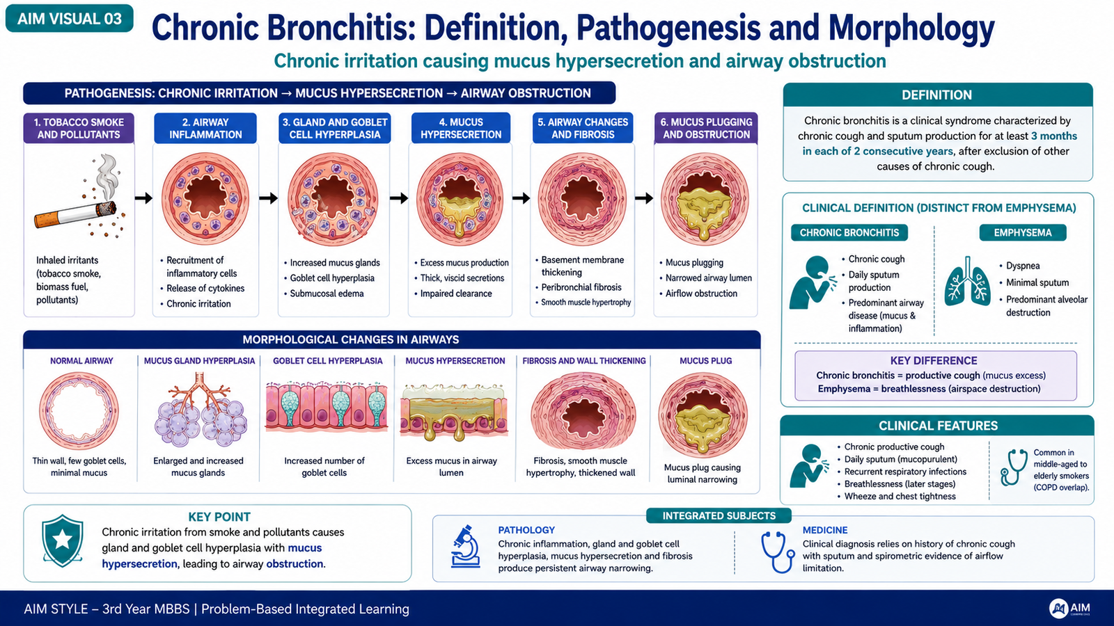
Chronic bronchitis is defined clinically as a productive cough lasting for at least three months in each of two consecutive years, when other causes of chronic productive cough have been excluded. Unlike emphysema, which is defined by structural destruction of distal airspaces, chronic bronchitis is identified clinically by chronic mucus-producing cough.
How chronic bronchitis develops
Long-term inhalation of cigarette smoke or other irritants stimulates excessive mucus production and produces persistent inflammation in the bronchial wall. Mucus-secreting glands enlarge, and goblet cells increase in the airway epithelium. At the same time, inflammation and fibrosis affect smaller bronchi and bronchioles. Mucus within the lumen together with small-airway narrowing obstructs airflow.
Morphology
Large airways: there is enlargement of mucus-secreting glands with increased mucus production. Goblet-cell increase contributes further to excessive secretion. Small airways: inflammation, mucus plugging and fibrotic narrowing are especially important in producing persistent airflow limitation. Repeated infection may worsen inflammation but infection is usually a complication and exacerbating factor rather than the primary initiating cause. The combination of excessive secretions and impaired clearance also creates conditions in which respiratory infections and acute exacerbations occur more easily.
D. Clinical Presentation and Diagnosis of COPD
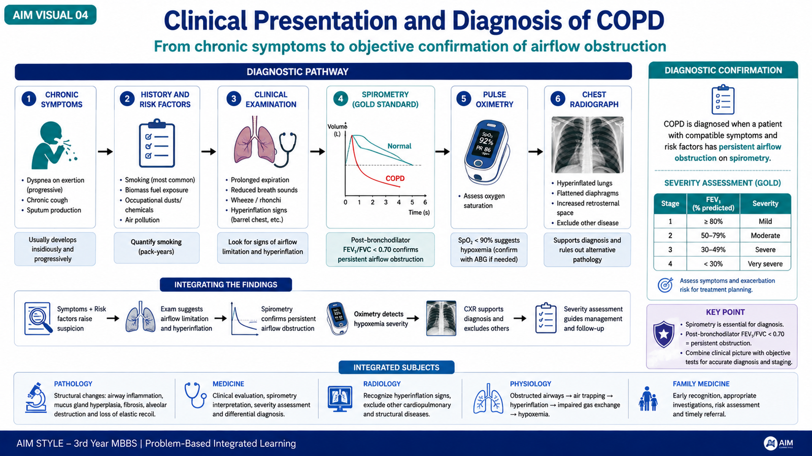
COPD usually develops gradually. A patient may initially notice symptoms only during exertion, but worsening airway obstruction and loss of pulmonary reserve eventually make ordinary activities more difficult. The typical clinical picture results from persistent airflow limitation, mucus production, air trapping and impaired gas exchange.
Clinical presentation
- Progressive dyspnea: often first noticed during exercise and later with less strenuous activity.
- Chronic cough: may be intermittent initially and persistent later.
- Sputum production: particularly prominent when chronic bronchitis is an important component.
- Wheeze or chest tightness: may occur because of narrowed airways.
- Recurrent exacerbations: periods of worsening respiratory symptoms may become more frequent as disease progresses.
Examination findings
Early disease may produce few obvious physical signs. With more advanced airflow obstruction, expiration may become prolonged and breath sounds may be reduced. Hyperinflation can increase the anteroposterior diameter of the chest. Accessory respiratory muscles may be used when breathing becomes difficult. Cyanosis indicates important impairment of oxygenation and should not be considered a harmless routine finding.
Investigations
Spirometry is the key test for demonstrating persistent expiratory airflow limitation. COPD produces a reduced ability to expire air rapidly, reflected by reduction in expiratory flow measurements and a persistently reduced relationship between forced expiratory volume and forced vital capacity.
- Spirometry: confirms persistent obstructive ventilatory abnormality and helps assess its severity.
- Pulse oximetry: provides a simple assessment of oxygen saturation.
- Arterial blood gases: are useful when significant hypoxemia, hypercapnia or respiratory failure is suspected.
- Chest radiograph: may show hyperinflation, flattened diaphragms or bullous change and can help identify alternative diagnoses or complications.
- Blood tests: may help identify problems such as anemia, polycythemia or infection according to the clinical situation.
E. Management of COPD and Long-Term Oxygen Therapy
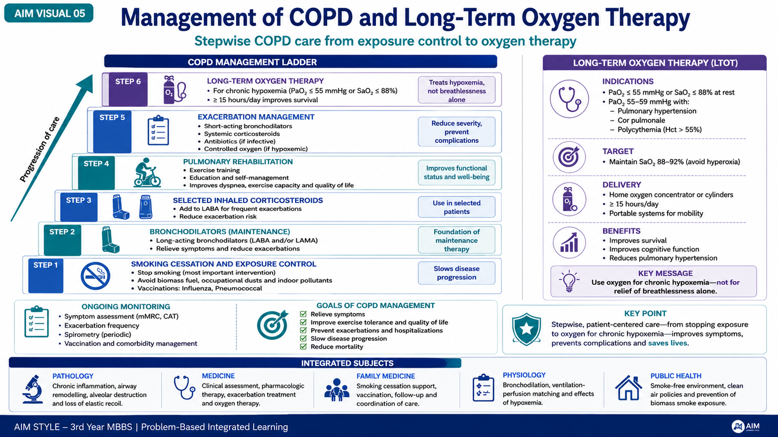
COPD management aims to reduce symptoms, improve activity and quality of life, reduce exacerbations and prevent complications. Because the structural damage of established emphysema is not simply reversed by medication, management combines removal of continuing harmful exposure, bronchodilation, appropriate anti-inflammatory treatment in selected patients, rehabilitation, preventive care and treatment of respiratory failure when present.
1. Remove the major continuing cause
Smoking cessation is one of the most important interventions because continuing exposure accelerates lung injury. Patients should also reduce avoidable occupational and domestic inhalational exposures.
2. Bronchodilator therapy
Bronchodilators reduce airway smooth-muscle tone, helping air move through narrowed airways more easily. Inhaled beta-2 agonists and antimuscarinic drugs are major bronchodilator classes used in COPD. Choice of short-acting or long-acting treatment depends on symptom pattern and the patient’s ongoing needs.
3. Inhaled corticosteroids in selected patients
Inhaled corticosteroids are not required for every patient with COPD. They may be added in selected patients, particularly when exacerbation risk remains important despite appropriate bronchodilator therapy and the clinical pattern suggests likely benefit. They are generally used as part of combination inhaled treatment rather than as routine stand-alone COPD therapy.
4. Pulmonary rehabilitation and physical activity
Pulmonary rehabilitation combines supervised exercise, education and strategies that help patients manage breathlessness and maintain functional capacity. Appropriate physical activity prevents unnecessary deconditioning and improves the patient’s ability to perform daily tasks.
5. Prevention and management of exacerbations
Patients should be educated to recognize significant worsening of breathlessness, sputum volume or sputum character. Acute deterioration requires assessment for causes such as respiratory infection, bronchospasm, pneumothorax or other cardiopulmonary disease. Bronchodilator treatment is usually intensified during an exacerbation, while additional therapy is selected according to the clinical findings.
Long-term oxygen therapy
Long-term oxygen therapy is intended for patients with chronic severe resting hypoxemia that persists when the patient is clinically stable. It should not be prescribed simply because a patient feels breathless. The purpose is to correct sustained inadequate oxygenation and reduce the physiological consequences of chronic hypoxemia. Before long-term oxygen is arranged, oxygenation should be assessed objectively and the patient should be clinically stable. Oxygen delivery is then individualized according to the degree of hypoxemia and the patient’s requirements.
F. General Practice Follow-Up, Prevention, Home Oxygen and Referral
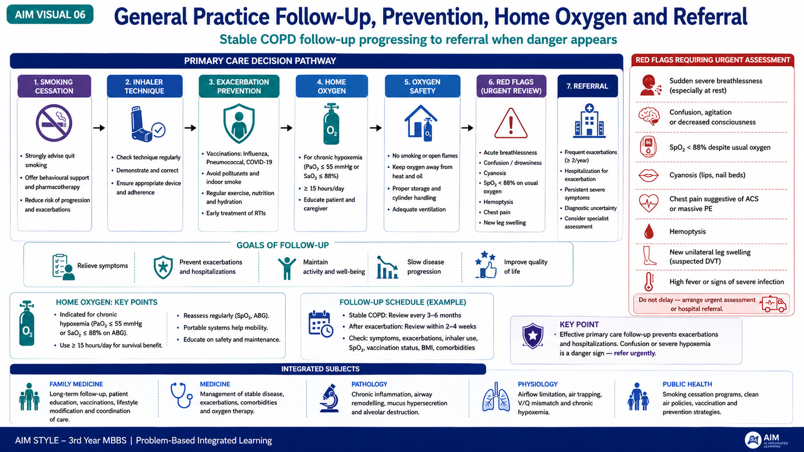
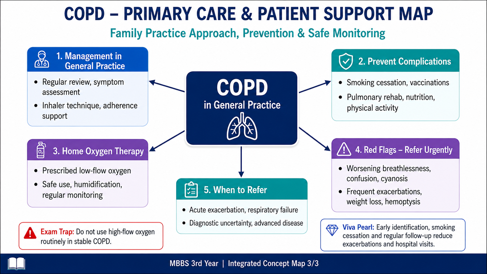
General practice has an important role because COPD requires continuous care rather than treatment only during acute attacks. The family physician helps reduce ongoing exposure, checks whether inhaled treatment is being used correctly, recognizes worsening disease, promotes prevention and identifies patients who need specialist or emergency assessment.
Routine COPD management in general practice
- Support smoking cessation and avoidance of respiratory irritants.
- Review severity and progression of breathlessness, cough and sputum production.
- Ask about exacerbations, hospital visits and limitation of daily activities.
- Review adherence and check inhaler technique.
- Encourage appropriate physical activity and pulmonary rehabilitation when indicated.
- Promote appropriate respiratory infection prevention and vaccination according to standard clinical practice.
- Assess oxygenation when disease becomes advanced or hypoxemia is suspected.
Prevention of complications
Complications are less likely when continuing lung injury is reduced and deterioration is recognized early. Smoking cessation, adherence to prescribed treatment, correct inhaler technique, physical conditioning, vaccination and early review during important symptom worsening all contribute to safer long-term care.
Home oxygen therapy
Patients who meet clinical indications for oxygen therapy may receive oxygen at home through an appropriate delivery system. The exact system depends on whether oxygen is needed mainly in the home or during mobility.
- Oxygen concentrator: extracts oxygen from room air and is suitable for regular home use when a continuous supply is required.
- Compressed oxygen cylinder: stores a finite oxygen supply and may be used as a stationary or portable source depending on the system.
- Portable oxygen system: allows selected patients requiring supplementary oxygen to remain mobile outside a fixed home location.
- Nasal cannula: is a commonly used interface for delivering low-flow supplementary oxygen.
Red flags requiring urgent assessment or referral
A patient with COPD should not automatically be assumed to have a routine exacerbation whenever symptoms worsen. Important warning features may indicate respiratory failure or another serious cardiopulmonary problem.
- Severe or rapidly worsening breathlessness.
- New or marked cyanosis.
- Confusion, drowsiness or reduced consciousness.
- Inability to speak normally because of breathlessness.
- New significant chest pain.
- Hemoptysis requiring evaluation.
- Suspected pneumothorax or other acute complication.
- Persistent or worsening hypoxemia.
- Failure to improve with appropriate initial treatment.
Specialist referral should also be considered when the diagnosis remains uncertain, symptoms remain poorly controlled despite appropriate treatment, exacerbations are recurrent, advanced respiratory impairment develops, or assessment for long-term oxygen or other specialized care is required.
Integrated Mechanism Flow
Important Comparison — Emphysema and Chronic Bronchitis
| Feature | Emphysema | Chronic Bronchitis |
|---|---|---|
| Basis of definition | Pathological | Clinical |
| Main lesion | Destruction and enlargement of distal airspaces | Mucus hypersecretion and inflamed narrowed airways |
| Important mechanism | Protease excess and oxidative injury | Chronic irritation causing mucus gland and goblet-cell changes |
| Major functional effect | Loss of elastic recoil and expiratory airway collapse | Mucus plugging and small-airway obstruction |
| Prominent symptom tendency | Progressive dyspnea | Chronic productive cough |
⭐ AIM High-Yield Review
- COPD produces persistent expiratory airflow limitation because of airway disease, alveolar destruction, or both.
- Important obstructive pulmonary diseases include chronic bronchitis, emphysema, asthma and bronchiectasis.
- ⭐ Cigarette smoking is the major preventable risk factor for COPD.
- Emphysema means permanent enlargement of distal airspaces with destruction of alveolar walls.
- Centriacinar emphysema is strongly associated with smoking; panacinar emphysema is classically linked with alpha-1 antitrypsin deficiency.
- ⭐ Emphysema develops through protease–antiprotease and oxidant–antioxidant imbalance.
- Loss of alveolar septa reduces elastic recoil, allowing small-airway collapse and air trapping during expiration.
- Chronic bronchitis is clinically defined by productive cough for at least three months in each of two consecutive years after other causes are excluded.
- Its major pathological changes are mucus gland enlargement, goblet-cell increase, inflammation and narrowing of small airways.
- ⭐ Progressive dyspnea, chronic cough and sputum production are major clinical features of COPD.
- Spirometry is the key investigation for objectively demonstrating persistent airflow obstruction.
- Management combines smoking cessation, bronchodilators, selected anti-inflammatory therapy, pulmonary rehabilitation and prevention of exacerbations.
- Long-term oxygen therapy is intended for appropriately assessed patients with persistent severe resting hypoxemia when clinically stable.
- Home oxygen requires strict avoidance of smoking, flames and ignition sources.
- ⭐ Marked cyanosis, confusion, severe respiratory distress or suspected respiratory failure requires urgent assessment.
Chronic Obstructive Pulmonary Disease and Emphysema
Watch the topic video to reinforce the major concepts of COPD, emphysema, chronic bronchitis, diagnosis and management.