
A. Pulmonary Thromboembolism, Hemorrhage and Infarction
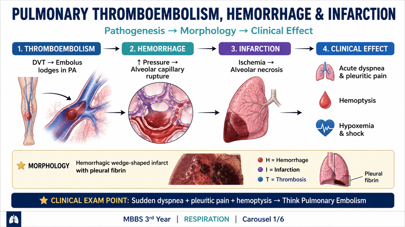
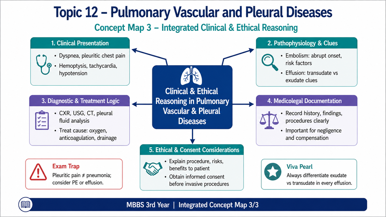
Pulmonary thromboembolism occurs when a thrombus formed elsewhere in the circulation travels to the lungs and obstructs a pulmonary artery or one of its branches. Most pulmonary thromboemboli arise from deep venous thrombosis of the lower limbs. Their effects vary greatly: a small embolus may produce little damage, while a large embolus can suddenly obstruct a major part of the pulmonary circulation and cause acute right ventricular failure or death.
Source and Pathogenesis
Most clinically important emboli originate from thrombi in the deep veins of the lower limbs, particularly the larger proximal veins. A portion of the thrombus may detach, pass through the inferior vena cava and right side of the heart, and finally enter the pulmonary arterial circulation.
Obstruction reduces perfusion to ventilated alveoli and therefore disturbs normal ventilation-perfusion matching. At the same time, obstruction of a large part of the pulmonary vascular bed suddenly increases the resistance against which the right ventricle must pump.
Effects of Pulmonary Emboli
The consequences depend mainly on the size and number of emboli, the patient’s cardiopulmonary reserve, and the adequacy of the bronchial arterial circulation.
- Small emboli may be clinically silent and may later undergo organization.
- Medium-sized emboli may cause pulmonary hemorrhage or pulmonary infarction.
- Large emboli may obstruct major pulmonary arteries and cause acute right-heart strain, cardiovascular collapse or sudden death.
- A large embolus lying across the bifurcation of the main pulmonary artery is called a saddle embolus.
- Repeated emboli can progressively reduce the pulmonary vascular bed and contribute to pulmonary hypertension.
Pulmonary Hemorrhage versus Pulmonary Infarction
The lung has a dual blood supply from the pulmonary and bronchial circulations. Therefore, blockage of a pulmonary arterial branch does not always cause tissue necrosis. Bronchial arterial blood may continue to supply the affected region. An embolus may therefore produce pulmonary hemorrhage without infarction. Blood enters the alveolar spaces, but the underlying lung tissue remains viable. Infarction is more likely when the additional bronchial blood supply is inadequate, particularly in patients with poor cardiovascular or pulmonary reserve.
Morphology of Pulmonary Infarction
Pulmonary infarcts are usually hemorrhagic and wedge-shaped. The wedge shape reflects the distribution of blood flow through an obstructed arterial branch. The broad base usually lies against the pleural surface, while the apex points toward the obstructed vessel.
- Usually peripheral.
- Wedge-shaped.
- Broad base toward the pleura.
- Initially dark red and hemorrhagic.
- Later becomes paler and may heal by fibrosis.
- Ischemic necrosis of alveolar walls.
- Marked hemorrhage into alveolar spaces.
- Inflammation develops at the margins.
- Organization and fibrosis occur during healing.
When the infarct extends to the pleural surface, pleural irritation produces pleuritic chest pain. Blood entering the airways may produce hemoptysis. Thus, the morphology of the infarct directly explains these clinical features.
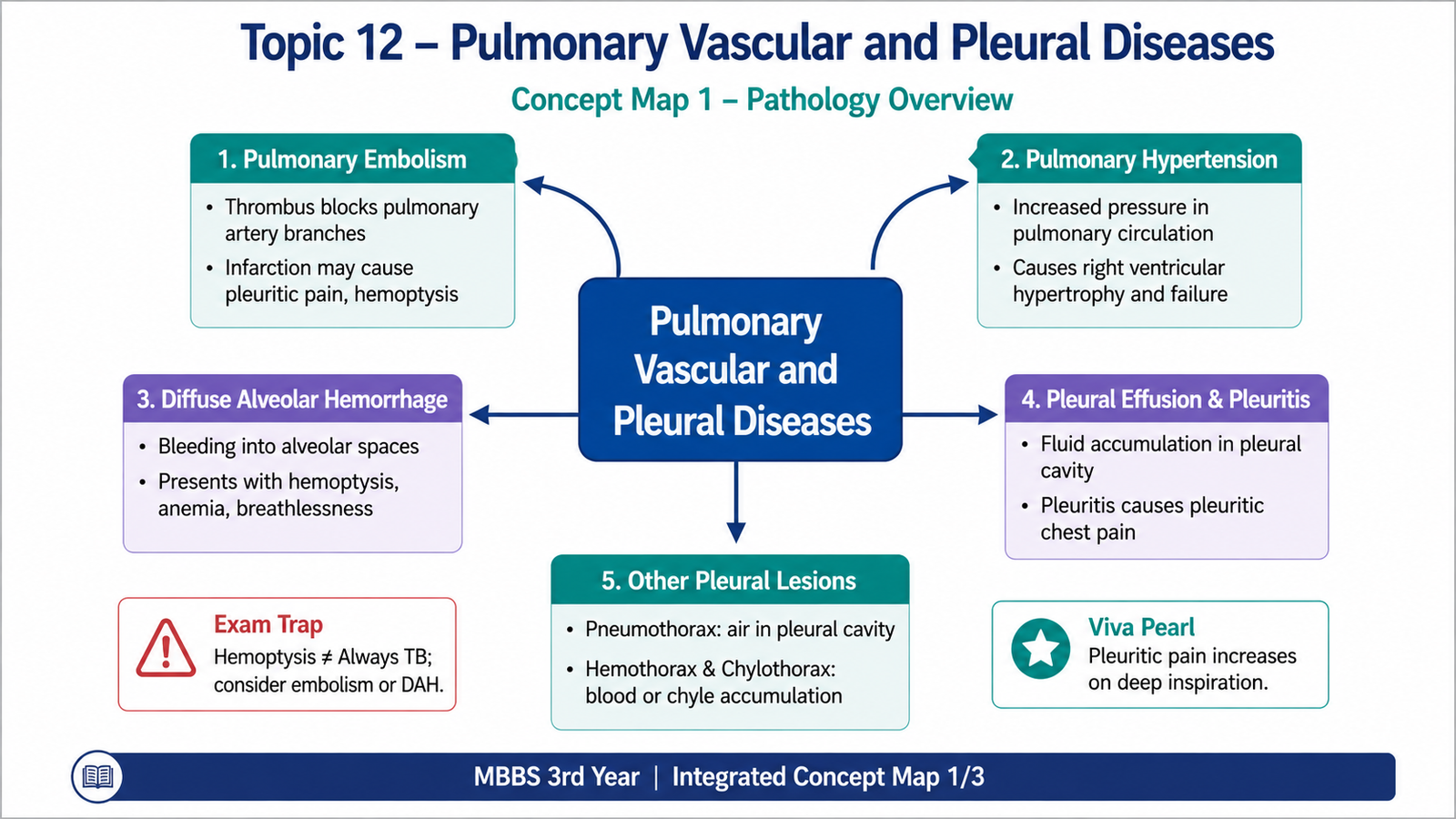
B. Pulmonary Hypertension
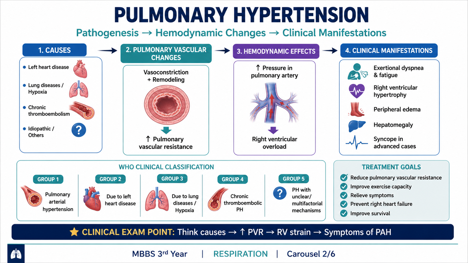
Pulmonary hypertension is an abnormal elevation of pressure within the pulmonary circulation. Because the normal pulmonary circulation is a low-pressure system, a persistent increase in pulmonary vascular resistance places an abnormal pressure load on the right ventricle. Over time, both the pulmonary arteries and the right side of the heart undergo structural changes.
How Pulmonary Hypertension Develops
Pulmonary arterial pressure can increase through several mechanisms. The common final pathway is an increase in resistance to blood flow through the pulmonary vascular bed or a sustained increase in pressure or flow within the pulmonary circulation.
- Chronic hypoxia causes pulmonary vasoconstriction and, when persistent, promotes vascular remodeling.
- Loss or obstruction of pulmonary vessels reduces the available vascular bed and increases resistance.
- Recurrent pulmonary thromboemboli may progressively obstruct pulmonary arteries.
- Chronic lung disease may produce hypoxia and destruction of pulmonary vessels.
- Increased pulmonary blood flow can place chronic stress on pulmonary arteries.
- Left-sided cardiac disease may increase pressure transmitted backward into the pulmonary circulation.
- Some disorders primarily affect the pulmonary arterial vessels themselves.
Vascular Morphology
Persistent elevation of pulmonary pressure causes structural remodeling of pulmonary arteries and arterioles. These changes initially develop as an adaptation to abnormal hemodynamic stress, but they eventually narrow the vascular lumen and further increase resistance.
- Medial hypertrophy of muscular pulmonary arteries and arterioles.
- Intimal thickening and fibrosis, producing progressive luminal narrowing.
- Muscularization of smaller arterioles that normally contain little smooth muscle.
- In severe pulmonary arterial hypertension, complex plexiform lesions may develop.
This creates a vicious cycle: increased pressure damages and remodels pulmonary vessels, while the narrowed vessels further increase pulmonary vascular resistance.
Effect on the Right Ventricle
The right ventricle must generate greater pressure to move blood through the high-resistance pulmonary circulation. Initially it responds by hypertrophy. If the pressure load continues, compensation may eventually fail and right-sided cardiac failure may develop.
C. Diffuse Alveolar Hemorrhage Syndromes
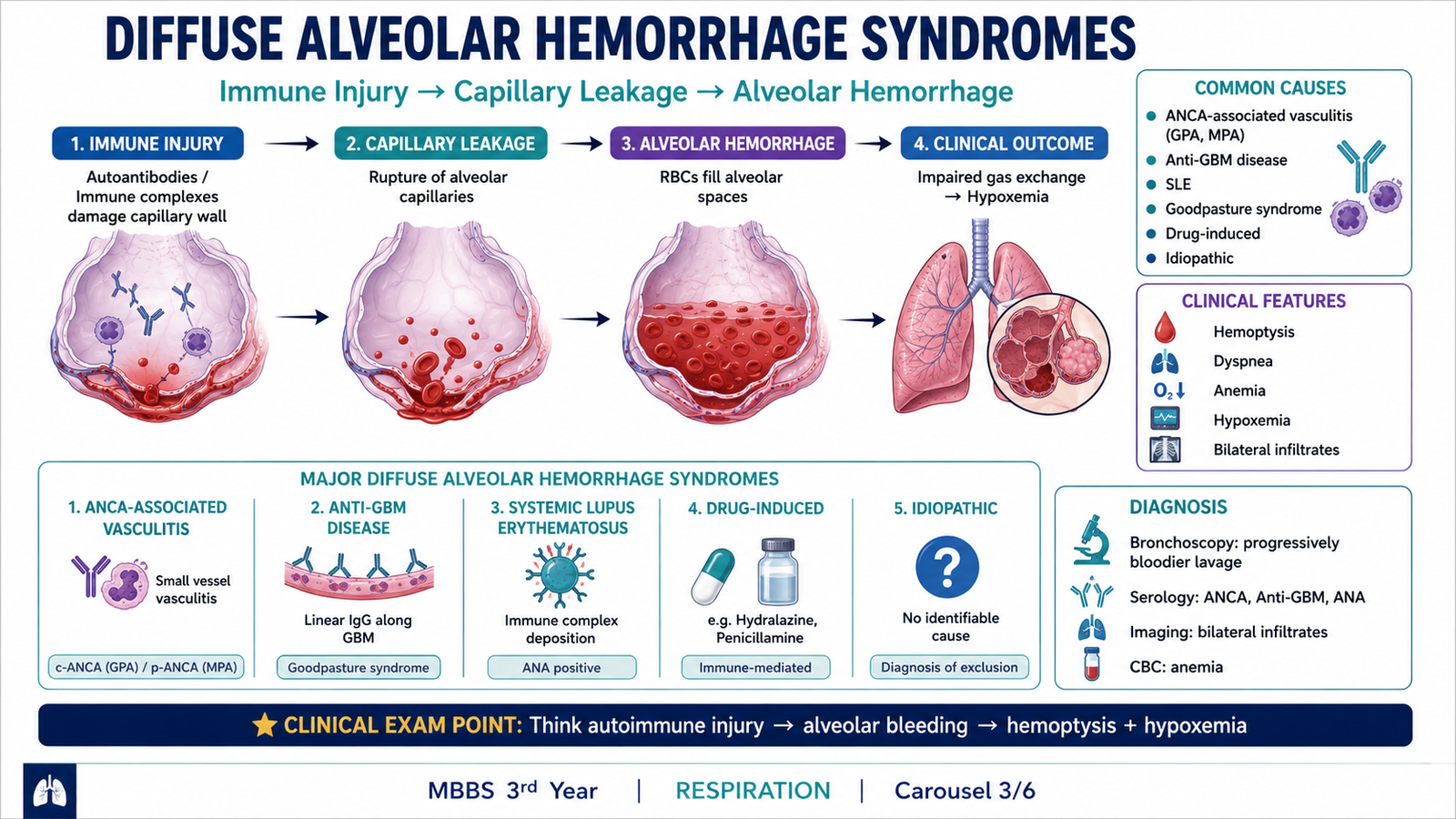
Diffuse alveolar hemorrhage refers to widespread bleeding into the alveolar spaces because of injury to the pulmonary microvasculature. Unlike a localized area of hemorrhage around a pulmonary infarct, diffuse alveolar hemorrhage may involve large areas of both lungs and can significantly interfere with gas exchange.
Pathogenesis
The key event is damage to small pulmonary vessels, particularly alveolar capillaries. In many important syndromes, the damage is immune-mediated. Inflammatory injury may produce pulmonary capillaritis, in which inflammatory cells damage capillary walls and allow red blood cells to escape into the alveolar spaces.
Important Disease Patterns
Diffuse alveolar hemorrhage may occur in immune-mediated diseases that injure pulmonary capillaries. Some of these disorders also affect the kidneys because pulmonary alveolar capillaries and renal glomerular capillaries may be targeted by related immune mechanisms. One important example is anti-glomerular basement membrane antibody disease. Antibodies directed against basement membrane components can damage both pulmonary alveolar capillaries and renal glomeruli. The patient may therefore develop pulmonary hemorrhage together with glomerular disease. Diffuse alveolar hemorrhage can also occur in systemic small-vessel vasculitic disorders in which inflammation directly damages pulmonary microvessels.
Morphology
- Numerous red blood cells fill alveolar spaces.
- Alveolar septa may show capillary injury and inflammation.
- Macrophages ingest extravasated red blood cells.
- Iron from hemoglobin is stored as hemosiderin within macrophages.
- Repeated hemorrhage therefore leads to hemosiderin-laden macrophages.
Because alveoli become filled with blood instead of air, the effective surface available for gas exchange decreases. This explains the development of breathlessness and impaired oxygenation.
D. Pleural Effusion and Pleuritis
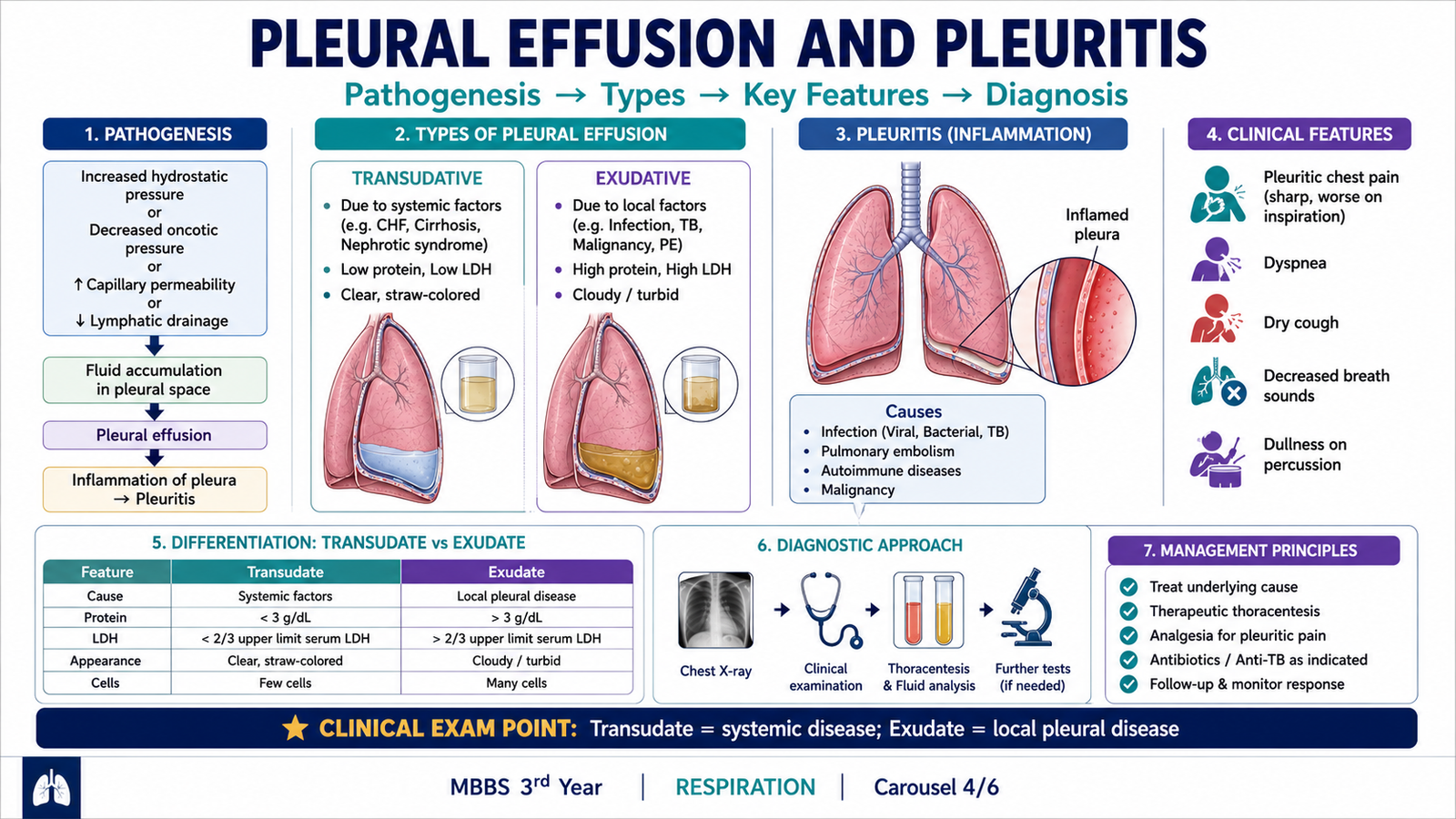
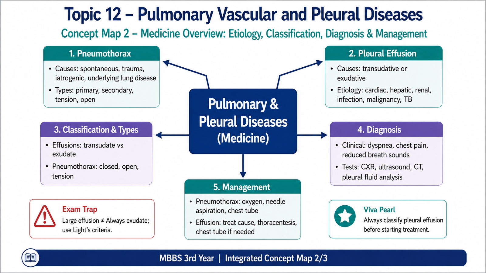
The pleural cavity normally contains only a thin film of fluid that allows the visceral and parietal pleura to move smoothly against each other during respiration. A pleural effusion occurs when an abnormal amount of fluid accumulates in this space. The most important first distinction is whether the fluid is a transudate or an exudate, because each develops through a different mechanism.
Transudative Pleural Effusion
A transudate develops because of a disturbance in systemic pressure forces controlling fluid movement, rather than primary inflammation of the pleura. The two major mechanisms are increased hydrostatic pressure and decreased plasma oncotic pressure.
Congestive cardiac failure is an important cause because increased vascular hydrostatic pressure pushes fluid out of the circulation. Conditions producing marked reduction in plasma protein can also favor movement of fluid into the pleural space because plasma oncotic pressure falls.
Exudative Pleural Effusion
An exudate develops when disease directly affects the pleura or nearby vessels. Inflammation increases vascular permeability, allowing protein-rich fluid and inflammatory cells to escape into the pleural cavity. Obstruction of lymphatic drainage can also cause an exudative effusion because pleural fluid cannot be removed normally.
Important causes include pleural infection, malignancy, inflammatory disease and pulmonary infarction.
Differentiating Transudates from Exudates
The basic distinction is based on the mechanism of fluid formation and the composition of the pleural fluid. A transudate is usually relatively low in protein because the pleural vessels are not primarily inflamed. An exudate contains more protein and cellular material because vascular permeability is increased or local lymphatic drainage is impaired.
| Feature | Transudate | Exudate |
|---|---|---|
| Main mechanism | Altered systemic hydrostatic or oncotic pressure | Local inflammation, vascular injury or impaired lymphatic drainage |
| Protein content | Relatively low | Relatively high |
| Cells | Usually few | Often increased |
| Typical settings | Heart failure; reduced plasma oncotic pressure | Infection; malignancy; inflammation; pulmonary infarction |
Pleuritis
Pleuritis means inflammation of the pleural surfaces. It may occur when inflammatory disease in the lung extends to the pleura, as can happen with pulmonary infection or infarction. The normally smooth pleural surfaces become inflamed and rough. As the visceral and parietal pleura move against each other during breathing, this irritation produces characteristic sharp pleuritic pain that worsens with inspiration. Pleural inflammation may be serous, fibrinous, purulent or hemorrhagic depending on its cause. Fibrinous inflammation may organize and produce fibrous adhesions between the pleural surfaces.
E. Pneumothorax — Etiology, Classification, Diagnosis and Management
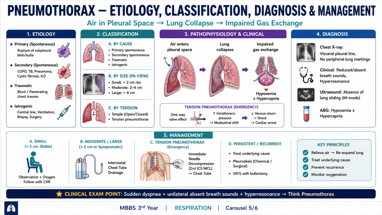
A pneumothorax is the presence of air within the pleural cavity. Normally, negative intrapleural pressure helps keep the lung expanded against the chest wall. When air enters the pleural space, this pressure relationship is lost and the elastic lung recoils inward. The resulting lung collapse may range from small and clinically mild to severe and immediately life-threatening.
Etiology and Classification
Pneumothorax is classified according to how air enters the pleural cavity and whether underlying lung disease is present.
- Primary spontaneous pneumothorax: occurs without clinically apparent underlying lung disease and is commonly associated with rupture of small subpleural blebs.
- Secondary spontaneous pneumothorax: occurs in a patient with underlying lung disease. Because pulmonary reserve is already reduced, it may cause more severe symptoms.
- Traumatic pneumothorax: follows blunt or penetrating chest injury that allows air to enter the pleural space.
- Iatrogenic pneumothorax: occurs as a complication of a medical procedure that inadvertently breaches the pleura.
Tension Pneumothorax
A tension pneumothorax develops when a one-way valve mechanism allows air to enter the pleural cavity but prevents adequate escape. With each breath, more air becomes trapped and intrapleural pressure rises progressively.
Increasing intrathoracic pressure compresses major veins and reduces the return of blood to the heart. Cardiac output falls, so an untreated tension pneumothorax can rapidly progress to severe hypotension and circulatory collapse.
Clinical Features
The usual presentation is sudden pleuritic chest pain accompanied by breathlessness. Physical findings occur because the affected lung has partially or completely separated from the chest wall.
- Sudden pleuritic chest pain.
- Acute shortness of breath.
- Reduced expansion of the affected side of the chest.
- Reduced or absent breath sounds on the affected side.
- Hyperresonant percussion note over the affected area.
In tension pneumothorax, respiratory distress becomes severe and may be accompanied by tachycardia, hypotension and other signs of impaired venous return and reduced cardiac output.
Diagnosis
Diagnosis is based on the clinical features and, in stable patients, appropriate chest imaging. On a chest radiograph, a visible visceral pleural line may separate the collapsed lung from the pleural air. Normal pulmonary vascular markings are absent beyond this line because this region represents pleural air rather than lung tissue.
Basic Management Principles
Management depends on the size of the pneumothorax, severity of symptoms, underlying lung disease and the presence or absence of tension physiology. The basic aim is to remove pleural air when necessary, allow the lung to re-expand and treat the underlying cause.
- Small and clinically stable pneumothorax: may be managed with observation in appropriately selected patients.
- Larger or symptomatic pneumothorax: generally requires evacuation of pleural air by an appropriate pleural drainage procedure.
- Tension pneumothorax: requires immediate emergency decompression followed by definitive pleural drainage.
- Secondary or recurrent pneumothorax: requires assessment and management of the underlying lung disorder or cause.
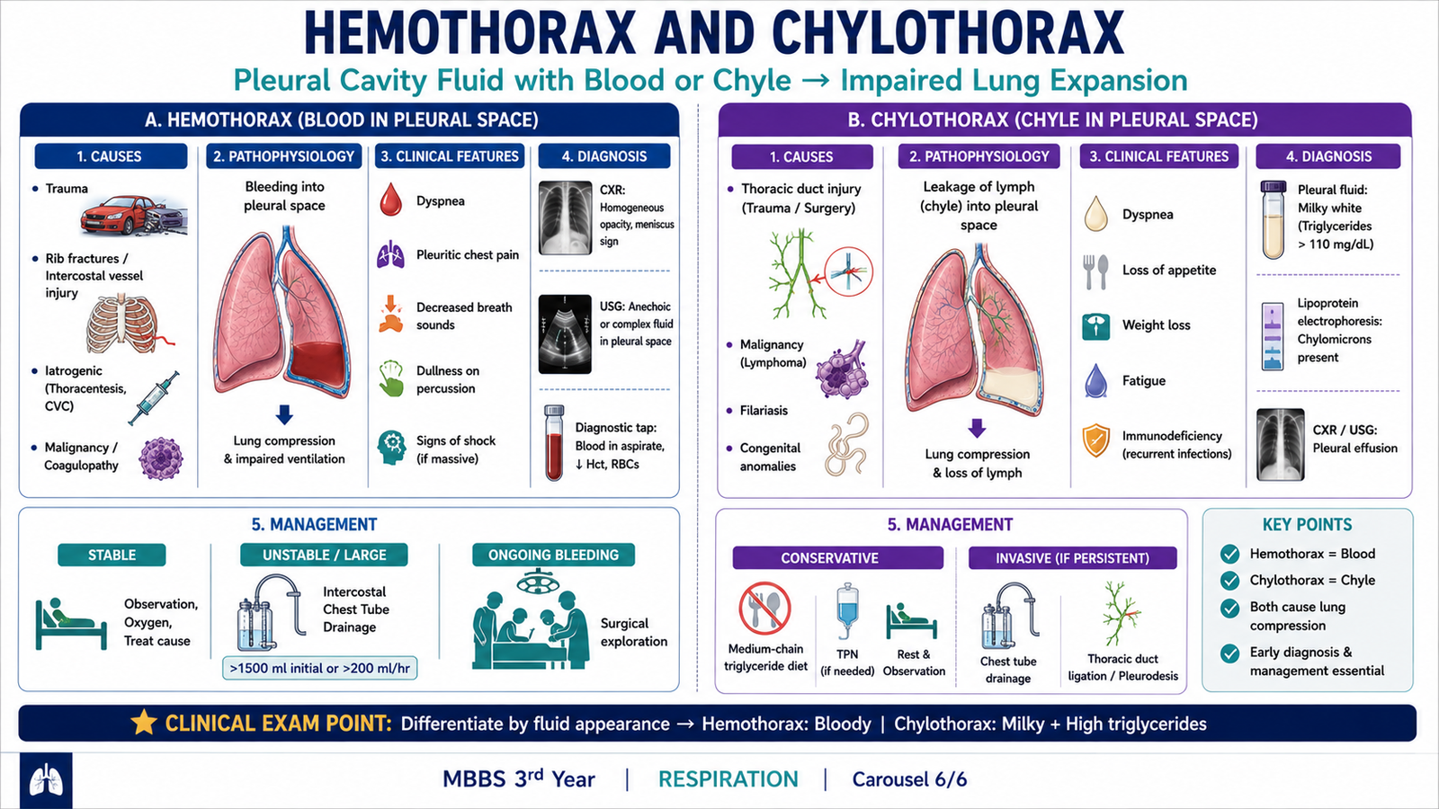
F. Hemothorax and Chylothorax
Pleural collections are not limited to serous transudates and exudates. Two important pathological collections are hemothorax, in which blood accumulates within the pleural cavity, and chylothorax, in which lymphatic chyle enters the pleural space. Recognizing what has accumulated helps identify the underlying mechanism.
Hemothorax
Hemothorax is the accumulation of blood within the pleural cavity. It commonly follows traumatic injury to the chest or disruption of an intrathoracic blood vessel.
A large volume of blood in the pleural cavity can compress the underlying lung and interfere with ventilation. At the same time, substantial intrathoracic blood loss may reduce circulating blood volume and produce hemodynamic instability.
Chylothorax
Chylothorax is the accumulation of chyle within the pleural cavity. Chyle is lymphatic fluid containing absorbed dietary lipids, particularly chylomicrons, and is transported through the thoracic duct. Disruption or obstruction of the thoracic duct allows chyle to escape into the pleural space. Traumatic damage to the thoracic duct and diseases that obstruct or involve the duct are therefore important mechanisms.
Because chyle contains a large amount of lipid in the form of chylomicrons, the pleural fluid classically has a milky appearance.
| Feature | Hemothorax | Chylothorax |
|---|---|---|
| Pleural content | Blood | Chyle |
| Basic mechanism | Bleeding into the pleural cavity | Thoracic duct disruption or obstruction |
| Appearance | Bloody | Classically milky |
| Major consequence | Lung compression and possible blood-volume loss | Lung compression with loss of lymphatic fluid |
Pleural Effusion & Pneumothorax
Use this video after reading the core learning material to reinforce pleural effusion, pneumothorax, clinical recognition and basic management concepts.