
📖 Step 2 — Learning Material
🔹 1️⃣ Introduction
Fetal circulation is a specialized circulatory system that allows the fetus to survive inside the uterus where lungs are nonfunctional. Oxygen and nutrients are supplied by the placenta through fetal blood vessels. To adapt to intrauterine life, the fetus possesses temporary vascular shunts that bypass the lungs and partially bypass the liver.
After birth, dramatic physiological circulatory changes occur when the newborn begins breathing. These changes convert fetal circulation into adult-type circulation by closure of fetal shunts. Failure of normal developmental processes may produce congenital heart diseases, many of which present with cyanosis, breathlessness, murmurs, or circulatory abnormalities.
Understanding fetal circulation is essential for understanding neonatal adaptation, congenital cardiac defects, and pediatric cardiovascular disorders. This topic integrates embryology, physiology, and clinical medicine and forms the basis for understanding many common congenital heart conditions.
🔹 2️⃣ Foundation Concepts
Key Definitions
- Fetal circulation: Specialized circulation present before birth.
- Placenta: Organ that exchanges oxygen, nutrients, and waste between mother and fetus.
- Shunt: A passage that diverts blood from one pathway to another.
- Foramen ovale: Opening between right and left atria in fetal heart.
- Ductus arteriosus: Vessel connecting pulmonary trunk to aorta.
- Ductus venosus: Vessel connecting umbilical vein to inferior vena cava.
- Congenital heart disease (CHD): Structural heart defect present at birth.
- Cyanosis: Bluish discoloration caused by reduced oxygenation.
- Tetralogy of Fallot: Cyanotic congenital heart disease with four abnormalities.
- Postnatal circulation: Adult-type circulation established after birth.
Essential Terminology
- Umbilical vein
- Umbilical arteries
- Right-to-left shunt
- Pulmonary circulation
- Systemic circulation
- Septal defects
- Cyanotic defects
- Acyanotic defects
Basic Overview
- Fetal lungs are nonfunctional before birth.
- Placenta acts as the organ of respiration.
- Three fetal shunts help bypass lungs and liver:
- Ductus venosus
- Foramen ovale
- Ductus arteriosus
- At birth:
- Lungs expand
- Pulmonary resistance falls
- Placental circulation stops
- Fetal shunts close
- Abnormal closure or abnormal development causes congenital heart disease.
🔹 3️⃣ Core Learning — Curriculum Coverage
Fetal Circulation and Fetal Shunts
🧠 CORE
- Placenta supplies oxygenated blood.
- Umbilical vein carries oxygenated blood to fetus.
- Umbilical arteries carry deoxygenated blood to placenta.
- Fetal lungs are collapsed and have high resistance.
- Three major fetal shunts:
- Ductus venosus
- Foramen ovale
- Ductus arteriosus
- Most oxygenated blood reaches brain and heart.
- Pulmonary blood flow is minimal before birth.
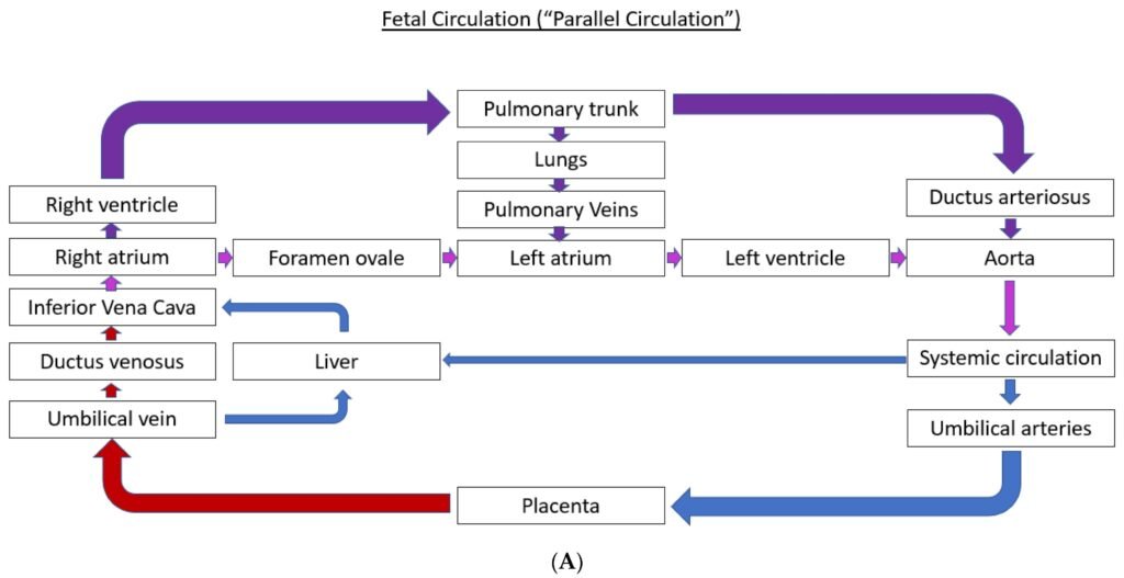
- Right and left ventricles work in parallel during fetal life.
🔬 CONCEPT EXPLAINED
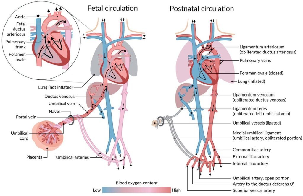
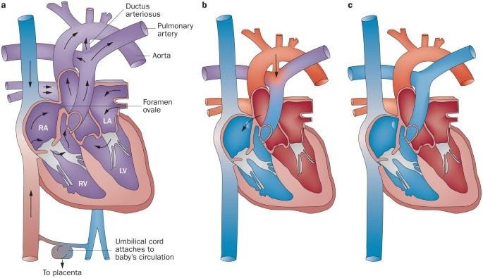
The fetus cannot use lungs for oxygen exchange because lungs are filled with fluid and alveoli are collapsed. Therefore, the placenta performs gas exchange. Oxygenated blood returns from placenta through the umbilical vein.
A portion of this blood bypasses the liver through the ductus venosus and enters the inferior vena cava. Blood then enters the right atrium.
Because pulmonary resistance is very high, most blood bypasses the lungs through the foramen ovale, moving from right atrium to left atrium. This allows oxygen-rich blood to enter systemic circulation and supply the brain and heart.
Blood entering the right ventricle is pumped into the pulmonary trunk. Since lungs are nonfunctional, most blood passes through the ductus arteriosus into the descending aorta instead of entering pulmonary circulation.
This arrangement ensures maximum oxygen delivery to vital organs while bypassing nonfunctional lungs.
⚠️ CLINICAL IMPORTANCE
- Failure of fetal shunts to close leads to congenital defects.
- Abnormal fetal circulation may cause neonatal cyanosis.
- Premature infants commonly develop patent ductus arteriosus.
- Understanding fetal circulation is essential in neonatal resuscitation and pediatric cardiology.
Postnatal Circulatory Changes
🧠 CORE
- First breath expands lungs.
- Pulmonary vascular resistance decreases.
- Pulmonary blood flow increases.
- Placental circulation stops after clamping umbilical cord.
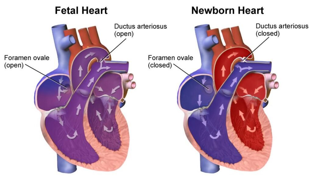
- Left atrial pressure becomes greater than right atrial pressure.
- Functional closure of foramen ovale occurs.
- Ductus arteriosus constricts after birth.
- Ductus venosus closes after birth.
- Adult circulation becomes established.
🔬 CONCEPT EXPLAINED
At birth, the newborn takes the first breath, causing lung expansion. Pulmonary vessels dilate and pulmonary resistance falls dramatically. Increased pulmonary blood flow returns more blood to the left atrium.
Simultaneously, clamping of the umbilical cord removes placental circulation and reduces venous return to the right atrium. Consequently, left atrial pressure exceeds right atrial pressure, pushing the septum primum against septum secundum and functionally closing the foramen ovale.
Increased oxygen concentration and reduced prostaglandins cause constriction of the ductus arteriosus. The ductus venosus also closes because umbilical blood flow ceases.
These changes transform fetal parallel circulation into adult serial circulation.
⚠️ CLINICAL IMPORTANCE
- Delayed closure of fetal shunts may produce murmurs and cyanosis.
- Persistent pulmonary hypertension prevents normal neonatal adaptation.
- Failure of circulatory transition can cause severe neonatal distress.
- Prematurity increases risk of patent ductus arteriosus.
Congenital Heart Disease (CHD)
🧠 CORE
- CHD results from abnormal embryological development.
- May involve septa, valves, vessels, or chambers.
- Classified into:
- Cyanotic defects
- Acyanotic defects
- Left-to-right shunts usually produce acyanotic disease.
- Right-to-left shunts commonly cause cyanosis.
- Symptoms include:
- Breathlessness
- Cyanosis
- Poor feeding
- Failure to thrive
- Murmurs
🔬 CONCEPT EXPLAINED
During embryonic development, the heart undergoes folding, septation, and vessel remodeling. Disturbance of these developmental steps causes congenital abnormalities.
Acyanotic defects usually allow oxygenated blood from the left side of the heart to pass into the right side, increasing pulmonary blood flow without initially causing cyanosis.
Cyanotic defects reduce oxygen delivery to systemic circulation due to right-to-left shunting or abnormal vessel arrangement.
Severity depends on the size of the defect and degree of circulatory disturbance.
⚠️ CLINICAL IMPORTANCE
- Congenital heart disease is among the most common birth defects.
- Severe defects present soon after birth.
- Chronic hypoxia may cause clubbing and developmental delay.
- Early diagnosis improves survival.
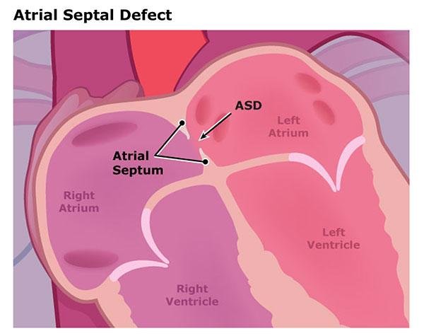
Atrial Septal Defect (ASD)
🧠 CORE
- Opening in interatrial septum.
- Usually due to defective septum formation.
- Produces left-to-right shunt.
- Commonly acyanotic.
- Increases right atrial and right ventricular workload.
- Pulmonary blood flow increases.
🔬 CONCEPT EXPLAINED
Normally, septum primum and septum secundum fuse after birth. Failure of proper fusion creates ASD.
Because left atrial pressure exceeds right atrial pressure, oxygenated blood moves from left atrium to right atrium. This increases pulmonary circulation and causes enlargement of right-sided chambers over time.
Small ASDs may remain asymptomatic, while large defects can produce exercise intolerance and recurrent respiratory infections.
⚠️ CLINICAL IMPORTANCE
- Fixed split second heart sound is characteristic.
- Long-standing ASD may cause pulmonary hypertension.
- Eisenmenger syndrome may develop in severe untreated cases.
Ventricular Septal Defect (VSD)
🧠 CORE
- Most common congenital heart defect.
- Opening in interventricular septum.
- Usually membranous septum affected.
- Produces left-to-right shunt.
- Pulmonary blood flow increases significantly.
- Large defects strain left ventricle.
🔬 CONCEPT EXPLAINED
During development, membranous and muscular septa normally fuse. Failure leads to VSD.
High-pressure blood from left ventricle enters right ventricle during systole. Increased pulmonary circulation returns excess blood to left atrium and left ventricle, causing volume overload.
Small VSDs may close spontaneously, while large defects produce heart failure symptoms.
⚠️ CLINICAL IMPORTANCE
- Harsh pansystolic murmur is typical.
- Large VSD causes heart failure in infancy.
- Pulmonary hypertension may eventually reverse shunt direction.
Patent Ductus Arteriosus (PDA)
🧠 CORE
- Persistence of ductus arteriosus after birth.
- Connects aorta and pulmonary trunk.
- Produces left-to-right shunt.
- More common in premature infants.
- Pulmonary circulation increases excessively.
🔬 CONCEPT EXPLAINED
Normally, increased oxygen tension after birth causes closure of ductus arteriosus. Failure of closure keeps abnormal communication between aorta and pulmonary artery.
Because aortic pressure is higher, blood flows into pulmonary circulation. This overloads pulmonary vessels and left side of the heart.
⚠️ CLINICAL IMPORTANCE
- Continuous “machinery murmur” is classic.
- Severe PDA causes heart failure and pulmonary hypertension.
- Indomethacin may help close PDA in premature infants.
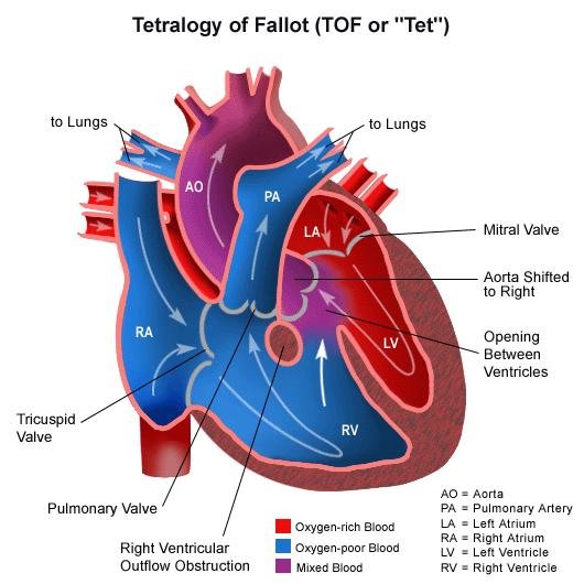
Tetralogy of Fallot
🧠 CORE
Tetralogy consists of four abnormalities:
- Pulmonary stenosis
- VSD
- Overriding aorta
- Right ventricular hypertrophy
- Most common cyanotic congenital heart disease.
- Causes right-to-left shunt.
- Produces severe cyanosis.
🔬 CONCEPT EXPLAINED
Abnormal neural crest migration causes unequal division of truncus arteriosus.
Pulmonary stenosis increases right ventricular pressure. Blood therefore passes through VSD into the overriding aorta, bypassing lungs and reducing oxygenation.
Chronic pressure overload causes right ventricular hypertrophy.
Children often squat after exertion because squatting increases systemic vascular resistance and temporarily reduces right-to-left shunting.
⚠️ CLINICAL IMPORTANCE
- Causes “blue baby” appearance.
- Cyanotic spells occur during crying or exertion.
- Severe hypoxia may impair growth and development.
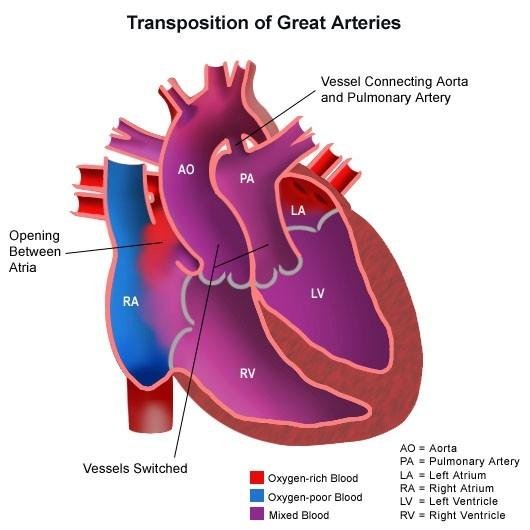
Transposition of Great Vessels
🧠 CORE
- Aorta arises from right ventricle.
- Pulmonary trunk arises from left ventricle.
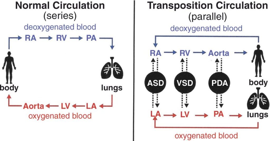
- Systemic and pulmonary circulations become separate.
- Severe cyanotic defect.
- Life depends on mixing of blood through shunts.
🔬 CONCEPT EXPLAINED
Failure of spiral septum formation causes abnormal arrangement of great vessels.
Deoxygenated blood continuously circulates through systemic circulation while oxygenated blood repeatedly circulates through lungs. Survival requires ASD, VSD, or PDA to permit mixing of blood.
This defect produces profound neonatal cyanosis soon after birth.
⚠️ CLINICAL IMPORTANCE
- Severe cyanosis appears immediately after birth.
- Requires urgent medical intervention.
- Prostaglandins may maintain PDA temporarily until surgery.
Vascular Developmental Anomalies
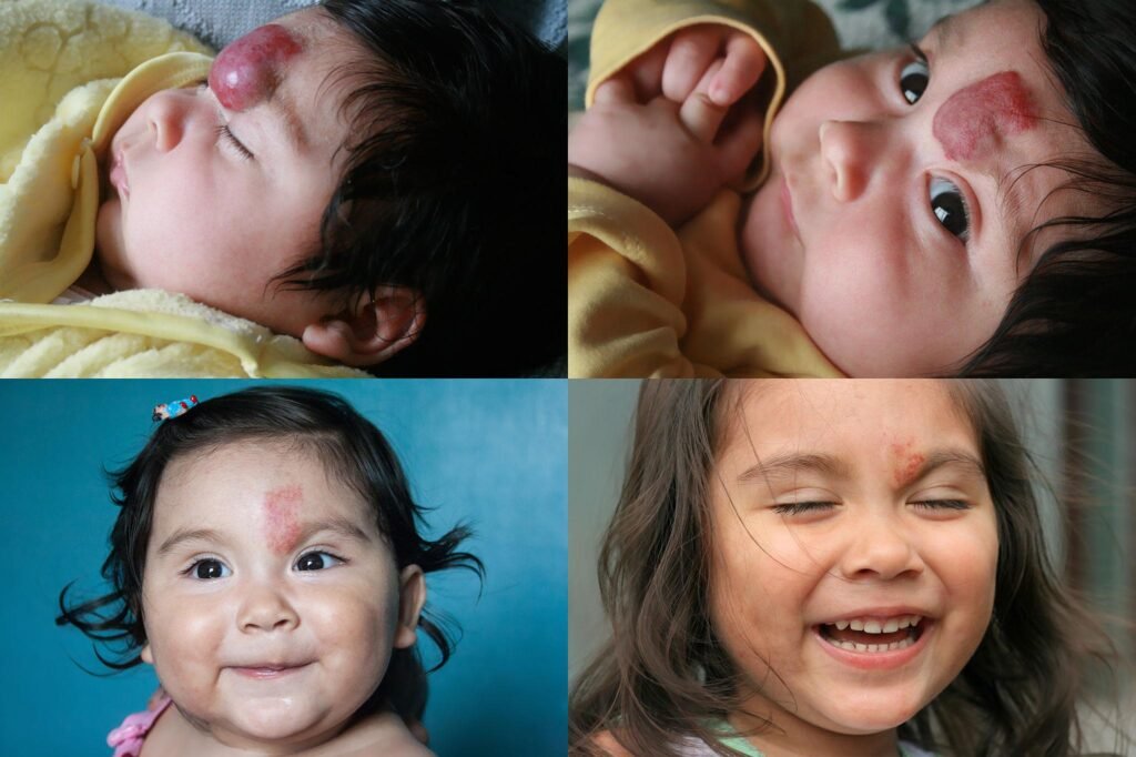
Hemangiomas
🧠 CORE
- Benign vascular tumors.
- Result from abnormal blood vessel proliferation.
- Common in infancy.
- Usually appear as red skin lesions.
🔬 CONCEPT EXPLAINED
Hemangiomas arise due to excessive endothelial cell proliferation during vascular development. Many enlarge during infancy and later regress spontaneously.
⚠️ CLINICAL IMPORTANCE
- Usually harmless.
- Large lesions may interfere with vision or breathing.
- Some require medical treatment.
Telangiectasia
🧠 CORE
- Persistent dilation of small blood vessels.
- Appears as red vascular markings.
- May be congenital or acquired.
🔬 CONCEPT EXPLAINED
Abnormal vessel wall development or dilation causes visible superficial vascular networks.
⚠️ CLINICAL IMPORTANCE
- May bleed easily.
- Can be associated with hereditary vascular disorders
⚙️ 4️⃣ Functional Flow
| Structure | Function | Outcome |
|---|---|---|
| Placenta | Gas exchange | Oxygen supply to fetus |
| Foramen ovale | Bypass lungs | Blood reaches systemic circulation rapidly |
| Ductus arteriosus | Diverts blood from lungs | Reduced pulmonary circulation |
| Ductus venosus | Bypass liver | Faster venous return |
| Postnatal lung expansion | Lowers pulmonary resistance | Establishes adult circulation |
| Septal integrity | Separates oxygenated and deoxygenated blood | Efficient oxygen delivery |
🩺 5️⃣ Clinical Correlation
Common Congenital Defects and Presentations
| Condition | Major Feature | Clinical Finding |
|---|---|---|
| ASD | Interatrial communication | Fixed split S2 |
| VSD | Interventricular communication | Pansystolic murmur |
| PDA | Persistent fetal shunt | Machinery murmur |
| Tetralogy of Fallot | Cyanotic defect | Blue baby |
| Transposition | Parallel circulation | Severe neonatal cyanosis |
Important Exam Correlations
- PDA is common in premature infants.
- VSD is the most common congenital heart defect.
- Tetralogy of Fallot is the most common cyanotic congenital heart disease.
- Squatting relieves cyanotic spells in Tetralogy of Fallot.
- Failure of fetal shunt closure causes abnormal circulation.
📌 6️⃣ Summary Points
- Placenta is the organ of respiration in fetus.
- Three fetal shunts are:
- Ductus venosus
- Foramen ovale
- Ductus arteriosus
- Pulmonary resistance is high before birth.
- First breath initiates postnatal circulatory changes.
- Closure of foramen ovale occurs due to pressure changes.
- PDA causes continuous machinery murmur.
- VSD is the most common congenital heart defect.
- Tetralogy of Fallot is the most common cyanotic congenital heart disease.
- Transposition of great vessels causes severe neonatal cyanosis.
- Right-to-left shunts produce cyanosis.
- Left-to-right shunts usually produce pulmonary overload.
- Congenital heart defects commonly present with breathlessness and cyanosis.